The Cytoplasmic Domain of L-Selectin Interacts with Cytoskeletal
Proteins Via ct-Actinin: Receptor Positioning in Microvilli Does Not
Require Interaction with et-Actinin
F r e d r i c k M. Pavalko,* D e n i s e M. Walker, * Lori G r a h a m * M i c h a e l Goheen,* Claire M. D o e r s c h u k , * a n d Geoffrey S. Kansas§
• Department of Physiology and Biophysics and ¢The Herman B. Wells Center for Pediatric Research, Section of Pulmonology, Department of Pediatrics, Indiana University School of Medicine, Indianapolis, IN 46202-5120; and § Department of
Microbiology-Immunology, Northwestern University Medical School, Chicago, IL 46202-5120
Abstract. The leukocyte adhesion molecule L-selectin mediates binding to lymph node high endothelial ven- ules (HEV) and contributes to leukocyte rolling on en- dothelium at sites of inflammation. Previously, it was shown that truncation of the L-selectin cytoplasmic tail by 11 amino acids abolished binding to lymph node HEV and leukocyte rolling in vivo, but the mo- lecular basis for that observation was not determined. This study examined potential interactions between L-selectin and cytoskeletal proteins. We found that the cytoplasmic domain of L-selectin interacts directly with the cytoplasmic actin-binding protein ot-actinin and forms a complex with vinculin and possibly talin. Solid phase binding assays using the full-length L-selectin cytoplasmic domain bound to microtiter wells demonstrated direct, specific, and saturable binding of purified ot-actinin to L-selectin (Kd = 550 nM), but no direct binding of purified talin or vincu- lin. Interestingly, talin potentiated binding of ot-actinin to the L-selectin cytoplasmic domain peptide despite the fact that direct binding of talin to L-selectin could not be measured. Vinculin binding to the L-selectin cytoplasmic domain peptide was detectable only in the
presence of ot-actinin. L-selectin coprecipitated with a complex of cytoskeletal proteins including ct-actinin and vinculin from cells transfected with L-selectin, consistent with the possibility that ot-actinin binds directly to L-selectin and that vinculin associates by binding to ot-actinin in vivo to link actin filaments to the L-selectin cytoplasmic domain. In contrast, a dele- tion mutant of L-selectin lacking the COOH-terminal
11 amino acids of the cytoplasmic domain failed to coprecipitate with ot-actinin or vinculin. Surprisingly, this mutant L-selectin localized normally to the microvillar projections on the cell surface. These data suggest that the COOH-terminal 11 amino acids of the L-selectin cytoplasmic domain are required for medi- ating interactions with the actin cytoskeleton via a complex of ct-actinin and vinculin, but that this por- tion of the cytoplasmic domain is not necessary for proper localization of L-selectin on the cell surface. Correct L-selectin receptor positioning is therefore insufficient for leukocyte adhesion mediated by L-selectin, suggesting that this adhesion may also re- quire direct interactions with the cytoskeleton.
YMPHOCYTE migration through lymphoid organs and leukocyte traffic into sites of inflammation are related processes that are both regulated principally at the level of leukocyte interactions with the lumenal surface of postcapillary venules. A number of leukocyte and endo- thelial cell surface molecules that participate in this interac- tion have been identified, including members of at least three
Please address all correspondence to E M. Pavalko; Department of Physi- ology and Biophysics, Indiana University School of Medicine, 635 Barn- hill Drive, Indianapolis, IN 46202-5120. Tel.: (317) 274-3140. Fax: (317) 274-3318.
gene families: the integrins, the immunoglobulin (Ig) super- family, and the selectins (for reviews see Springer, 1994; Butcher, 1991; Arnaout, 1993). Selectins mediate the initial phase of leukocyte recognition of endothelium, which usu- ally takes the form of rolling along the vessel wall at veloci- ties much slower than freely flowing blood elements (Law- rence and Springer, 1991; Bevilacqua and Nelson, 1993). In contrast, integrins mediate the subsequent firm arrest of the leukocytes to the endotheliurn, spreading on the endothelial surface, and transendothelial migration into the tissues (Smith, 1992). While the integrin and Ig families consists of many members and are broadly expressed by numerous cell types, there are only three known selectins, each of which
© The Rockefeller University Press, 0021-9525/95/05/1155/10 $2.130
exhibits a much narrower range of expression. In particular, L-selectin is expressed exclusively on leukocytes (Kansas et al., 1985; Tedder et al., 1990), E-selectin is expressed only on endothelium activated in vivo or in vitro by inflammatory stimuli such as TNF-ct, IL-1, or LPS (Bevilacqua et al.,
1987), and P-selectin is selectively expressed on both plate- lets and endothelium which have been activated in vitro by thrombin, histamine, or phorbol esters (Berman et al., 1986; McEver et al., 1989). This pattern of expression of the selec- tins is consistent with their specialized role in regulating the interaction of circulating elements of the blood with the vas- cular endothelium.
Although the selectins exhibit considerable homology in their extracellular regions, no homology exists between the cytoplasmic domains of different selectins, suggesting that distinct functions are encoded within these regions. Consis- tent with this idea, each of the selectin cytoplasmic tails is well conserved between different species (for review see Bevilacqua and Nelson, 1993). In addition, direct functional evidence supports this view. Sorting of newly synthesized P-selectin to Weibel-Palade bodies and t~-granules of plate- lets and endothelial cells and to granules of transfected AtT- 20 cells is mediated by the P-selectin cytoplasmic tail, and endocytosis of E-selectin by human endothelial cells may in- volve the E-selectin cytoplasmic tail (Disdier et al., 1992; Koedam et al., 1992; Eckhardt et al., 1992).
A precise function for the L-selectin cytoplasmic tail has not yet been established. However, truncation of the pre- dicted 17-amino acid cytoplasmic domain of L-selectin by 11 residues abolishes both binding to lymph node high en- dothelial venules (HEV) t and leukocyte rolling in vivo (Kansas et al., 1993). Because the lectin/ligand recognition activity of this mutant was preserved, the molecular basis for this phenotype was unclear. At least two possibilities must be considered. The observation that cytochalasin B, which blocks the function of actin microfilaments, also abolishes L-selectin-mediated adhesion without affecting lectin activ- ity (Kansas et al., 1993), suggests that interactions between one or more cytoskeletal proteins and the L-selectin cyto- plasmic tail are necessary for L-selectin function. A second, not mutually exclusive possibility is that the cytoplasmic do- main of L-selectin mediates the preferential localization of this receptor to the microvilli and ruffles on the surface of normal leukocytes (Picker et al., 1991; Erlandsen et al.,
1993; Bruce and Doerschuk, 1994), analogous to the sorting function of the P-selectin cytoplasmic domain, and that inap- propriate subcellular positioning is responsible for the defect exhibited by the L-selectin cytoplasmic domain truncation mutant.
We have investigated whether L-selectin binds to cyto- skeletal proteins using solid phase binding assays of purified cytoskeletal proteins to a peptide corresponding to the L-selectin cytoplasmic domain and coimmunoprecipitation experiments in vivo using cells transfected with wild-type and truncated L-selectin. In addition, the distribution of wild-type and truncated L-selectin has been examined by im- munoelectronic microscopy. The results indicate that the L-selectin cytoplasmic domain interacts with the cytoskele-
1. Abbreviation used in this paper: HEV, high endothelial venule.
ton via the actin-binding protein a-actinin, and that this in- teraction may also involve two other cytoskeletal proteins, vinculin and talin. These interactions, however, are not re- quired for localization of L-selectin to the microvillar projections.
Materials and Methods
Cell Culture
The mouse pre-B cell line 300.19 was transfected by electroporation with either full-length (LAM-1) or truncated human L-selectin cDNA (LAcyto, lacking the amino terminal 11 residues of the cytoplasmic domain) in the pZIPneoSV(X) vector as previously described (Kansas et al., 1993). Stable transfectants and mock-transfected cells (300.19) were maintained in sus- pension culture in RPMI-1640 medium containing 0.5 mg/ml G418 (geneti- cin, Sigma Immunochemicals, St. Louis, MO) and supplemented with 10% FBS and antibiotics at 37°C in a humidified 5% CO2 atmosphere. Cells were passaged approximately every 2 d at 1:10 dilution into fresh media.
Coimmunoprecipitation and Immunoblotting
300.19, 300.19/LAM-1, and 300.19/LAcyto cells were harvested during log phase of growth and washed in I-IBSS. Cells at a concentration of 5 × 10⁶/ml were extracted with 1 ml of coimmunoprecipitation lysis buffer (1% TX-100, 150 mM NaC1, 10 mM Tris-HCl, pH 7.6, 1 mM CaCi2, 1 mM MgC12, 0.01% NAN3, 20 mM DNaseI, 1 mM aprotinin, 10 mM benzami- dine, and 1 mM PMSF) on ice for 5 rain. Extracts were clarified by centrifu- gation at 15,000 g for 10 rain, and the supernatants transferred to a fresh tube. Cellular proteins which bind to protein A were precleared by incuba- tion with 50 #1 of a 10% wt/vol solution of protein-A positive S. aureus cells for 30 rain at 4°C. The S. aureus cells were sedimented, and an excess of primary antibody (monoclonal antibody [mAb] LAMI-14 against the ex- traceilular domain of L-selectin or antibody against talin, vinculin, ~-acti- nin, paxillin, filamin, or tensin) was added to the supernatant, and then in- cubated for 1 h at 4°C. 100 #1 of a 10% vol/vol suspension of protein A-Scpharose CL4B (or a protein A-Sepharose-rabbit anti-mouse Ig con- jugate when the primary antibody was a monoclonal) was subsequently added for an additional 1 h. Immunocomplexes were washed 4 x in coim- munoprecipitation lysis buffer containing 0.1% TX-100, released from the Sepharose beads by addition of SDS-PAGE sample buffer, and boiled for 5 min. Samples were electrophoresed on 10% polyacrylamide gels and transferred to nitrocellulose which was blocked with 2 % gelatin in Tris- buffered saline-Tween (TBS-T, 20 mM Tris-HCl, pH 7.6, 137 mM NaCI, 0.1% Tween-20) for 2 h. Depending upon the experiment, blots were probed with LAMI-14 or with antibody against talin, vinculin, ot-actinin, paxillin, tensin, or filamin in TBS-T for 1 h, washed in TBS-T, and then incubated with either goat anti-mouse Ig or goat anti-rabbit Ig conjugated to horse- radish peroxidase for I h. After a final wash in TBS-T, blots were visualized by chemiluminescence generated upon addition of LumiGLO TM and ex-
posure to X-ray film.
Analysis of L-Selectin Surface Expression
Expression of the full-length and truncated L-selectin was evaluated by in- direct immunofluorescence microscopy. Harvested cells were washed in HBSS and suspended in PBS. Cell suspensions were incubated with LAM1- 14 (5 #g/ml) for 30 rain, washed in PBS, and then incubated in FITC- conjugated anti-mouse Ig (Jackson Immunoresearch, West Grove, PA) for an additional 30 rain. Ceils were again washed in PBS and analyzed by flow cytometry on a FACStar Plus (Becton Dickinson, San Jose, CA). Surface expression was also measured by surface iodination using lactoperoxidase catalyzed iodination followed by immunoprecipitation with mAb LAMI-14. Iodinated full-length or truncated L-selectin was located by SDS-PAGE and autoradiography of LAMI-14 immunoprecipitates and quantitaaxi by gamma- counting of the excised bands from the gel,
Protein Purification and Solid Phase Binding Assays
a-Actinin, talin, and vinculin were purified from chicken gizzard as previ- ously described (Feramisco and Burridge, 1980). Proteins were judged to be >95 % pure by SDS-PAGE. Purified proteins were iodinated using the
Iodogen method (Fraker and Speck, 1978). Iodogen (50/~1 of 1 mg/ml in CHCI3; Pierce Chem. Co., Rockford, IL) was dried onto the inside of a microfuge tube under a gentle nitrogen stream. Protein, dialyzed into PBS, was added to the tube followed by 0.5 mCi of NaJ25I (Amersham) and the reaction was carried out at 4"C for 3 min. Saturated tyrosine was added to terminate the reaction and labeled protein was separated from free iodine and iodo-tyrosine by chromatography on Sephadex G-50 in 50 mM Tris, ~ 20 mM NaCI, 0.1% NAN3, and 0.1% /~-mereaptoethanol (buffer B) plus 0.2% gelatin as a carrier. The proteins were labeled to a specific activity of 0.5 x 10⁷ cpm/#g for ot-actinin, 1.0 x 10⁷ cpm/#g for vinculin, and 0.3 X 10⁷ cpm/#g for talin. Proteolytic fragments of c~-actinin were gener- ated using thermolysin as previously described (Pavalko and Burridge, 1991).
The L-selectin cytoplasmic domain peptide (RRLKKGKKSKRSMND- PY) was synthesized by Multiple Peptide Systems, Inc. (San Diego, CA), purified by reverse phase HPLC on a C18 column, and subjected to amino acid analysis and partial sequence analysis to confirm that it contained the correct sequence. The scrambled peptide consisted of the sequence KMY- PKRSKDNKRLSKGR. A polyclonal antibody against the L-selectin cyto- plasmic domain did not recognize this scrambled peptide in solid phase radioimmunoassays (not shown). The O2-cytoplasmic domain peptide cor- responded to residues 726-744. We have previously shown that this 02(726- 744) peptide interacts with c~-actinin (Pavalko and LaRoche, 1993). The E-selectin cytoplasmic domain peptide corresponded to the complete E-selectin cytoplasmic domain and was shown previously not to interact with ot-actinin (Pavalko and LaRoche, 1993). Removable microtiter wells (Dynatech Laboratories, Inc., Alexandria, VA) were coated with 50 #1 of cytoplasmic domain peptide (1 mg/ml in PBS) for 2 h at 37°C. The wells were rinsed briefly with wash buffer (0.1% BSA, 0.1% NAN3, in PBS), blocked with 2% BSA for 30 mln at 37°C, and then rinsed again with wash
buffer. Control wells were coated with 2 % BSA alone. Iodinated protein and unlabeled competitor protein were added to the wells and diluted to a final volume of 110 #1 with PBS. The wells were incubated for 2 h at 37°C, and then washed four times with wash buffer. Individual wells were removed and bound radioactivity measured in a Packard-Bell 7-counter. Each data point is the average of duplicate or triplicate wells. Background binding to BSA was not subtracted from the binding to peptide but is plotted separately for each experiment.
Immunoelectron Microscopy
Lymphoblasts expressing either the intact or the truncated L-selectin were cultured at a low density. The cells were suspended at a concentration of 107/ml in PBS and incubated with 10 #g/ml anti-L-selectin antibody, Leu-8 (Becton Dickinson) or Laml.14 (gift from Dr. T. Tedder), or with control mouse IgG for 60 min at room temperature. After centrifugation, the cells were suspended in PBS-I% FCS and incubated with goat anti- mouse IgG antibody bound to 10 nm colloidal gold (final concentration 10%, Sigma Chem. Co.) for 30 min at room temperature. After washing, the lymphoblasts were fixed in 0.5% glutaraldehyde in PBS for 10 min. The cell pellet was embedded in 2% agarose. After sectioning into 1 mm 3 cubes, the cells were postfixed in osmium tetroxide, embedded in epoxy resin, sectioned using an ultrarnicrotome at 90 nm, collected on formvar- coated grids, stained with uranyl acetate, and examined using transmission electron microscopy.
Cell profiles were randomly selected, photographed at ll,500x or 15,500x magnification, and printed at 20,125 or 27,125 magnification. Each gold particle was counted and categorized as on microvillar projec- tions or flat intervening regions. The fraction of gold particles that were expressed on mlcrovillar projections was calculated. The distribution of in-
A B
25.0 22.5 ,.1 t.-, 20.0
5 175
e~ z 15.o m
mO 12.5
z
• z 10.0 7.5
?
- 5.0 2.5
o
z
I
a-ACTININ BINDING TO THE L-SELECTIN CYTOPLASMIC DOMAIN PEPTIDE
0.0
• BINDING TO L-SELECTIN °°
v BINDING TO BSA "~.
o=
m
I I I I I I I
0 5 10 15 20 25 30 35
U N L A B E L E D a - A C T I N I N (nM)
V
I I 510 40 45
2 5 . 0 22.5 2 0 . 0 17.5 15.0 12.5 10,0 7.5 5.0 2.5 0.0
TALIN BINDING TO THE L-SELECTIN CYTOPLASMIC DOMAIN PEPTIDE
• BINDING TO L-SELECTIN
v BINDING TO BSA
i i i i |
0 5 10 15 20 25 30 35 40
UNLABELED TALIN (nil)
o
D
o
z
S C A T C H A R D A N A L Y S I S O F a - A C T I N I N BINDING
TO T H E L - S E L E C T I N C Y T O P L A S M I C D O M A I N P E P T I D E
0,30 0.25 0.20 0.15 0.10 0.05 0.00 0.0
• K d = 5 . 5 • 1 0 - T M
I I I I "i I
2.5 5.0 7.5 10,0 12.5
Bound ( x 10 - t S m o l e s ) 15.0
D
25.0 22,5 2 0 . 0 17.5 15.0 12.5 10.0 7.5 5.0
VINCULIN BINDING TO THE L-SELECTIN CYTOPLASMIC DOMAIN PEPTIDE
• BINDING TO L-SELECTIN v BINDING TO BSA
2.5
0.0 ~ , , ~ , , V , , V - ,
0 5 10 15 20 25 30 35 40 45 50
U N L A B E L E D VINCULIN (nM)
Figure L Solid phase binding assays measuring the binding of three purified cytoskeletal- membrane linker proteins to the L-selectin cytoplasmic do- main peptide. Binding of iodi- hated ~x-actinin (A), talin (C), and vinculin (D) in the pres- ence of increasing concentra- tion of unlabeled competitor protein to microtiter wells coated with the Gill-length L-selectin cytoplasmic do- main peptide (closed circles),
or to control wells coated with
BSA (open triangles). (B)
Scatchard analysis of the bind- ing of (x-actinin to the L-selec- tin cytoplasmic domain pep- tide. Each point represents the average of triplicate wells.
tact L-selecdn was compared to that of the truncated form using a Student's t test.
Results
The L-Selectin Cytoplasmic Domain Interacts with the Actin-binding Protein ot-Actinin
Solid phase binding assays were performed to determine if talin, vinculin, and o~-actinin, which have been shown to participate in linking the actin cytoskeleton to the cytoplas- mic tails of integrins, might also participate in linking the cytoskeleton to L-selectin cytoplasmic domains. Talin, vin- culin, and ot-actinin were purified from smooth muscle for these in vitro studies. The purified proteins were labeled with z25I-Na and assayed for their ability to interact with a peptide corresponding to the cytoplasmic domain of L-selec- tin that was used to coat plastic microtiter wells (Fig. 1). ~Actinin bound to the L-selectin cytoplasmic domain pep- tide, and this binding was inhibited by an excess of unlabeled u-actinin (Fig. 1 A). These data were subjected to analysis by Scatchard plot, and a dissociation constant (Kd) of 5.5 X
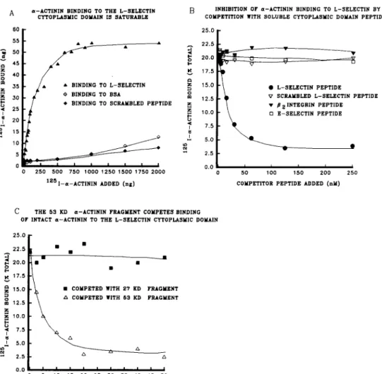
10 -7 M was calculated (Fig. 1 B). Neither talin (Fig. 1 C) nor vinculin (Fig. 1 D) were able to bind dire, cOy to the L-selectin cytoplasmic domain peptide. Binding of 12sI- o~-actinin to the L-selectin cytoplasmic domain peptide was saturable (Fig. 2 A). lZ~I-a-actinin did not bind to a scram- bled 17-amino acid peptide with the same amino acid com- position as the L-selectin peptide but arranged in a random sequence (Fig. 2 A) further suggesting that the u-actinin- L-selectin interaction was specific. Furthermore, binding of 125I-ot-actinin to the L-selectin cytoplasmic domain was competed by excess soluble peptide, but was not competed by the scrambled peptide (Fig. 2 B). Peptides corresponding to either the complete E-selectin cytoplasmic domain or an 18-amino acid region within the /32 integrin cytoplasmic domain that was previously shown to interact with o~-actinin (Pavalko and LaRoche, 1993) did not compete for binding of ec-actinin to L-selectin (Fig. 2 B).
To determine the L-selectin-binding region on o~-actinin, 27-kD and 53-kD proteolytic fragments of o~-actinin were generated using the enzyme thermolysin and purified on a FPLC Mono Q column. The 27-kD fragment binds to actin but not to integrin/3 subunit cytoplasmic domains, whereas
A
6 0 5 5
~ 5o
45 D 40 35 ~ 30
~ 25
~ 20
~ 15
10
a-ACTININ BIHDING TO THE L-SELECTIN CYTOPLASMIC DOMAIN IS SATURABLE
B INHIBITION OF a-ACTIHIN BINDING TO L-SELECTIN BY
COMPETITION WITH SOLUBLE CYTOPLASMIC DOMAIN PEPTIDE
25,0
• •
•
•
~ 22.5
~ N
2 0 . 0E 17.5
• • BINDING TO L-SELECTIN ~ 1 5 . 0
DING
TO BEA
m°
1 2 . 5t e BINDING TO SCRAMBLED PEPTIDE ~ 1 0 . 0
i 7.5
* " s.o
~ 2.5
0.0
2 5 0 5 0 0 7 5 0 1 0 0 0 1 2 5 0 1 5 0 0 1 7 5 0 2 0 0 0 0
1 2 5 1 - a - A C T I N I N ADDED ( n g )
• L-SELECTIN PEPTIDE
V SCRAMBLED L-SELECTIN PEPTIDE • ~8 2 INTEGRIN PEPTIDE
SELECTIN PEPTIDE •
i I I I
5 1 0 0 1 5 0 2 0 0 2 5 0
COMPETITOR PEPTIDE ADDED (nM)
C THE 53 KD a-ACTININ FRAGMENT COMPETES BINDING
OF INTACT a-ACTININ TO THE L-SELECTIN CYTOPLASMIC DOMAIN
25.0
22.5
A
2 0 . 0
E 17.5
1 5 . 0 D o 1 2 . 5
z
z 10.0 ~1 7.5 I 5.0 2.5 0.0
~ C m
COMPETED WITH 27 KD FRAGMENT OMPETED WITH 53 KD FRAGMENT' ' ' ' ' ' ' I I I 5 1 0 1 5 2 0 2 5 3 0 3 5 4,0 4 5 5 0
UNLABELED a-ACTININ FRAGMENT (nM)
Figure 2. Characterization of the interaction between u-ac- tinin and the L-selectin cyto- plasmic domain peptide. (A) Binding of purified a-actinin to the L-selectin cytoplasmic domain (closed triangles) is sa- turable. No binding of c~-acti- nin to a scrambled 17-amino acid version of the L-selectin cytoplasmic domain peptide
(closed diamonds) or to BSA
(open diamonds) could be measured. (B) Addition of soluble L-selectin cytoplas- mic domain peptide (closed circles) inhibits binding of c~-actinin to microtiter wells coated with the L-selectin cy- toplasmic domain peptide. Three other soluble peptides: the scrambled L-selectin pep- tide (open triangles), the/32- integrin cytoplasmic domain peptide (closed triangles), or the E-selectin cytoplasmic do- main peptide (open squares)
failed to inhibit a-actinin binding. (C) Binding of a-ac- tinin to the L-selectin cytoplas- mic domain peptide is com- peted by the 53-kD proteolytic fragment of a-actinin (open triangles), but not by the 27- kD c=-actinin fragment (closed squares), suggesting that the L-selectin binding region lies within the 53-kD rod domain of a-actinin and not in the 27-kD globular actin-binding region. Each point represents the average of triplicate wells.
"2
E-, 0 [,~
0 Izl
t - , r ~ .,,¢
I I tt~ t%/
A
70
60
50
4-0
30
20
10
u - A C T I N I N BINDING TO L - S E L E C T I N IS E N H A N C E D B Y TALIN
2 0 . 0
,--- 1 7 . 5 ,,-1 ,,¢
0 1 5 . 0
" " 1 2 . 5 I=1
1 0 . 0 m Z 7.5
t,~ Z 5 . 0
I
~ 2.5
L~t
r._ ~ "
I >
~3
UNLABELED COldPETITOR
ADDED ~qTH 1~'5 I - a - A C T I N I N
a - A C T I N I N PROMOTES BINDING OF VINCULIN TO THE L-SELECTIN CYTOPLASMIC DOMAIN
0.C
/
I ~,.
o
PROTEIN ADDED WITH 1251_VINCULIN
Figure 3.
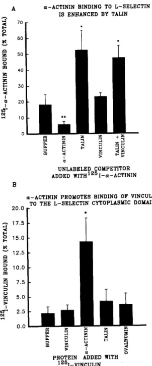
Solid phase assays characterizing the effects of talin and vinculin on the interaction between c~-actinin and the L-selectin cy- toplasmic domain peptide. (A) Binding of iodinated c~-actinin to the L-selectin cytoplasmic domain peptide was significantly enhanced in the presence of talin, and talin plus vinculin, compared to buffer only controls,(*) p > 0.05. Unlabeled ct-actinin significantly in- hibited binding compared to buffer only controls, (**) p > 0.05. Vinculin alone did not affect binding of iodinated a-actinin to L-selectin. (B) Binding of iodinated vinculin to the L-selectin cyto- plasmic domain peptide was significantly higher in the presence of excess unlabeled c~-actinin, (*) p > 0.05, compared to buffer only controls. The addition of excess unlabeled vinculin, talin, or oval- bumin did not elevate vinculin binding to the L-selectin peptide suggesting that c~-actinin forms a bridge between vinculin and the L-selectin peptide.the 53-kD fragment binds to integrin # subunit cytoplasmic domains but not to actin (Mimura and Asano, 1987; Otey et al., 1990; Pavalko and Burridge, 1991). When the 27- and 53-kD ot-actinin fragments were used as potential competi- tors of intact t2~I-ot-actinin binding to the L-selectin pep- tide, only the 53-kD fragment was able to inhibit binding (Fig. 2 C), suggesting that the L-seleetin binding site on a-actinin is located within the 53-kD domain of ot-actinin, and that the 27-kD actin-binding domain does not directly bind to L-selectin.
Because ot-actinin, vinculin, and talin are each found in sites of actin-membrane interaction, we examined the effect of mixtures of these proteins on binding of c~-actinin to the L-selectin cytoplasmic domain. When talin and vinculin were both added to L-selectin-coated microtiter wells in the presence of '25I-a-actinin, ot-actinin binding to the peptide increased "o2.5-fold (Fig. 3 A). Addition of vinculin alone to labeled ot-actinin had no effect on ot-actinin binding, while addition of talin alone increased ot-actinin binding (Fig. 3 A). These results indicated that u-actinin binding to L-seleetin was enhanced in the presence of talin, despite the fact that talin does not appear to interact directly with L-selectin. When '2~I-vinculin was added to L-selectin-coated wells, ~actinin, but not talin, promoted vinculin binding (Fig. 3 B). This result was consistent with the possibility that vincu- lin may associated indirectly with L-selectin cytoplasmic do- mains by binding to o~-actinin; vinculin binding to o~-actinin has previously been characterized (Otto, 1983; Wachsstock et al., 1987).
Coimmunoprecipitation of L-Selectin
with Cytoskeletal Proteins
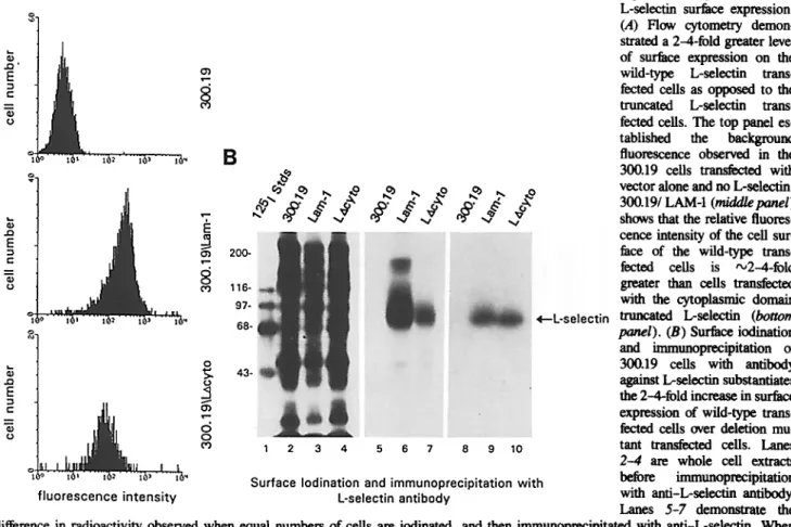
The pre-B lymphocyte cell lines, 300.19, transfected with wild-type L-selectin cDNA or with cDNA lacking a large portion (11 of 17 residues) of the L-selectin cytoplasmic do- main were used to assess the ability of L-selectin to interact with cytoskeletal proteins in vivo. The wild-type transfectant has been shown previously to bind HEV (Kansas et al., 1993) and roll along endothelium in vivo (Ley et al., 1993). Cells transfected with the cytoplasmic domain deletion mutant (LAcyto) failed to bind HEV or roll in vivo (Kansas et al., 1993). Before making a qualitative evaluation of L-selectin- cytoskeletal interactions, the levels of L-selectin surface expression on cells transfected with the wild-type and truncated L-selectin were assessed using both indirect immunofluorescence followed by flow cytometry and surface iodination followed by immunoprecipitation. Flow cytome- try of the surface labeled transfectants and nontransfeeted 300.19 cells revealed 2-3-fold higher levels of L-selectin sur- face expression on the wild-type transfectants when com- pared to the truncated protein (Fig. 4 A). Immunoprecipita- tion after surface iodination also indicated approximately threefold higher levels of surface expression by the wild-type transfectant (Fig. 4 B). Therefore, the number of cells used in subsequent coimmunoprecipitation experiments was cor- respondingly adjusted.
Antibodies against the cytoskeletal proteins c~-actinin, vinculin, talin, paxillin, tensin, and filamin were used to im- munoprecipitate from detergent extracts of 300.19 L-selectin transfectants under nondenaturing conditions to investigate whether L-selectin interacts with these proteins in vivo. Western blots of whole cell extracts confirmed that all of the
Figure 4. Quantitation of L-selectin surface expression. (,4) Flow cytometry demon- strated a 2-4-fold greater level of surface expression on the wild-type L-selectin trans- fected cells as opposed to the truncated L-selectin trans- fected cells. The too panel es- tablished the background fluorescence observed in the 300.19 cells transfec~ with vector alone and no L-seleetin. 300.19/LAM-I (m/dd/epanel) shows that the relative fluores- cence intensity of the cell sur- face of the wild-type trans- fected cells is "~2-4-fold greater than cells transfected with the cytoplasmic domain truncated L-selectin (bottom panel). (B) Surface iodination and immunoprecipitation of 300.19 cells with antibody against L-selectin substantiates the 2-4-fold increase in surface expression of wild-type trans- feeted cells over deletion mu- tant transfected cells. Lanes 2--4 are whole cell extracts before immunoorecipitation with anti-L-selectin antibody.
Lanes 5-7 demonstrate the difference in radioactivity observed when equal numbers of cells are iodinated, and then immunoorecipitated with anti-L-selectin. When cell numbers are adjusted so that there are a 3-4 greater number of the LAcyto cells, expression as quantitated by t25I, appears to be com- parable (lanes 8-10).
antibodies used in these studies recognized the correspond- ing protein in 300.19 cells (not shown). Coimmunoprecipi- tates using these antibodies were subjected to SDS-PAGE, transferred to nitrocellulose, and probed for L-selectin that had coprecipitated with cytoskeletal complexes. L-selectin was easily detected in coimmunoprecipitates with both a-actinin and vinculin (Fig. 5 A), but not with any of the other cytoskeletal proteins tested including talin, paxillin, tensin, and filamin (not shown). In addition, when the ex- periment was done in reverse, i.e., by immunoprecipitating with L-selectin antibody and immunoblotting for cytoskele- tal proteins, both a-actinin and vinculin were detected in L-selectin immunoprecipitates (Fig. 5 B) while talin, paxil- lin, tensin, and filamin were not detected (not shown). To visualize the total protein precipitated with anti-L-selectin antibodies, cells were labeled with [35S]methionine (Fig. 5 C). A band migrating at the appropriate molecular weight for L-selectin was the most prominent band that could be de- tected. In combination with the in vitro solid phase binding assays, these data suggest that L-selectin forms a complex with both a-actinin and vinculin in vivo and that the interac- tion of L-selectin with vinculin is indirect, probably through binding to ot-actinin.
In contrast to the results obtained with the full-length L-selectin transfectant, no L-selectin was detected in coim- munoprecipitates of c~-actinin or vinculin from the LAcyto transfectant (Fig. 5 A). Similarly, these cytoskeletal proteins were not detected in L-selectin coimmunoprecipitates from
the LAcyto transfectant (Fig. 5 B). These data indicate that L-selectin interacts with the cytoskeleton through a direct in- teraction between .-actinin and the 11 carboxy terminal amino acids of the L-selectin cytoplasmic tail. Despite the abundance of talin in 300.19 cells and the ability of talin to potentiate ot-actinin binding to the L-selectin peptide in vitro, coprecipitation of L-selectin with talin was never de- tected from cells transfected with either the full-length or truncated L-selectin.
Cell Surface Localization of Vdld-~pe and Truncated L-Selectin by Immunoelectron Microscopy
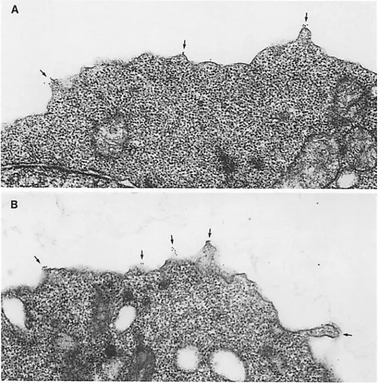
The role of the L-selectin cytoplasmic domain on the local- ization of L-selectin on the surface of transfected cells was examined by immunoelectron microscopy. L-selectin is nor- mally localized predominantly to the microvillar projections which cover the surface of neutrophils (Picker et al., 1991; Erlandsen et al., 1993; Burns and Doerschuk, 1994). Cells transfected with wild-type or truncated L-selectin were la- beled with an antibody against the extracellular domain of L-selectin, followed by a secondary antibody conjugated to a gold particle, and processed for immunoelectron micros- copy (Fig. 6). Quantitation of gold particles revealed that =90% of the label was localized to the microviUar projec- tions of cells transfected with wild-type L-selectin (Table I). The subcellular localization of L-selectin in these transfected
300.19 cells was therefore quite similar to that in neutrophils. Interestingly, the LAcyto mutant also localized almost exclu-
Figure 5. ~-Actinin and vinculin form a complex with L-selectin through the cytoplasmic domain that is detected by coimmunoprecipitation. 300.19, 300.19/LAM-I, and 300.19/LAcyto cells were lysed with detergent and these extracts were used to determine if specific cytoskeletal proteins formed a complex with the cytoplasmic domain of L-selectin. (A) The first three lanes are whole cell detergent extracts before immuno- precipitation with cytoskeletal proteins and are immunoblotted for anti-L-selectin. The next lanes, corresponding to the immunoprecipitates with anti-ct-actinin and anti-vinculin followed by immunoblotting with anti-L-selectin, demonstrate the in vivo association of the wild-type L-selectin (Lam-/), but not the cytoplasmic domain truncation mutant (LAcyto), with the cytoskeletal proteins c~-actinin and vinculin. (B) Full-length L-selectin (I.am-1), but not the deletion mutant (LAcyto) coimmunoprecipitates c~-actinin and vinculin. Antibody against L-selectin was used to immunoprecipitate from cell extracts and the cytoskeletal proteins that coprecipitated were detected by immunoblotting with antibodies against c~-actinin and vinculin. Antibodies against four other cytoskeletal proteins: talin, paxillin, tensin, and filamin, failed to detect the corresponding protein coprecipitating with L-selectin (not shown). (C) To visualize the total protein precipitated with L-selectin under coimmunoprecipitation conditions, cells were labeled with [35]methionine before lysis. L-selectin (arrow) is the only major band visi- ble from LAM-1 and LAcyto cells immunoprecipitated with anti-L-selectin when the total protein is visualized by autoradiography of the immunoprecipitates.
Figure 6. Immunoelectron microscopic localization of L-selectin on the surface of cells transfected with truncated (A) or full-length L-selectin cDNA (B). Cells were labeled with a monoclonal antibody (Leu8) against the extracellu- lar domain of L-selectin, fol- lowed by 10-rim gold particle conjugated goat anti-mouse Ig antibody conjugated to 10- run gold particles. Labeling is limited to the plasma mem- brane and occurs predomi- nantly on the small microvil- lar projections (see also Table I).
sively to the microvilli (Fig. 6; Table I). Lymphoblasts that were exposed to control IgG instead of anti-L-selectin showed no labeling. These results indicated that an associa- tion with the cytoskeleton via ot-actinin involving the COOH- terminal 11 residues of the L-selectin cytoplasmic domain was not necessary to maintain the selective localization of L-selectin to the microvilli of transfected 300.19 cells.
Discussion
This study represents the first demonstration of a direct inter- action between the cytoskeleton and a member of the selectin family of cell adhesion molecules. This interaction is medi- ated by a region within the carboxy terminal 11 residues of
the 17-amino acid cytoplasmic domain and occurs via a di- rect interaction with the rod domain of the actin-binding pro- tein u-actinin. In contrast to selectins, cytoskeletal interac- tions with integrins are better defined (for review see Sastry and Horwitz, 1993). Talin was shown to interact with the cy- toplasmic domain of integrin heterodimers (Horwitz et al., 1986), and a-actinin mediates cytoskeletal attachment to the cytoplasmic domains of members of at least two other fami- lies of cell adhesion molecules: ~ (Otey et al., 1990, 1993; Pavalko and Burridge, 1991) and/~2 (Pavalko and LaRoche, 1993) integrins, and an Ig family member, ICAM-1 (Carpen et al., 1992). The interaction between ct-actinin and inte- grins occurs through binding of a defined portion of the cyto- plasmic domain of integrin 8, (Otey et al., 1990, 1993) and
Table L Distribution of L-Selectin on the Plasma Membrane
Intact L-selectin Truncated L-selectin
(n = 18 cells) (n = 15 cells)
Percent gold particles on membrane of:
Microvillar projections 87 + 2 89 ± 2
Flat regions 13 + 2 11 + 2
Data expressed as mean ± SEM
f12 (Pavalko and LaRoche, 1993) subunits in vitro to a site within the 53-kD rod domain of ot-actinin. Similarly, binding of ot-actinin to L-selectin in vitro was also inhibited by the 53-kD fragment of ot-actinin (Fig. 2 C). ot-Actinin binding to L-selectin and to integrins appears to occur through dis- tinct regions of the ot-actinin rod domain, however, since the f12 cytoplasmic domain peptide failed to inhibit binding of a-actinin to the L-selectin peptide. The association of ot-actinin with members of the f12 integrin subfamily in vivo can be induced upon activation of neutrophils with chemo- tactic peptides (Pavalko and LaRoche, 1993) or by activation of T cells by cross-linking of the T cell receptor (Pardi et al., 1993). In contrast, the interaction between t~-actinin and L-selectin is constitutive in these transfected cell lines. The results of this study indicate that L-selectin can be added to the list of cell adhesion molecules that use ct-actinin as a pri- mary linker molecule to the actin cytoskeleton.
Our results also suggest that two additional cytoskeletal proteins, vinculin and talin, might also be involved in the in- teraction between L-selectin and the cytoskeleton, perhaps as part of a multiprotein complex. Although a direct interac- tion between either vinculin or talin and the L-selectin cyto- plasmic domain peptide could not be detected in the in vitro solid phase binding assays, vinculin was detected in im- munoprecipitates from L-selectin transfectants but not from the LAcyto transfectants. Solid phase binding experiments indicated that vinculin bound to wells coated with L-selectin cytoplasmic domain peptide only in the presence of ct-actinin. Coimmunoprecipitation of vinculin with L-selectin is there- fore probably indirect, and is likely to be mediated through the high affinity interaction of oe-actinin with vinculin (Wachsstock et al., 1987).
Our results with talin are somewhat more puzzling. The ability of talin to potentiate the binding of ct-actinin to the L-selectin cytoplasmic domain peptide is difficult to recon- cile with a lack of binding of talin to this peptide in vitro and the lack of coimmunoprecipitation of L-selectin and talin in vivo. Furthermore, no evidence exists that talin can directly interact with ot-actinin, although talin does bind to vinculin in vitro (Otto, 1983; Burridge and Mangeat, 1984). This potentiation effect appears to be specific for talin, inasmuch as vinculin does not exhibit any ability to modulate the bind- ing of c~-actinin to the L-selectin cytoplasmic domain pep- tide. It is possible that talin does not interact with L-selectin or ~-actinin with sufficient affinity to be detected in our as- says, but that it can nonetheless facilitate interactions be- tween L-selectin and a-actinin, perhaps by lowering the free energy barrier of the L-selectin/c~-actinin interaction. Ex- periments are in progress to determine the mechanism of ac- tion by which talin potentiates the binding of L-selectin to c~-actinin.
We found that the eleven carboxy terminal amino acids of the L-selectin cytoplasmic tail were not required for local- ization of L-selectin to the microvillar projections of the plasma membrane. Interaction of L-selectin with ct-actinin or vinculin is therefore not required for correct receptor positioning. The few remaining amino acids of the L-selectin cytoplasmic tail may be sufficient for this function, through alternative interactions with other cytoskeletal or cytoplas- mic proteins that are not sufficient for L-selectin's adhesive function. In this regard, it will be interesting to examine the possibility that the membrane proximal region of the L-selec-
tin cytoplasmic domain interacts with members of the ERM (ezrin/radixin/moesin) family of cytoskeletai proteins (Sato et al., 1991), which have recently been shown to be required for the maintenance of microvilli and ruffles (Takeuchi et ai., 1994). A less likely explanation for the unique distribution of L-selectin inmicrovillar projections may involve a role for proteins other than cytoskeletal molecules, such as interac- tions with molecules that are located within the plasma membrane. In either case, the normal localization of the LAcyto mutant argues strongly that L-selectin positioning alone is not sufficient to promote L-selectin-mediated adhe- sion to endothelium, and hence that the unique association between L-selectin and cytoskeletal proteins fulfills a dis- tinct requirement for adhesion by L-selectin.
The results of this study highlight the potential functional importance of a link between L-selectin and the actin cytoskeleton. The LAcyto mutant exhibits normal carbohy- drate ligand recognition (Kansas et al., 1993) and normal receptor positioning (this report), and yet it is unable to mediate leukocyte rolling or adhesion to HEV (Kansas et al., 1993). Ongoing studies are aimed at determining the factors necessary for normal L-selectin function, in addition to car- bohydrate ligand recognition and microvillar localization, and the precise role of cytoskeletal interactions with the L-selectin cytoplasmic domain in regulating leukocyte roll- ing and adhesion to HEV.
We thank Dr. Thomas Tedder for the gift of L-selectin antibody and Dr. Denis English and Kevin Harvey for help with FACS analysis.
This work was supported by National Institutes of Health grants GM47333 (E M. Pavalko) and HL48160 (C. M. Doerschuk), by research grants from the American Lung Association (E M. Pavalko), the American Heart Association, Indiana Affiliate (E M. Pavalko), and a Career Investi- gator Award from the American Lung Association (C. M. Doerschuk).
Received for publication 22 September 1994 and in revised form 12 January 1995.
References
Arnaout, M. A. 1993. Dynamics and regulation of leukocyte-endothelial cell interactions. Curr. Opin. Hematol. 1 : 113-122.
Berman, C. L., E. L. Yeo, J. D. Wencel-Drake, B. C. Furie, M. H. Ginsberg, and B. Furie. 1986. A platelet alpha granule membrane protein that is as- sociated with the plasma membrane after activation. J. Clin. Invest.
78:130-137.
Bevilacqua, M. P., J. S. Pober, D. L. Mendrick, R. S. Cotran, and M. A. Gim- brnne. 1987. Identification of an inducible endothelial-leukocyte adhesion molecule. Proc. Natl. Acad. Sci. USA. 84:9238-9243.
Bevilacqua, M. P., and R. M. Nelson. 1993. Selectins. J. Clin. Invest. 91: 379-387.
Burns, A. B., and C. M. Doerschuk. 1994. L-selectin and CDI8 expression on rabbit neutrophils during CD18-independent and CD18-dependent emigration in the lung. J. lmmunol. 153:3177-3188.
Burridge, K., and P. Mangeat. 1984. An interaction between vinculin and talin.
Nature (Lond.). 308:744-746.
Butcher, E. C. 1991. Leukocyte-endothelial cell recognition: three (or more) steps to specificity and diversity. Cell. 57:1033-1036.
Carpen, O., P. Pallal, D. E. Staanton, and T. A. Springer. 1992. Association of intercellular adhesion molecule-1 (ICAM-I) with actin-containing cyto- skeleton and a-actinin. J. Cell Biol. 118:1223-1234.
Disdier, M., J. H. Morrissey, R. D. Fugate, D. F. Bainton, and R. P. McEver. ! 992.. Cytoplasmic domain of P-selectin (CD62) contains the signal for sort- mg into the regulated secretory pathway. Mol. Biol. Cell. 3:309-321. Eckhardt, J. U. A., E. F. Smeets, L. A. Ginsel, J. J. M. Onderwater, J. F. M.
Lecuwenberg, and W. A. Buurman. 1992. Evidence for endocytosis of E-selectin in human endothelial cells. Fur. J. lmmunol. 22:2519-2526. Erlandsen, S. L., S. R. Hasslin, and R. D. Nelson. 1993. Detection and spatial
distribution of the f12 integrin (mac-l) and L-selectin (LECAM-I) adher- ence receptors on human neutrophils by high resolution field emission SEM.
J. Histochem. Cytochem. 41:327-334.
Feramsico, J. R., and K. Burridge. 1980. A rapid purification of a-actinin, filamin and a 130,000-dalton protein from smooth muscle. J. Biol. Chem.
255:1194-1199.
Fraker, P. J., and J. C. Speck. 1978. Protein and cell membrane iodinations with a sparingly soluble chloroamide 1,3,4,5-tetrachloro-3a-6a-diphenyl.
Biochem. Biophys. Res. Commun. 80:849-855.
Horwitz, A. F., K. Duggan, C. Buck, M. C. Beckerle, and K. Burridge. 1986. Interaction of plasma membrane fibronectin receptor with talin-a trans- membrane linkage. Nature (Lond.). 320:531-533.
Kansas, G. S., K. Ley, I. M. Munro, and T. F. Tedder. 1993. Regulation of leukocyte rolling and adhesion to high endothelial venules through the cyto- plasmic domain of L-selectin. J. Exp. Med. 177:833-838.
Kansas, G. S., G. S. Wood, D. M. Fishwild, and E. G. Engleman. 1985. Func- tional analysis of human T lymphocyte subsets defined with anti-Leu-8. J.
Immunol. 134:2995-3000.
Koedam, J. A., E. M. Cramer, E. Briend, B. Furie, B. C. Furie, and D. A. Wagner. 1992. P-selectin, a granule membrane protein of platelets and en- dothelial cells, follow the regulated secretory pathway in AtT-20 cells. J.
Cell Biol. 116:617-625.
Lawrence, M. B., and T. A. Springer. 1991. Leukocytes roll on a selectin at physiologic flow rates: distinction from and prerequisite for adhesion through integrins. Cell. 65:859-873.
Ley, K., T. R. Tedder, and (3. S. Kansas. 1993. L-selectin can mediate leuko- cyte roiling in untreated mesenteric venules in vivo independent of E- or P-selectin. Blood. 82:1632-163g.
McEver, R. P., J. H. Beckstead, K. L. Moore, L. Marshall-Carlson, and D. R. Bainton. 1989. GMP-140, a platelet alpha granule membrane protein, is also synthesized by vascular endothelial cells and is localized in Weibel-Palade bodies. J. Clin. Invest. 84:92-99.
Mimura, N., and A. Asano. 1987. Further characterization of a conserved actin-binding 27 kDa fragment of actinogelin and ct-actinins and mapping of their binding sites on the actin molecule by chemical cross-linking. J. Biol. Chem. 262:4717--4723.
Otey, C. A., F. M. Pavalko, and K. Burridge. 1990. An interaction between ct-actinin and the 31 integrin subunit in vitro. J. Cell Biol. 111:721-730. Otey, C. A., G. B. Vasques, K. Burridge, and B. W. Erickson. 1993. Mapping
of the ~-actinin binding site within the ~l integrin cytoplasmic domain. J.
Biol. Chem. 268:21193-21197.
Otto, J. J. 1983. Detection of vinculin-binding proteins with an '251-vinculin gel overlay technique. J. Cell Biol. 97:!283-1287.
Pardi, R., L. Inverardi, C. Rugarli, and J. R. Bender. 1992. Antigen-receptor complex stimulation triggers protein kinase C-d~endent CD 11 a/CD 18 cytoskeleton association in T lymphocytes. J. Cell Biol. 116:1211-1220. Pavalko, F. M., and K. Burridge. 1991. Disruption of the actin cytoskaleton
after microinjection of proteolytic fragments of t~-actinin. J. Cell Biol. 114: 481-491.
Pavalko, F. M., and S. M. LaRoche. 1993. Activation of human nentrophils induces an interaction between the integrin /~2-subunit (CDI8) and the actin-binding protein ct-actinin. J. lmmunol. 151:3795-3807.
Picker, L. J., R. A. Waruock, A. R. Burns, C. M. Doerschuk, E. L. Berg, and E. C. Butcher. 1991. The neutrophil selectin LECAM-1 presents carbo- hydrate ligands to the vascular selectins ELAM-1 and GMP-140. Cell. 66:
921-933.
Sastry, S. K., and A. F. Horwitz. 1993. Integrin cytoplasmic domains: media- tors of cytoskeletal linkages and extra- and intracellular initiated transmem- brahe signaling. Curt. Opin. Cell Biol. 5:819-831.
Sato, N., S. Yomemurs, T. Obinata, Sa. Tsukita, and Sh. Tsukita. 1991. A gene family consisting of ezrin, radixin, and moesin. Rs specific localization at actin filament/plasma membrane association sites. J. Cell Sci. 103:131-143. Smith, C. W. 1992. Transendothelial migration, in Adhesion: Its Role in Inflammatory Disease. J. M. Garlan and D. Y. Liu, editors. W. H. Freeman and Co., NY. 83-115.
Springer, T. A. 1994. Traffic signals for lymphocyte recireulation and leuko- cyte emigration: the multistep paradigm. Cell. 76:301-314.
Takeuchi, K., N. Sato, H. Kasahara, N. Funayama, A. Nagafuchi, S. Yone- mura, Sa. Tsukita, and Sh. Tsukita. 1994. Perturbation of cell adhesion and microvilli formation by antisense oligonucleotides to ERM family members.
J. Cell Biol. 125:1371-1384.
Tedder, T. F., A. C. Penta, H. B. Levine, and A. S. Freedman. 1990. Expres- sion of the leukocyte adhesion molecule LAM-I. Identity with the TQI and Leu-8 differentiation antigens. J. lmmunol. 144:532-540.
Wachsstock, D. J., J. A. Wilkins, and S. Lin. 1987. Specific interaction of vin- culin with ct-actinin. Biochem. Biophys. Res. Commun. 146:554-560.