iii
ABSTRACT
Jolien Suzanne Tyler: The Dynamic Chromatin Landscape in Saccharomyces cerevisiae (Under the direction of Kerry Bloom)
The accurate and faithful segregation of chromosomes during mitosis is essential for cellular
survival. Current paradigms of chromosome segregation focus on the mechanical contributions of
the mitotic spindle without considering the biomechanical properties of the chromatin itself. In
order to further our understanding of how the inherent physical properties of chromatin contribute
to cellular processes like chromosome segregation, we have examined both histone and chromatin
dynamics in the budding yeast Saccharomyces cerevisiae. During mitosis, the mitotic spindle exerts
force on the pericentromeric chromatin, which adjusts structurally to accommodate this force. By
measuring the fluorescence recovery after photobleaching (FRAP), we found that histones are more
dynamic in the pericentromeric region as compared to the chromosome arm, and these increased
recovery rates are dependent on spindle‐based tension. The tension‐dependent histone dynamics
in the pericentromere are dependent on the chromatin remodeling activities of the Remodels the
Structure of Chromatin (RSC) and Imitation Switch (ISWI) ATP‐dependent chromatin remodeling
complexes. Thus, balanced histone removal and reincorporation in the pericentromere provide a
mechanism for accommodation of spindle‐based tension and the maintenance of chromatin
packaging. Having measured the dynamic nature of histone turnover within the chromatin polymer
in response to spindle‐based tension, we subsequently examined the spatio‐temporal fluctuations
of the chromatin polymer. We combined in vivo chromatin motion analysis and mathematical
fluctuations. The range of chromatin motion and its effective spring constants are found to vary
along the length of the chromosome, in a manner dependent on tethering at the centromere. These
polymer properties of the chromatin are dependent on both histone occupancy and cohesin
packaging. As a whole, the work detailed in this dissertation contributes valuable insights into the
importance of dynamic histone occupancy and chromatin motion in defining and maintaining the
biomechanical polymer properties of chromosomes in vivo.
v
ACKNOWLEDGEMENTS
It is bittersweet to finally reach this point in my life. I’ve spent much time and effort to get here and am excited for the next challenge, but am sad that this chapter is almost over. My
graduate experience at UNC Chapel Hill has been filled with both endless excitement and
frustrations, but I would not change any single moment.
To my committee – Greg, Kevin, and Brian – Thanks for agreeing to be on my committee and thanks
for all your enthusiastic suggestions and comments throughout my graduate years. I always went
into committee meetings with both a healthy fear and enthusiasm to get awesome feedback.
To the Bloom lab – I am lucky to have landed where I did. It is a joy to be surrounded by such
passionate scientists and good friends every day. Julian, thanks for always making sure we had what
we needed to focus on the science and taking the time to help a lost grad student out. Drew, it was
tremendously helpful to have a fellow grad student to share the ups and downs of grad school.
Thanks for always pushing me to do better science and tell the story right. Besides being great
friends in lab, thanks for letting me vent over a beer or showing me how poorly I play video games.
To Elaine Yeh – Thanks for all your patient wisdom regarding science, life, and my future. I am lucky
to have had the privilege of working for two great bosses. Even on the most frustrating days, your
express how tremendously helpful it has been for me as a student and scientist to have had so much
encouragement in lab.
To Kerry Bloom
“The most exciting phrase to hear in science, the one that heralds new discoveries, is not
‘Eureka!’ but ‘That’s funny…” – Issac Asimov
Thank you. Thanks for taking convincing me UNC was the right grad school for me, and for taking
me on as a grad student. I am so grateful to have had a PI who gave me just the right amount of
guidance while also letting me follow my own ideas. Thanks for always having an open door to chat
about experiments and data, and for encouraging me to always do good science. To this day I have
no idea how you manage to wander over just as I am having a “That’s funny…” moment regarding
my latest experiment. The excitement and drive you have for science creates a great atmosphere in
lab, and I know I am the scientist I am because of your advice and support.
To my family – I could not ask for more supportive and patient parents. Mama en Papa, thank you
for always encouraging me to “just to my best.” Thank you for always being my biggest fans. You
always set an example in persistence and the value in doing something right (otherwise, why
bother?) that I carry with me every day. I hope I can make you both proud. Marjan, you are
everything a good older sister should be ‐ my partner in crime growing up, you now set the example
I strive to emulate. I’m glad you and Philip, my favorite brother‐in‐law, have created such a
wonderful life together. I have two loving grandmothers who always know just what to say and
when to say it. Opa Wim, I often think back on when you shared you passion for science with me
vii
To Dan – With a husband, comes a new family. Patty and Danny, thanks for being such great in‐
laws. Thanks for all your love and support, and thanks for raising such a great guy.
Dan, not a day goes by that I am not eternally grateful I have you in my life. You’ve always stood by
my side and held me up when I was just too tired, stressed, or scared to stand alone. We both know
that none of this would be possible without your unfailing love and support. Although I just don’t
TABLE OF CONTENTS
LIST OF TABLES ... xii
LIST OF FIGURES ... xiii
LIST OF ABBREVIATIONS ... xv
CHAPTER 1: INTRODUCTION ... 1
CHAPTER 2: CENTROMERES: UNIQUE CHROMATIN STRUCTURES THAT DRIVE CHROMOSOME SEGREGATION ... 4
Introduction ... 4
The centromere ... 6
DNA at the centromere ... 6
Centromere‐associated proteins ... 7
Histones at the centromere ... 9
Patterns of histone modifications at the centromere ... 9
CENPA, an H3 variant unique to the centromere ... 12
The H2A.Z variant at the centromere ... 15
Assembling the centromere ... 16
CENPA loading ... 16
Chromatin remodelling at the centromere ... 18
Architecture of centromeric chromatin ... 19
ix
Polymer physics at the centromere ... 24
Chromosome segregation by entropy ... 24
Maintaining chromatin‐spindle force balance ... 26
The future ... 27
Appendix 2.1: Box – The kinetochore ... 37
Appendix 2.2: Glossary terms ... 39
CHAPTER 3: TENSION‐DEPENDENT NUCLEOSOME REMODELING AT THE PERICENTROMERE IN YEAST ... 41
Introduction ... 41
Results ... 44
Histone dynamics differ in the pericentromere and chromosome arm during metaphase ... 44
Histone dynamics in the pericentromere are reduced upon loss of spindle‐based tension ... 45
Increased histone dynamics are the result of increased histone removal ... 46
Loss of Sth1p/Nps1p or Isw1p leads to reduced histone turnover in the pericentromere ... 47
Chromatin packaging contributes to kinetochore organization ... 49
Discussion ... 50
Patterns of histone dynamics in metaphase ... 50
Redefining the pericentromere ... 52
Materials and Methods ... 54
Yeast strains and imaging ... 54
Fluorescence recovery after photobleaching ... 55
Photoactivation ... 57
CHAPTER 4: BENDING THE RULES: WIDEFIELD MICROSCOPY AND THE
ABBE LIMIT OF RESOLUTION ... 77
Introduction ... 77
Deconvolution – putting light (back) in its proper place ... 80
Model convolution – blurring the lines between reality and simulation ... 82
Gaussian fitting – cutting through the haze ... 85
Conclusion ... 89
Acknowledgments ... 89
CHAPTER 5: CENTROMERE TETHERING CONFINES CHROMOSOME DOMAINS ... 97
Introduction ... 97
Results ... 99
Chromatin confinement varies along the length of the chromosome ... 99
Chromatin dynamics in interphase are dictated by tethering ... 100
The chromatin polymer behaves like an elastic filament during interphase ... 101
Modeling the chromatin spring as a doubly‐tethered, confined bead‐spring chain with excluded volume interactions can recapitulate experimental dynamics ... 102
The effective spring constant along the entropic chromatin spring can be measured in vivo ... 104
Cohesin contributes to local clamping of chromatin ... 105
Nucleosome depletion results in a stiffer chromatin fiber ... 106
Dynamic fluctuations underlie chromosome territories ... 108
xi
Examining chromosome territories to understand cellular
behaviors like repair ... 110
Experimental Procedures ... 111
Image analysis ... 111
Calculating Rc from experimental data ... 112
Entropic bead‐spring chain model ... 113
Effective spring constant in a double‐tethered Rouse chain ... 114
Defining model variables ... 114
Calculating effective ks from experimental data ... 115
Statistical analysis ... 116
Acknowledgments ... 116
Appendix 5.1: Supplemental Experimental Procedures ... 129
Yeast strains and cell preparations ... 129
Microscopy ... 129
Statistical analysis ... 130
Appendix 5.2: Formulae ... 140
LIST OF TABLES
Table 2.1 – Centromere‐associated proteins ... 36
Table 3.1 – Saccharomyces cerevisiae strains ... 70
Table 3.S1 – Summary of all data for FRAP of histone H2B ... 73
Table 3.S2 – Summary of all data for FRAP of histone H4 ... 74
Table 3.S3 – Summary of all data for photoactivation of H2B ... 75
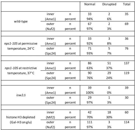
Table 3.S4 – Summary of all data for kinetochore organization ... 76
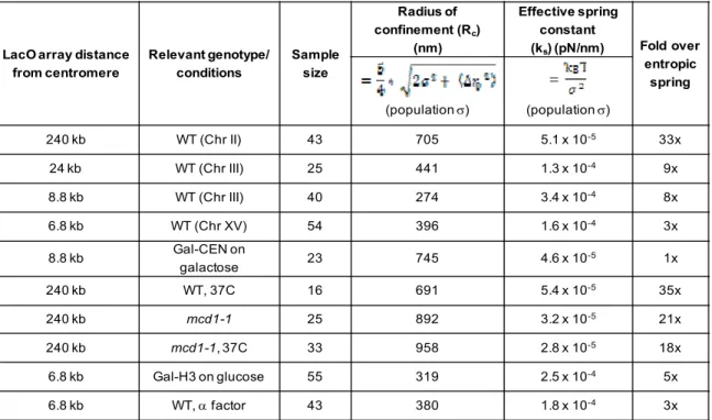
Table 5.1 – Summary of Rc and ks measurements from population variance ... 128
Table 5.S1 ‐ Summary of WT results from equations used to calculate Rc and effective ks, Related to Figures 1 and 4 ... 138
Table 5.S2 ‐ Saccharomyces cerevisiae strains used in this paper, Related to Experimental Procedures ... 139
xiii
LIST OF FIGURES
Figure 2.1 – Chromosome segregation in the cell cycle ... 28
Figure 2.2 – Characteristics of point and regional centromeres ... 30
Figure 2.3 – The CENPA‐containing nucleosome ... 31
Figure 2.4 – Chromatin geometry at the centromere ... 33
Figure 2.5 – Applying the principles of polymer physics to chromosome segregation ... 35
Figure 2.6 – The kinetochore ... 38
Figure 3.1 – In vivo photobleaching of the pericentromere and chromosome arm using spindle pole bodies as fiduciary markers ... 59
Figure 3.2 – Histones in the pericentromere are more dynamic than those of the chromosome arm ... 61
Figure 3.3 – Loss of spindle tension or chromatin remodeling activity results in reduced histone dynamics primarily at the pericentromere ... 62
Figure 3.4 – Loss of spindle tension results in reduced dispersal of photoactivated histone H2B ... 64
Figure 3.5 – Disruption of the underlying chromatin platform results in disruption of the kinetochore ... 66
Figure 3.6 – Model diagram of histone occupancy in the pericentromere and arm under various experimental conditions ... 68
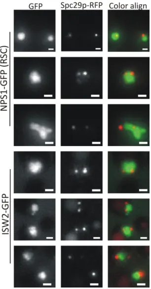
Figure 3.S1. RSC and ISW2 both localize throughout the budding yeast nucleus ... 71
Figure 3.S2. Loss of RSC function (nps1‐105) does not alter metaphase spindle length at permissive (24°C) temperature ... 72
Figure 4.1 ... 90
Figure 4.2 ... 92
Figure 4.3 ... 93
Figure 5.1 – Rc of chromatin varies along the length of the chromosome ... 118
Figure 5.2 – Rc is dictated by the attachment at the centromere ... 120
Figure 5.3 – Interphase chromatin is dynamic ... 121
Figure 5.4 – Modeling interphase chromatin dynamics as a doubly
tethered bead‐spring polymer chain ... 122
Figure 5.5 – Loss of cohesin or nucleosome depletion during interphase
result in altered confinement and chromatin stiffness ... 124
Figure 5.6 – Modeling the chromatin spring as a doubly tethered, confined bead‐spring chain with excluded volume interactions can recapitulate chromosome territory formation as observed
by chromosome interaction heat maps ... 126
Figure 5.S1 – Statistical comparison of equations used to calculate Rc
and effective ks, Related to Figures 5.1 and 5.4 ... 131
Figure 5.S2 – Statistical comparisons of data sets, Related to
Figures 5.1, 5.2, 5.4, and 5.5 ... 133
Figure 5.S3 – Additional information regarding chromatin elasticity
measurements, Related to Figure 5.3 ... 134
Figure 5.S4 – Additional information regarding aspect ratio measurements upon nucleosome depletion, Related
to Figure 5.5 ... 136
Figure 5.S5 ‐ ATP‐dependent chromatin motion, Related to Experimental
Procedures ... 137
xv
LIST OF ABBREVIATIONS
Cr compaction ratio
CEN centromere
Gal galactose
GFP green fluorescent protein
Glu glucose
ks spring constant
Lc contour length
Lp persistence length
MSD mean square displacement
Noc nocodazole
paGFP photoactivatable GFP
Rc radius of confinement
RFP red fluorescent protein
SPB spindle pole body
WT wild type
CHAPTER 1: INTRODUCTION
The integrity of the genome must be maintained throughout fundamental cellular processes
including chromosome segregation. The canonical paradigms for investigating mitosis are centered
on developing a “parts list” or examining the motion of the spindle. While these provide important
insights into the molecular basis for faithful chromosome segregation, in this view the chromosomes
are generally considered passive components of the segregation machinery. It is clear, however,
that chromosomes are complex polymers whose behaviors and properties are essential components
to the larger mitotic chromosome segregation apparatus (Bloom, 2008; Stephens et al., 2011;
Stephens et al., 2013). My graduate work has focused on expanding our understanding of the
importance of dynamics, both of histones and chromatin, in the study of cellular processes such as
chromosome segregation and DNA repair.
Current and previous work from the Bloom lab described the geometric organization of the
pericentromere and found it to be under tension (Bouck and Bloom, 2007; Haase et al., 2012;
Stephens et al., 2011; Yeh et al., 2008). Building on that framework, chapter 2 starts with a review
of mitosis and the composition of the centromere, moves into a discussion of the organization of the
pericentromeric chromatin flanking the centromere, and discuss how the polymer properties of the
chromatin contribute to segregation (Verdaasdonk and Bloom, 2011). Histone proteins are the
fundamental level of chromatin compaction, and thus likely contribute in large part to the polymer
properties of the chromosome. Chapter 3 explores the nature of histone dynamics in the
2
(Verdaasdonk et al., 2012). We find that histones are more dynamic in the pericentromere as
compared to the chromosome arm, and the increased removal and reincorporation is due to
spindle‐based tension. In pioneering work measuring photoactivation kinetics performed by Ryan
Gardner, an undergraduate student I mentored, we show that the increased dynamics in the
pericentromere are the result of increased histone removal. Further experiments reveal that the
observed dynamics are, in part, the result of ATP‐dependent chromatin remodeling activities of
Sth1p/Nps1p (RSC complex) and Isw2p (ISWI complex). We propose that these dynamics are
essential for the chromatin to accommodate spindle‐based tension while maintaining proper
packaging.
The second half of my graduate work shifted from histone dynamics to the dynamic motion
of the chromatin polymer as a whole. As described in chapter 4, we utilized Gaussian fitting to
obtain sub‐pixel precision (Verdaasdonk et al., 2013a). By examining the motion of lacO/lacI‐GFP
labeled chromatin spots at various distances from the centromere, and comparing these to
literature values, we find that the radius of confinement varies along the length of the chromosome
(Verdaasdonk et al., 2013b) Chromatin close to the centromere tether point is more confined, and
this confinement is dependent on tethering at the centromere. Utilizing mathematical modeling, we
show that the motion of a confined and tethered bead‐spring chain can capture the observed
experimental dynamics. In addition, we provide novel mathematical basis for calculation of an
effective spring constant from the in vivo motion of chromatin. In order to examine the molecular
basis for chromatin confinement, we measured motion upon depletion of nucleosome occupancy or
cohesin. Nucleosome depletion results in increased confinement and relative stiffening of the
chromatin fiber. Cohesin depletion results in reduced confinement, likely the result of reduced
properties of chromatin and how these contribute to chromosome segregation, the work here
presents a basis for understanding the dynamic chromatin landscape in Saccharomyces cerevisiae.
4
CHAPTER 2: CENTROMERES: UNIQUE CHROMATIN STRUCTURES THAT DRIVE
CHROMOSOME SEGREGATION1
INTRODUCTION
The canonical cell cycle is divided into interphase and mitosis (Figure 2.1). During
interphase, cells undergo growth (G1 phase) and DNA replication (S phase). After S phase, cells
undergo another phase of growth (G2) and prepare to enter mitosis (M phase). During mitosis the
sister chromatids need to be accurately segregated to each daughter cell, thereby ensuring survival
from one generation to the next. This is facilitated by a complex array of proteins that regulate the
timing and accuracy of chromosome segregation. Chromosome segregation is directed by the
centromere, a chromosomal locus that is required for mitosis and acts as the site of kinetochore
formation. The proteinaceous kinetochore (Appendix 2.1) ensures proper segregation by linking the
chromosome to the dynamic microtubules (composed of tubulin dimers), thereby forming the
mitotic spindle. Because the centromere mediates chromosome segregation, it is essential that the
cell form only one centromere and associated kinetochore attachment per chromosome to prevent
breakage, although organisms with holocentric chromosomes, such as Caenorhabditis elegans, have
attachment sites spread throughout the length of the chromosome.
Upon entry into mitosis, chromosomes condense, and the primary constriction forms at the
centromere, the region of the chromosome defined by the incorporation of a histone H3 variant,
1 This chapter previously appeared as an article in Nature Reviews Molecular Cell Biology. The original
CENPA (Moore and Roth, 2001; Sullivan and Karpen, 2004). The kinetochore is recruited to the
centromere, and signalling proteins, such as cyclin‐dependent kinases (CDKs), signal to microtubules
originating at the centrosomes to form a bipolar spindle and attach to the kinetochores. This links
the microtubules and chromosomes mechanically and through signalling processes that sense
attachment and force to ensure that all chromosomes are bioriented and bound before anaphase
onset (for a more detailed review of mitosis, see (Bouck et al., 2008; Cheeseman and Desai, 2008;
Fukagawa and De Wulf, 2009; Santaguida and Musacchio, 2009)).
Although much is known about the organization and structure of the kinetochore, the
physical structures of centromeric chromatin and pericentromeric chromatin, and their contribution
to fidelity during chromosome segregation, are not as clear. Recent studies have shed new light on
a range of topics, including the composition of the centromeric nucleosome, the histone
modifications and variants that are unique to the centromeric chromatin, and the physical
organization of this region during mitosis. These studies have expanded our understanding of how
the centromere functions to ensure segregation fidelity and prevent errors that lead to aneuploidy,
which can ultimately lead to cancer and diseases such as Down’s syndrome, Edwards’ syndrome and
Patau’s syndrome.
In this Review we describe the distinct properties that define centromeric chromatin,
including associated proteins, epigenetics and histone variants. We also examine the physical
architecture of the chromatin and its spatial geometry, and discuss the mechanical and physical
properties of the pericentric chromatin and the importance of maintaining the balances of forces
during mitosis. From this work, it is evident that the centromere does not only serve passively as a
site (or “landing pad”) for kinetochore formation, but that the underlying chromatin itself dictates,
6
essential role in mitosis. We do not focus on the kinetochore, which has been reviewed extensively
elsewhere (Cheeseman and Desai, 2008; Fukagawa and De Wulf, 2009; Przewloka and Glover, 2009).
THE CENTROMERE
The centromere is a unique region on the chromosome that is required for attachment to
the mitotic spindle and chromosome segregation. Simple eukaryotic organisms, such as budding
yeast, have point centromeres, defined by a specific DNA sequence found on all chromosomes (see
below), whereas more complex eukaryotes have larger regional centromeres that are defined by
hierarchical arrays of satellite repeats. Many similarities between centromeres in different species
have been observed that highlight the unique nature and essential functions of the centromere.
DNA AT THE CENTROMERE
The Saccharomyces cerevisiae centromere DNA was the first eukaryotic centromere to be isolated(Clarke and Carbon, 1980) and is now known to have three conserved DNA sequences
common to all chromosomes, a feature not seen in higher eukaryotes (Figure 2.2) (Clarke and
Carbon, 1983). These are termed centromere DNA element I (CDE I) CDE II, and CDE III, and
together form a 116‐120 bp sequence that is sufficient to confer mitotic stability when introduced
into plasmids (Clarke and Carbon, 1980; Fitzgerald‐Hayes et al., 1982; Hieter et al., 1985). CDE I is 8
bp and is required for high fidelity chromosome segregation; CDE II is a 78‐86 bp AT‐rich region that
is required for chromosome segregation; and CDE III is 26 bp and contains seven invariant
nucleotides conserved across all chromosomes, the mutation of which abolishes centromere
attachment per chromosome, the centromeres of budding yeast and of the yeast Kluyveromyces
lactis have been termed point centromeres.
By contrast, the regional centromeres of the fission yeast Schizosaccharomyces pombe are longer (between 40‐100 kb), have multiple microtubule attachment sites per centromere and do not
contain a conserved DNA sequence common to all chromosomes (Baum et al., 1994; Chikashige et
al., 1989; Clarke et al., 1986; Joglekar et al., 2008; Sullivan, 2009). Instead, these centromeres are
composed of core region 1 (cnt1), cnt2 and cnt3 bordered on either side by inverted repeat
sequence L (imrL) and imrR, which are flanked by outer repeat L (otrL) and otrR (Pidoux and Allshire,
2004). These regions do not show DNA sequence homology to the centromere of budding yeast.
Centromeres have been identified in Candida albicans (Sanyal et al., 2004), Neurospora
crassa (Centola and Carbon, 1994), Arabidopsis thaliana (Copenhaver et al., 1999), Drosophila
melangaster (Sun et al., 2003) and Homo sapiens (Schueler et al., 2001). The regional centromeres
of higher eukaryotes are more difficult to study because they are large and contain arrays of tandem
repeats. Indeed, human centromeres can be up to 5 Mb and contain 1‐4 Mb of 171 bp α‐satellite
repeats (Choo, 2001; Maio, 1971; Pidoux and Allshire, 2004).
CENTROMERE‐ASSOCIATED PROTEINS
Despite large divergence in centromere DNA sequences across organisms, CENPs are highly
conserved (see (Malik and Henikoff, 2009) for a discussion of the evolutionary characteristics of
centromeres). CENPs are not the only centromere‐associated proteins; indeed, many others, for
example the budding yeast proteins chromosome transmission fidelity 3 (Ctf3) and Mif2, were
discovered and named prior to identifying their centromeric associations. However, we focus on
8
(CCAN) proteins are briefly described (TABLE 1). The CCAN proteins are generally classified as part
of either the CENPA‐containing nucleosome‐associated complex (NAC) or the CENPA‐containing
nucleosome distal (CAD) proteins (Foltz et al., 2006; Panchenko and Black, 2009).
CENPA is a centromere‐associated protein that acts as a histone variant and is required to
build a fully functioning kinetochore (Blower and Karpen, 2001; Howman et al., 2000; Oegema et al.,
2001; Sullivan et al., 1994). CENPA was shown to co‐purify with nucleosome core particles, which
indicated that it probably forms a complex with the core histones (Palmer et al., 1987). Homologues
of CENPA have been identified in species from yeast to mammals. Complete loss of CENPA has been
found to be lethal in every organism studied to date, although mammalian cells can tolerate a 90%
reduction in the level of CENPA.
CENPC is a DNA‐binding protein that associates with the inner‐kinetochore plate(Saitoh et al., 1992). CENPC homologues have been identified in many model organisms, including yeasts,
flies, plants and mammals, and it has been shown to be essential for proper progression through
mitosis and chromosome segregation (Dawe et al., 1999; Erhardt et al., 2008; Fukagawa et al., 2001;
Moore and Roth, 2001; Ogura et al., 2004; Schuh et al., 2007; Tomkiel et al., 1994). Indeed, loss of
CENPC at the centromere has been shown in human cells to result in small or absent kinetochores
(Tomkiel et al., 1994). CENPC localizes to CENPA‐containing chromatin, and this requires CENPA;
reciprocally, CENPA localization requires CENPC (Erhardt et al., 2008; Oegema et al., 2001). CENPC
binds two different groups of proteins that serve distinct functions: to the Mis12 complex, which is
part of the KMN network (which comprises the Knl1 complex, Mis12 complex, and Ndc80 complex)
of the outer kinetochore that is also needed for recruitment of checkpoint proteins (Przewloka et
al., 2011; Screpanti et al., 2011); and other CCAN components such as CENPH, CENPI, CENPK and
CENPT (Carroll et al., 2010). Furthermore, CENPC, together with CENPN, binds distinct domains in
The CENPT‐CENPW subcomplex is recruited to H3‐containing centromeric chromatin and
has been proposed to be an alternative to CENPC for the connection between the centromere and
the kinetochore. The CENPT‐CENPW subcomplex functions upstream of the CENPH subcomplex
(Hori et al., 2008a), which comprises CENPH, CENPI and CENPK. These proteins are recruited to the
centromere by CENPC (Carroll et al., 2010) . They are essential for kinetochore function in
vertebrates and have been found to play a part in CENPA loading and the recruitment of other,
more distal centromere complexes (Okada et al., 2006). The CENPO subcomplex (made up of
CENPO, CENPP, CENPQ, and CENPR) and the similar CENP‐U protein (also called CENP50) are needed
to prevent premature separation upon spindle damage (Hori et al., 2008b). The CENPS subcomplex
(comprising CENPS and CENPX) is required for proper and stable formation of the outer kinetochore,
and localization of these proteins to the centromere requires CENPT or CENPK (Amano et al., 2009).
HISTONES AT THE CENTROMERE
Histone modifications and histone variants serve to demarcate unique regions of the
chromosome, including the centromere. The centromeric DNA sequence is rapidly evolving and, as
such, centromere function does not depend solely on primary DNA sequence, but also on the
presence of essential proteins, such as histone variants (including CENPA and H2A.Z) and the local
chromatin context as defined by distinct histone modifications.
PATTERNS OF HISTONE MODIFICATIONS AT THE CENTROMERE
The epigenetic specification of centromeres was first postulated by Earnshaw and Migeon,
10
(Earnshaw and Migeon, 1985). This indicated that additional proteins at the centromere are
required to facilitate segregation. Position effect variegation studies have shown that genes placed
adjacent to centromeres lead to stochastic inheritance of gene expression (Ekwall, 2007).
Subsequent studies have identified epigenetic factors required for centromere function in S.
cerevisiae (Mythreye and Bloom, 2003), C. albicans (Mishra et al., 2007), S. pombe (Folco et al.,
2008) and H. sapiens(Morris and Moazed, 2007).
Chromatin can be flagged up for transcriptional activation (euchromatin) or repression
(heterochromatin) through the modification of the amino‐terminal tails of canonical histones by
methylation, acetylation and phosphorylation (Glynn et al., 2010; Sullivan, 2009). Heterochromatin
surrounding the centromere is known to contribute to sister chromatid cohesion and condensation
(Bernard et al., 2001; Giet and Glover, 2001; Hagstrom et al., 2002; Hendzel et al., 1997; Jager et al.,
2005; Maddox et al., 2007). At regional centromeres in organisms such as humans, mice, flies and
fission yeast, the nucleosomes containing the canonical H3 histone (as opposed to the variant
CENPA, discussed below) are dimethylated at Lys4 (H3K4me2) (Figure 2.2), a modification associated
with euchromatin. This modification is thought to be important for the physical organization of the
centromere (Dunleavy et al., 2005); indeed, depletion of H3K4me2 has been shown to result in a
lack of recruitment of Holliday junction recognition protein (HJURP; see below), leading to failed
incorporation of CENPA (Bergmann et al., 2011).
The pericentric chromatin can also be defined by methylation (Greaves et al., 2007; Guenatri
et al., 2004). For example, H3 and H4 in heterochromatic regions surrounding the centromere can
be methylated at Lys9 (H3K9me2 and H3K9me3) (Guenatri et al., 2004; Sullivan and Karpen, 2004)
and Lys20 (H4K20me3) (Martens et al., 2005; Peters et al., 2003), and these modifications serve to
recruit proteins such as cohesin (Nonaka et al., 2002) and to maintain the structure of pericentric
recently been shown to require the nucleolar remodelling complex (NoRC) in mice (Guetg et al.,
2010). Interestingly, loss of the heterochromatic modification H4K20me3 is thought to result in
aberrant centromere function, as in humans it has been associated with the presence of cancer
cells, which are characterized by a high degree of aneuploidy (Fraga et al., 2005).
In addition to methylation (correlating with silenced chromatin), centromeric histones lack
acetylation, which denotes actively transcribed chromatin. The hypoacetylation of the histones and
the methylation of the DNA at the centromere alter the chemical interactions of the histones and
DNA and define a region distinct from traditional euchromatin and heterochromatin (Dunleavy et
al., 2005; Sullivan and Karpen, 2004; Zhang et al., 2008). These modifications maintain the
heterochromatic nature of centromeric chromatin, which is thought to contribute to the physical
structure of the centromere as well as to sister chromatid cohesion and condensation (Bernard et
al., 2001; Giet and Glover, 2001; Hagstrom et al., 2002; Hendzel et al., 1997; Jager et al., 2005;
Maddox et al., 2007). Histone modifications are also essential for defining and maintaining the
centromeric region. Recent evidence has shown that it is possible to engineer a human artificial
chromosome (HAC) to assess the contribution of chromatin state adjacent to the centromere
without perturbing all the centromeres of the cell (Cardinale et al., 2009; Nakano et al., 2008).
These experiments reveal that altering the chromatin to a more closed state results in loss of
essential centromere proteins as well as depletion of the H3K4me2 modification and accumulation
of the H3K9me3 (found in heterochromatin adjacent to the centromere) (Nakano et al., 2008). The
loss of centromere function is not solely due to loss of CENPA; instead the authors observed a
hierarchical loss of various components of the inner centromere (including CENPC and CENPH)
(Cardinale et al., 2009).
12
CENPA, AN H3 VARIANT UNIQUE TO THE CENTROMERE
CENPA is a variant of histone H3, and the homology is found mainly at the α‐helical carboxy‐
terminal histone‐fold domain (Luger et al., 1997). The amino‐terminal tail of CENPA is highly
variable between species and is required to recruit kinetochore proteins to the centromere
(Henikoff and Dalal, 2005; Van Hooser et al., 2001). The CENPA centromere targeting domain
(CATD) within the histone fold domain is known to be required for centromere targeting and
function (Black et al., 2004; Black et al., 2007). Work in S. cerevisiae has shown that specific residues
within the CATD are required for interaction of CENPA with suppressor of chromosome
missegregation 3 (Scm3) (Zhou et al., 2011). At point centromeres, such as those of budding yeast, a
single CENPA‐containing nucleosome forms the basis for kinetochore formation and microtubule
attachment (Furuyama and Biggins, 2007). Larger regional centromeres have multiple CENPA‐
containing nucleosomes interspersed between canonical H3‐containing nucleosomes (Blower et al.,
2002). Tetrasomes of CENPA and H4 have been found to be more rigid than the H3–H4 tetramer, a
feature inherent in the structure of the histones (Black et al., 2004). Furthermore, CENPA‐
containing nucleosomes are more prone to unwrapping and releasing the H2A–H2B dimer (Conde e
Silva et al., 2007; Sekulic et al., 2010), which suggests that CENPA nucleosomes may be pliable,
possibly to allow for easier removal from non‐centromeric chromatin. These findings indicate that
CENPA‐containing nucleosomes are structurally distinct from canonical H3‐H4‐containing
nucleosomes, and that they may facilitate the exposure of CENPA‐containing nucleosomes on the
outer surface at the centromere, as opposed to becoming buried within the bulk chromatin.
Since the structure of the canonical nucleosome octamer was published (Luger et al., 1997),
much work has been done to determine the structure of the centromeric nucleosome. A wide range
2009; Chen et al., 2000; Foltz et al., 2006; Sekulic et al., 2010; Shelby et al., 1997), a nucleosome
with only four histones (tetrasome (Williams et al., 2009) or hemisomes (Dalal et al., 2007a; Dalal et
al., 2007b; Furuyama and Henikoff, 2009)) or an alternative structure containing the non‐histone
protein Scm3 (Furuyama and Henikoff, 2009; Mizuguchi et al., 2007) (Figure 2.3A). These different
theories have been proposed in various model organisms, and more work will be needed to further
understand differences in centromeric structure and to develop a cohesive model to reconcile these
observations. For example, human centromeric nucleosomes are thought to be composed of eight
histones, and most are homotypic octamers containing two copies of CENPA (Shelby et al., 1997).
More recent work, however, has shown that about 10% of human centromeric nucleosomes form a
heterotypic octamer containing one CENPA histone and one canonical H3 histone (Foltz et al., 2006).
Furthermore, work in D. melanogaster has found that different compositions of the centromeric nucleosome are present: a nucleosome containing at least two copies of Centromere identifier (CID;
the D. melanogaster homologue of CENPA) (Erhardt et al., 2008) and a hemisome structure
composed of one copy each of H2A, H2B, H4 and CID (Dalal et al., 2007a; Dalal et al., 2007b). These
experiments found that purified cross‐linked centromeric nucleosomes had a molecular mass equal
to half of an octamer, and were calculated by atomic force microscopy to be half as high as would be
expected for an octamer (Dalal et al., 2007a; Dalal et al., 2007b). It is possible that these represent
different stages of assembly or variations found at different stages of the cell cycle.
More controversial is the recent work in S. cerevisiae that has revealed a new hexameric nucleosome structure at the centromere that lacks H2A and H2B and instead contains Scm3
(Mizuguchi et al., 2007). This protein was first identified in a screen for suppressors of chromosome
14
shown that Scm3 is a kinetochore protein that is needed to target Cse4 to the centromeric DNA and
to ensure kinetochore function (Camahort et al., 2007; Camahort et al., 2009; Pidoux et al., 2009;
Stoler et al., 2007; Williams et al., 2009). As such, it seems likely that the observed hexameric
nucleosome represents perhaps a transitional state during assembly or that Scm3 is associated with,
but not part of, the centromeric nucleosome, especially in light of recent work in budding yeast
suggesting that binding of DNA to a Cse4‐containing nucleosome is incompatible with Scm3 binding
(Zhou et al., 2011). A homologue of Scm3 has not been identified in species other than S. cerevisiae and S. pombe, but functional and sequence analysis has shown that the mammalian HJURP and
yeast Scm3 are orthologues that share the same functional domain and are required for proper
deposition of CENPA (Aravind et al., 2007; Dunleavy et al., 2009; Foltz et al., 2009; Sanchez‐Pulido et
al., 2009; Shuaib et al., 2010). Interestingly, Scm3 was shown to prevent ubiquitylation of CENPA by
Psh1 (POB3 and SPT16 histone‐associated), an E3 ubiquitin ligase that is thought to control the level
of CENPA and remove misincorporated protein (Hewawasam et al., 2010; Ranjitkar et al., 2010).
The physical properties of centromeric nucleosomes are starting to emerge. Intriguing
findings were obtained by the crystallization of a protein fragment of CENPA (Sekulic et al., 2010).
These experiments found that CENPA nucleosomes formed canonical octameric nucleosomes that
wrap DNA in a left‐handed manner (Figure 2.3A). However, the region of CENPA cleaved prior to
crystallization is the flexible, and highly variable, amino‐terminal region, and it is unclear what effect
the removal of this portion of the protein has on the subsequent structure of the nucleosome
(Sekulic et al., 2010). Furthermore, these experiments were carried out using a plasmid lacking
centromeric DNA sequence and without any histone chaperones. Histone chaperones, such as
RbAp48 (also known as RBBP4) or Scm3, are needed to assemble CENPA‐containing nucleosomes in
vitro and that this assembly is known to induce positive supercoiling (Dalal et al., 2007a; Furuyama
the centromeric nucleosome is not exclusively dictated by the histone particles themselves, but by
the underlying DNA and histone chaperones as well.
By contrast, other studies have indicated that CENPA‐containing nucleosomes induce
positive supercoils, which result from right‐handed wrapping of the DNA around the nucleosome
(Dalal et al., 2007a; Furuyama and Henikoff, 2009). Positive supercoiling would serve to
differentiate the centromere from the negatively supercoiled bulk chromatin. The experiments
examining the effects of CENPA‐containing nucleosomes on chromatin were carried out by in vitro reconstitution of Drosophila proteins using circular minichromosomes. DNA wrapping around nucleosomes can be detected by changes in plasmid supercoiling following protein removal. The
supercoiling is topologically defined by linking number, which is the sum of twist and writhe. In this
study, the linking number was changed in the presence of CENPA, which could be explained by
altered supercoiling; however, this could also be explained by loss of a nucleosome. If the
centromeric nucleosome is positively supercoiled, under tension the centromere nucleosome may
split into two hemisomes or become more tightly wrapped in response to the applied force
(Furuyama and Henikoff, 2009; Lavelle et al., 2009). The positive supercoiling of this nucleosome
would focus the spindle tension on the centromere, and could serve as a place for the checkpoint to
monitor attachment (Figure 2.3B).
THE H2A.Z VARIANT AT THE CENTROMERE
CENPA‐containing nucleosomes contain canonical H2A, whereas the variant H2A.Z is
associated with nucleosomes containing H3K4me2 (and to a smaller extent H3K9me3) in the
centromere of human and mouse cells (although the H2A.Z variant is not unique to the centromere).
16
containing nucleosome (Greaves et al., 2007; Luger et al., 1997; Suto et al., 2000). Interestingly,
however, the interaction between H2A.Z and the H3‐H4 tetramer is destabilized owing to the
differences in amino acid sequence between H2A and H2A.Z. Furthermore, an acidic region on the
surface of the H2A.Z‐containing nucleosomes allows interactions with non‐histone proteins and
serves as a signpost to direct chromatin‐remodelling factors (Suto et al., 2000). H2A.Z also functions
as a boundary between heterochromatin and euchromatin by antagonizing silencing. The H2A.Z‐
containing nucleosomes are more resistant to condensation and form a boundary between
heterochromatin and euchromatin (Abbott et al., 2001; Bruce et al., 2005; Fan et al., 2002;
Meneghini et al., 2003). Indeed, loss of the H2A.Z homologue in yeast (Htz1) leads to spreading of
the silencing factors silent information regulator (Sir2), Sir3, and Sir4, which affects centromere
function by perturbing the structural organization of the chromatin and aberrant gene expression
throughout the genome.
ASSEMBLING THE CENTROMERE
Various hypotheses have been proposed to explain how the cell identifies and maintains the
centromeric region, including the presence of CENPA on the parental chromatid (Shelby et al.,
1997), chromatin tension and conformation (Mellone and Allshire, 2003), and heterochromatin
modifications in the pericentromere (Folco et al., 2008; Henikoff et al., 2000).
CENPA LOADING
The timing of CENPA loading varies across different species. In animals such as humans and
D. melanogaster, CENPA is loaded between anaphase and G1 (Ahmad and Henikoff, 2001; Jansen et
2007; Lermontova et al., 2006), and fission and budding yeast load CENPA in S‐G2 (Pearson et al.,
2004; Takahashi et al., 2005). Much work recently has centered on identifying the proteins
responsible for loading and maintaining CENPA at the centromere (Mellone et al., 2009).
In addition to the CENPA targeting domain, various proteins have known roles in CENPA
loading, including the human proteins RbAp46 (also known as RBBP7) and RbAp48 (which is similar
to S. pombe Mis16 (Hayashi et al., 2004)), CENPH and CENPI (Fujita et al., 2007; Okada et al., 2006). Furthermore, Scm3 and its mammalian orthologue, HJURP (which is part of the CENPA
prenucleosomal complex (Foltz et al., 2006; Kato et al., 2007)), are also thought to have a role in
CENPA loading. Work in S. pombe has shown that Scm3 localization to the centromere requires Mis16 and Mis18, and it is thought that Scm3, Mis16 and Mis18 act as assembly factors to bring and
load CENPA at the centromere (Pidoux et al., 2009; Williams et al., 2009). These data have led to a
multistep model for centromere histone loading, distinguishing between licensing and loading
(Mellone et al., 2009; Perpelescu et al., 2009). Licensing is proposed to occur in humans by the
recruitment of MIS18 (Mis16‐Mis18 in S. pombe), CENPH, CENPI, and RbAp46 and RbAp48 (Foltz et
al., 2009; Fujita et al., 2007), followed by the recruitment of loading factors, such as KNL2 (Maddox
et al., 2007) and HJURP (Scm3 in yeast) (Dunleavy et al., 2009; Foltz et al., 2009) to load new CENPA
into centromeric chromatin, and proper spacing by the remodeling and spacing factor (RSF) complex
(Perpelescu et al., 2009). Further work into the targeting, loading and proper incorporation of
CENPA will continue to develop our understanding in this field.
Histone chaperones serve many functions, including recruiting, loading and removing
histone proteins from chromatin, but do not form part of the chromatin itself. The histone
chaperone complexes chromatin assembly factor 1 (CAF1) and histone regulator (HIR) are known to
18
have been implicated in preventing extra‐centromeric incorporation of CENPA by regulating histone
eviction (removal of inappropriate incorporation of histones) (Lopes da Rosa et al., 2010). Another
histone chaperone, anti‐silencing function (ASF1), works together with CAF1 and HIR to deposit
histone H3 variants in human cells, although it is not clear whether this interaction is needed for
CENPA loading (Galvani et al., 2008). Interestingly, Sir1 has been found to be present at budding
yeast centromeres to and bind to a component of CAF1, helping retain it there (Sharp et al., 2003).
This role for Sir1 in budding yeast indicates that, although point and regional centromeres have
different mechanisms for forming and maintaining heterochromatin, both form unique chromatin
architecture to maintain fidelity in chromosome segregation.
CHROMATIN REMODELLING AT THE CENTROMERE
Various chromosome‐remodelling complexes function at the centromere to maintain the unique chromatin architecture that underlies the proper functioning of the centromere.
Topoisomerase II is thought to function at the centromere in an ATP‐dependent manner to remove
topological linkages (decatenation), to create these linkages to ensure sister chromatids stay
together (catenation) and to maintain chromatin packaging when the pericentric chromatin is under
tension to provide balancing inwards force (discussed below) (Lee and Bachant, 2009). The CCAN
(TABLE 1) has been shown to affect chromatin structure in the pericentromere independently of
CENPA (Hori et al., 2008a). The RSC (remodels the structure of chromatin) complex in budding yeast
is known to localize to the pericentromere and to maintain the chromatin structure flanking the
centromeric nucleosomes (Hsu et al., 2003). The loss of this remodelling activity results in loss of
fidelity in chromosome segregation and suggests that the architecture found at the centromere is
needed to facilitate chromosome segregation. This activity is also found in humans: the switch‐
yeast and also localizes to kinetochores during mitosis (Xue et al., 2000). Further research into the
roles of remodelling and chromatin‐modifying complexes at the centromere will advance our
understanding of the structure of the centromeric chromatin and how this is maintained.
ARCHITECTURE OF CENTROMERIC CHROMATIN
The physical organization of the chromatin plays an important part in preventing aneuploidy
during mitosis. The cohesin and condensin complexes, which are enriched in the pericentric region
(Glynn et al., 2004), have been identified as major contributors to the physical organization and
packaging of the mitotic chromosome.
TENSION AND BI‐ORIENTATION OF CENTROMERIC CHROMATIN
Packaging proteins, including cohesin and condensin, serve to organize the large amounts of
DNA inside the nucleus into manageable and discrete units that can be successfully segregated. In
addition, these proteins allow the cell to sense bi‐orientation of the chromosomes and generate
tension across the centromeric DNA. Cohesin is composed of structural maintenance of
chromosomes 1 (SMC1), SMC3, SCC1 and SCC3, and is needed to maintain sister chromatid
cohesion. Condensin is composed of SMC2, SMC4, and other non‐SMC proteins, and is needed to
compact the chromosome and prevent tangles of duplex DNA, termed catenations, which can
impede accurate chromosome segregation. Cohesin and condensin have been extensively reviewed
recently, and we will focus on the contributions of these proteins to organizing the centromeric
chromatin (Hirano, 2006; Nasmyth and Haering, 2009; Wood et al., 2010).
20
(Glynn et al., 2004). How could proteins that specifically bind DNA together be enriched in a region
where they appear to be separated and transiently together? Two models have been proposed to
reconcile these observations: transient disassociation of cohesin from pericentric chromatin, and
the formation of intramolecular loops in the pericentromere, as shown in S. cerevisiae (Ocampo‐
Hafalla et al., 2007; Yeh et al., 2008).
Bi‐orientation of sister chromatids during mitosis and tension generation by the mitotic
spindle are both thought to require cohesin. It is essential that the chromatin not break when under
tension from the mitotic spindle, this tension is accommodated by cohesin, together with the
chromatin itself (Dewar et al., 2004; Tanaka et al., 2000). In addition to balancing tension, cohesin is
important to organize the geometry of the centromere, ensuring that sister centromeres (and
associated kinetochores) face opposite spindle poles and bi‐orient (Sakuno et al., 2009). Proper bi‐
orientation is essential for survival to ensure that the sister chromatids are equally segregated to
daughter cells. Two probably overlapping methods have been proposed to promote amphitelic
attachment: error correction and geometric bias (Indjeian and Murray, 2007; Stumpff and Asbury,
2008). However, the relative contributions of these mechanisms remain unclear.
Error correction promotes detachment in the absence of tension, and this is accomplished
by Aurora B (increase‐in‐ploidy (Ipl1) in S. cerevisiae). It is thought that Aurora B is spatially confined
and that phosphorylation by Aurora B facilitates detachment of incorrect attachments, whereas the
tension generated across correct attachments physically separates them from Aurora B activity
(Maresca and Salmon, 2009, 2010; Santaguida and Musacchio, 2009).
The underlying geometry of the centromere (see below) is thought to facilitate segregation
by exposing the centromere on the outer surface of the chromosome. Given the high level of
conservation of proteins across different species, it is likely that this geometry displays similar
GEOMETRIC ORGANIZATION OF THE CENTROMERE
Various models have been proposed for the geometric organization of the eukaryotic
centromere, including the looping model (Dalal et al., 2007a; Yeh et al., 2008; Zinkowski et al.,
1991), the solenoid model (Birchler et al., 2009; Blower et al., 2002; Sullivan and Karpen, 2004) and
the sinusoidal patch model (Ribeiro et al., 2010) (Figure 2.4A). The looping model proposes that the
pericentric chromatin is looped out from the bulk chromatin towards the spindle pole, whereas the
solenoid model proposes that the pericentric chromatin forms a coil with the CENPA‐containing
nucleosomes facing the spindle pole. The sinusoidal patch model attempts to explain the observed
location of various CCAN proteins and the unfolding of the vertebrate kinetochore (Ribeiro et al.,
2010).
These models all propose an organization that would favour CENPA‐containing nucleosomes
(and therefore kinetochore formation) facing the spindle pole to facilitate attachment to the
kinetochore at the centromere. The sinusoidal patch model also allows for H3‐containing
nucleosomes to be present on the surface of the centromeric chromatin, where they have been
shown to interact with the CENPT‐CENPW subcomplex (Hori et al., 2008a). The heterochromatin
would face inwards towards the sister chromatid, which would provide the physical basis for
generating tension across chromosomes and serve as a geometric bias for biorientation.
Although geometric orientation increases the likelihood of correct amphitelic attachment, it
is not essential and is dispensable for bi‐orientation (Dewar et al., 2004; Indjeian and Murray, 2007).
By contrast, tension is needed to correct erroneous microtubule‐kinetochore attachments (Dewar et
al., 2004). Tension is generated across the centromeres of sister chromatids when they are attached
22
H3 and heterochromatin, and that CENPA must be aligned outwards towards the spindle pole
bodies to allow kinetochore formation and microtubule attachment, which supports the three
models mentioned above.
We propose that the whole budding yeast mitotic spindle serves as a model for a single
regional centromere with multiple microtubule attachments per chromosome, and the cruciform
structure found at budding yeast centromeres is analogous to the looping model for more complex
centromeres (Figure 2.4B) (Zinkowski et al., 1991). The cruciform structure of the pericentromere
places the centromeres at the apex of the intramolecular loop loaded with cohesin, maximizing the
distance between sister centromeres and thus reconciling increased cohesin and maximal spot
separation during mitosis (Yeh et al., 2008). Further work has revealed that the formation of this
structure is promoted by the DNA‐binding components of the kinetochore (Anderson et al., 2009).
This function is likely inherent in the structure of the proteins. For example, the S. cerevisiae protein Ndc10 (also known as Cbf3a) is needed to form the looping cruciform structure; it is thought to bind
as a dimer, and it is possible that these dimers serve to bring two regions of chromatin together to
form a loop. Given the high level of conservation in composition between yeast and higher
eukaryotic kinetochores, one view is that multiple binding site kinetochores of regional centromeres
are repeats of the basic kinetochore of budding yeast, as proposed by the repeated subunit
hypothesis (Joglekar et al., 2009; Joglekar et al., 2008; Joglekar et al., 2006; Johnston et al., 2010;
Wan et al., 2009; Zinkowski et al., 1991). However, electron microscopy work has suggested that
the mammalian kinetochore is disorganized and lacks the recurring subunits proposed by the
repeated subunit hypothesis (Dong et al., 2007; McEwen and Dong, 2010). A view that reconciles
these perspectives is the inner‐kinetochore‐centromere interface resembles a woven fabric, rather
In addition to the effects of the kinetochore, histone variants at the centromere also
contribute to the geometry of the centromere. Work in human cell lines has shown that the
incorporation of H2A.Z and epigenetic modifications contribute to the spatial organization of the
centromere, although the authors do not distinguish between a solenoid and a looping model
(Greaves et al., 2007). Furuyama and Henikoff showed that reconstituted CENPA‐containing
nucleosomes from D. melanogaster wrap DNA in a right‐handed manner, opposite of canonical H3‐ containing nucleosomes (Figure 2.3A) (Furuyama and Henikoff, 2009). The reverse‐wrap
nucleosome and the proposed hemisome structure combine to suggest that centromeric chromatin
is packaged fundamentally differently from underlying heterochromatin, promoting the idea that
these regions are excluded from the normal packaging of the chromosome (Birchler et al., 2009;
Dalal et al., 2007a; Dalal et al., 2007b). This exclusion would serve to separate the centromere from
the bulk chromatin to allow the formation of the kinetochore and increase the likelihood of
microtubule capture. In addition, the relative enrichment of cohesin and condensin at the
centromere suggests a unique physical architecture that allows stable attachment to the mitotic
spindle and movement to maintain force balance during metaphase. As the microtubules exhibit
dynamic instability and are always in a state of lengthening or shortening, the pericentric chromatin
must allow for this movement while remaining attached. The geometry of the centromere would
promote interactions between the centromere and kinetochore, beyond serving simply as a fixed
site of attachment.
Genome‐wide assays for chromatin looping have uncovered a high level of nuclear
organization and have shown that centromeric regions tended to not interact over longer ranges
and formed a cluster of centromeres lasting throughout, and facilitating progression of, the cell
24
chromatin forms a geometry that promotes clustering of the centromeres at the surface of the
chromosome, which reduces the region that the microtubules must ‘search’ to attach to the
centromeres.
POLYMER PHYSICS AT THE CENTROMERE
The molecular architecture of the centromere – the ‘parts list’ – and the in vivo interactions
between the many protein complexes that function to load and remodel the centromere, as well as
aid in chromosome segregation, continue to be comprehensively studied. In addition to the
valuable data these studies provide, polymer physics can be applied to the mitotic spindle to
understand chromosome segregation (Bloom, 2007). Often these concepts seem counterintuitive,
and it is essential to remember that the scale and forces at work inside the cell are not immediately
obvious to us in our everyday lives.
CHROMOSOME SEGREGATION BY ENTROPY
Entropy (S) is a measure of the distribution of energy in a system, and can be defined by the
following equation:
S = kB ln(W),
in which kB is Boltzmann’s constant (4.1 pN x nm at room temperature), ln is the natural logarithm and W is the number of possible ways the molecule or polymer can occupy the space (Grosberg and
Khokhlov, 1997). From this equation, we see that, as the number of possible conformations for a
polymer increases, the entropy will also increase, which is energetically favourable and pushes the