Discovery of Enterovirus A71-like
nonstructural genomes in recent circulating
viruses of the
Enterovirus A
species
Kuo-Ming Lee
1, Yu-Nong Gong
1, Tzu-Hsuan Hsieh
2, Andrew Woodman
3, Nynke H. Dekker
4, Craig E. Cameron
3and
Shin-Ru Shih
1,2,5,6Abstract
Enterovirus A71 (EV-A71) is an important nonpolio enterovirus that causes severe neurological complications. In 1998, Taiwan experienced an EV-A71 outbreak that caused 78 deaths. Since then, periodic epidemics of EV-A71 associated with newly emerging strains have occurred. Several of these strains are known to be recombinant; however, how these strains arose within such a short period of time remains unknown. Here, we sequenced 64 full-length genomes from clinical isolates collected from 2005 to 2016 and incorporated all 91 Taiwanese genomes downloaded from the Virus Pathogen Resource to extensively analyze EV-A71 recombination in Taiwan. We found that the B3 subgenotype was a potential recombinant parent of the EV-A71 C2-like and C4 strains by intratypic recombination. Such B3-similar regions were also found in many cocirculating coxsackieviruses belonging to
Enterovirus Aspecies (EV-A) through a series of intertypic recombinations. Therefore, locally enriched outbreaks of
cocirculating viruses from different genotypes/serotypes may facilitate recombination. Most recombination breakpoints we found had nonrandom distributions and were located within the region spanning from the
boundary of P1 (structural gene) and P2 (nonstructural) to thecis-acting replication element at P2, indicating that
specific genome reassembly of structural and nonstructural genes may be subject to natural selection. Through
intensive recombination, 11 EV-A71-like signatures (including one in 3A, two in 3C, and eight in 3D) were found to be present in a variety of recently cocirculating EV-A viruses worldwide, suggesting that these viruses may be targets for wide-spectrum antiviral development.
Introduction
Enterovirus A71 (EV-A71), a member of the nonpolio enterovirus family, belongs to theEnterovirus Aspecies (EV-A) of Picornaviridae1. Notably, infection with EV-A71 in children under 5 years of age sometimes leads to severe neurological complications (e.g., brainstem encephalitis, meningitis, and acuteflaccid paralysis) and
even death owing to cardiopulmonary failure1–3. EV-A71 is a nonenveloped small RNA virus with a positive,
single-stranded RNA genome1. The viral genome can
be directly translated into a polyprotein consisting of the structural (P1) and nonstructural (P2 and P3) regions; this polyprotein is subjected to a series of proteolytic cleavages to generate functional proteins, including structural proteins (VP1–4) functioning in
capsid assembly and nonstructural proteins (2A–C,
3A–D) required for viral replication1. Based on
the nucleotide sequence of the VP1 protein, EV-A71 is classified into seven genotypes (A–G)4–6. Genotypes B and C can be further divided into subgenotypes
© The Author(s) 2018
Open AccessThis article is licensed under a Creative Commons Attribution 4.0 International License, which permits use, sharing, adaptation, distribution and reproduction in any medium or format, as long as you give appropriate credit to the original author(s) and the source, provide a link to the Creative Commons license, and indicate if changes were made. The images or other third party material in this article are included in the article’s Creative Commons license, unless indicated otherwise in a credit line to the material. If material is not included in the article’s Creative Commons license and your intended use is not permitted by statutory regulation or exceeds the permitted use, you will need to obtain permission directly from the copyright holder. To view a copy of this license, visithttp://creativecommons.org/licenses/by/4.0/.
Correspondence: Shin-Ru Shih ([email protected])
1Research Center for Emerging Viral Infections, College of Medicine, Chang
Gung University, Taoyuan, Taiwan
2Department of Medical Biotechnology and Laboratory Science, College of
Medicine, Chang Gung University, Taoyuan, Taiwan
Results
Molecular epidemiological analysis of EV-A71 from 2005 to 2016 in Taiwan
We summarized epidemiological reports of enterovirus infections from the Taiwan Centre for Disease Control in Fig. 1. More than 1000 enterovirus infection cases have been reported annually since 2005 in Taiwan, and both
EV-A and EV-B viruses were common (Fig. 1a). No
dominant serotype has been observed since 2005, and EV-A71 infections (Fig. 1a, marked in red) had a lower pre-valence than those of the other cocirculating types, except in 2012. However, most severe cases were associated with EV-A71 infections (Fig.1b), and much higher numbers of severe cases were reported in 2005, 2008, 2011, and 2012, correlated with EV-A71 outbreaks12,13.
Sixty-four clinical isolates collected from 2005 to 2016 were sequenced to investigate EV-A71 recombination. Yearly counts are presented in Table1. For the purpose of recombination analysis, all of the 91 full-length genomes isolated from Taiwan were downloaded from the Virus Pathogen Resource (ViPR) and subjected to the following examinations. A maximum likelihood (ML) tree was inferred using all Taiwanese genomes and published reference sequences with known subgenotypes (EV-A71 A, B0–B5, and C1–C5; Fig.1c). The genotype distribution of the Taiwanese strains is summarized in Table1. Except for one C5 strain collected in 2007, all our clinical isolates belonged to the B5 and C4 subgenotypes. The B5 and C4 subtrees are shown in Fig. 1d, e, respectively. The B5 strain showed a ladder-like distribution in the phylo-genetic tree (Fig. 1d). The strains collected in each year formed a distinct clade, and the consecutive replacement of the older clades with the more recent clades indicated the continuous evolution of the B5 strain after its diver-gence from the B4 strain. However, the most recent B5 strains seemed to represent a new branch (Fig. 1d, upper), which is further discussed in Fig.2b. The EV-A71 C4 strains collected in different years also belonged to different clades (Fig.1e). In contrast, these clades showed a scattered distribution, meaning that multiple lineages of the C4 strain may coexist and be transmitted over time.
Interestingly, a periodic switching between the EV-A71 C4 and B5 strains occurred in Taiwan during the years from 2005 to 2016 (Table1). To elucidate whether this genotype switching may be caused by the transmission of different EV-A71 strains from other countries, the VP1 genes of genomes collected worldwide were obtained from ViPR and phylogenetically analyzed (Fig. 2a). The details of each analyzed sequence can be found in Supplementary Table 1. Since B5 and C4 were the dominant circulating EV-A71 strains in Taiwan, we examined their relationships with the same strains circulating in nearby countries, including China, Cambodia, Thailand, and Vietnam (Fig.2b, c). In the B5 tree, most sequences came from Taiwan; indeed, Taiwan from B0 toB5 and fromC1 toC5, respectively7.
Out-breaks caused by these variable genotypes have been
reported8.
In 1969 and the early 1970s, EV-A71 genotype A
caused outbreaks in the United States of America
(USA). However, genotype A did not recur until 2008, andtheinterveningworldwideepidemicswerefoundto be caused by other genotypes8. From the1970s to the late 1980s, genotypeBdominatedandledto outbreaks in the USA, Japan, Australia, and Europe. In the late 1980s, the prevalent genotype changed to genotype C, whichiscurrentlyactiveoutsidetheAsia-Pacific region.
However, after the 1975 Bulgaria and 1978 Hungary
outbreaks, no severe outbreaks occurred until 19977,8; sincethen,severalEV-A71outbreakshaveaccompanied fatal hand–foot–mouth disease (HFMD) cases in var-ious countries in the Western Pacific region, and the threat continues8. Thus, EV-A71 has been selected by the World Health Organization as one of the top five viruses inthe post-polioeradicationera9.These recent outbreaks wereassociatedwith newlyemerging strains,
including the recombinant B3, B4, C2, and
C4 subgenotypes7,8.Notably, recombination isbelieved toplayamoreimportantrolethanthatofmutationsin the evolutionof EV-A717,10. Although clinicaltrials of
the EV-A71 vaccine are currently ongoing11, novel
vaccine-resistant strains might appear as a result of
recombination. Therefore, characterization of the
mechanisms ofviral recombinationremainsessential.
InTaiwan, EV-A71has becomealong-term pathogen
andcanbetracedbacktoasearlyas19803;however,the
first severe outbreak occurred in 1998, and Taiwan
experiencedthemostsevereEV-A71outbreakonrecord. Inasubsequentoutbreakin2000–2002,846severecases and 129 deaths were reported2,3. Thus, enterovirus infection has been evaluated as animportant infectious disease in Taiwan. From 1989 to 2009, physicians and hospitals used sentinel surveillance systems to monitor highly infectious diseases; now, computerized systems carrythis responsibility. Additionally,alaboratory-based virological surveillance system was established in 2000 to focus on influenza virus and enterovirus infections3,
and reporting of severe cases is now mandatory in
Taiwan.
Here, we evaluated the evolutionand recombination of different genotypes of EV-A71 and several
cocircu-lating EV-A viruses in Taiwan based on full-genome
sequenceanalyses.Our resultsprovideinsightsintothe crucial role of the similar nonstructural regions via a series of recombination events associated with various serotypes,which maybe triggered by theemergence of
the temporal EV-A71 B3 strain. These results may
a
b
c
d
e
has been the only country to suffer from EV-A71 outbreaks caused by the B5 strain. Sequences from other countries collected at similar times formed clusters with Taiwanese sequences (Fig.2b, marked in red). For example, Taiwanese strains in 2003 (TW/2003) were closely related to strains from Malaysia in the same year. TW/2011-13 were clus-tered together, but they also clusclus-tered with two Vietnam/ 2012-13 strains and one China/2009 strain. In contrast, the new B5 branch may be derived from recent sequences found in Thailand. Two TW/2015 strains were closely related to the Thailand/2012–14 strains (Fig. 2b, upper), and the older Thailand strains (before 2012) were within the other cluster containing the TW/2003 strains rather than
that containing the TW/2007-11 strain (Fig. 2b, lower). Thus, the B5 strain may havefirst been transmitted from Taiwan to Thailand, where the virus independently evolved. Later, the locally evolved Thailand B5 strain was transferred back to Taiwan. Regarding the C4 strain, Taiwanese strains isolated after 2005 showed higher similarity to those iso-lated from China (Fig.2c). Unlike that of the B5 strains, a scattered distribution of the C4 strains was observed (Fig.2c). This result indicated that their genetic clades were not correlated with isolation year, despite the close rela-tionships between strains from Taiwan and China in similar isolation years. Considering the different anti-genicities of the B5 and C4 strains, which could alter herd immunity13, genotype switching in Taiwan may be related to the frequent transmission of different strains outside the Taiwan region.
Interconnection of circulating EV-A71 by intratypic recombination
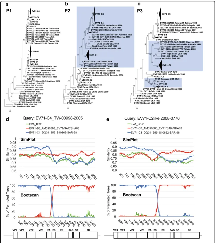
Recent EV-A71 outbreaks have been characterized by an association with newly emerging subgenotypes8. Both the C2 and C4 strains are recombinogenic and carry partial genomes derived through intertypic recombination with coxsackievirus (CV)-A8 and CV-A16, respectively13,14. Furthermore, the B4 strain is an example of intratypic recombination within EV-A71 genotype B13. The emer-gence of various EV-A71 strains within a decade may be attributable to regionally enriched, large-scale outbreaks that can increase the risk of coinfection, a key requirement for recombination15. To examine this possibility, we eval-uated Taiwanese full-genome sequences that consisted of a variety of genotypes/subgenotypes (Table 1) and explored the relationships among these newly emerging strains. EV-A71 and prototype CV (including types A2–A8, A10, A12, A14, and A16) sequences of EV-A were compared to reveal their recombinogenic properties. Breakpoints in EV-A71 recombination are usually located in the P2 and P3 regions16. Therefore, we first reconstructed ML phyloge-netic trees of the P1, P2, and P3 regions (Fig. 3a–c, respectively). Possible recombination events were revealed by changes in the tree positions of analyzed sequences in the subgenomic phylogenies17.
In the phylogenetic tree constructed via the P1 region and rooted with the oldest strain (CVA2-Fleetwood), distinct clades representative of each genotype/sub-genotype were observed, and all sequences of prototype
Table 1 Taiwanese genomes of EV-A71 analyzed in this study
Year Counts (genotype) of sequences acquired in this study
Counts (genotype) of sequences downloaded from ViPR
1986 6 (B1)
1998 6 (C2)
1999 1 (B4)
2000 1 (B4)
2001 1 (B4)
2002 1 (C4)
2003 1 (B5)
2004 11 (C4)
2005 9 (C4) 5 (C4)
2007 1 (C5) 1 (B5), 1 (C5)
2008 9 (B5) 19 (B5), 3 (C2-like), 1 (C4)
2009 2 (B5)
2010 5 (C4) 1 (C4)
2011 2 (B5), 2 (C4) 8 (B5), 6 (B4), 4 (C4)
2012 28 (B5), 2 (C4) 10 (B5), 1 (C2)
2013 1 (B5) 1 (C2)
2014
2015 2 (B5), 1 (C4)
2016 2 (C4)
Total 64 91
(seefigure on previous page)
a
b
a
b
c
d
e
Taiwan for the current study), some differences may still exist between the published full-length genomes and the actual viral population.
Extensive genomic recombinations among the cocirculating enteroviruses
Various viruses belonging to EV-A continuously cocir-culate with EV-A71 in Taiwan (Fig.1a), and many of these non-EV-A71 viruses are recombinant with unknown parents16. Thus, we next examined whether EV-A71 may be involved in the recombination of non-EV-A71 viruses of EV-A. To prevent sampling bias, we collected all of the historical EV-A full-genome sequences worldwide for the following analyses. Since most recombinations in other EV-A viruses also occur outside the P1 region16, we evaluated EV-A71 and CV sequences in the P2/P3 coding region. We utilized Bayesian evolutionary analysis to specify the spatial-temporal relationships among these sequences. When sequences spanning from P2 to the 3′ end of the viral genome were analyzed, several clusters were observed, most of which contained clades corre-sponding to different serotypes of EV-A and subgenotypes of EV-A71 (Fig. 4a). One cluster of particular interest contained the EV-A71 B3, C2-like, and C4 strains as well as several currently circulating viruses (Fig. 4a, red rec-tangle). The details of this cluster are shown in Fig. 4b. Notably, this cluster was proximal to a second cluster containing the prototype sequences of CV-A4, CV-A14, and CV-A16, indicating a potential role of these viruses as recombination parents. Because the evolutionary paths of the circulating strains in phylogenies might be biased by a series of recombination events, we emphasized the detection of incongruous genetic clusters20. Considering the times at which the viruses distributed in this cluster arose, EV-A71 B3 represented the oldest strain among all the branches and may represent the possible origin of the other viruses (Fig. 4b). However, more genomes (parti-cularly of historical EV-A strains) are required to strengthen this conclusion.
We next compared the locations of these recombinant viruses within the phylogenies constructed using either the P2 or P3 sequence. In the P2 phylogeny, the prototype strains of CV-A4, CV-A14, and CV-A16 were redis-tributed to another cluster consisting of most strains of genotype B (Fig.4c, lower), indicating the high sequence similarity among these viruses, which may have promoted the emergence of the EV-A71 B3 strain through intertypic recombination. In the EV-A71 B3-containing cluster, currently circulating rather than prototype strains of CV-A2, CV-A6, CV-A8, and CV-A12 were found. The majority of currently circulating CV-A4 was distributed to another EV-A71 C4-containing cluster, suggesting that additional recombination events involving EV-A71 C4 may occur (Fig.4c, upper). In contrast, all viruses mixed CVappearedasanoutgroup(Fig.3a).Incontrast,
cross-genotypic patterns in the CV-A and EV-A71 sequences
were identified in the P2 and P3 phylogenetic trees (Fig. 3b, c). The inconsistency of these phylogenies reflectedrecombinationevents. Forexample,CV-A8was reported as the recombination parent of EV-A71 C213, andthisresultisreflectedbytheircoclusteringintheP3 region (Fig. 3c, highlighted in blue), indicating that the
high sequence similarity between CV-A8 and EV-A71
genotype C may have facilitated the recombination and emergenceoftheEV-A71C2strain. Similarreclustering also occurred in the C2-like and C4 strains. In the P2 phylogenetic tree, the C2-like strain was an outlier of genotype C of EV-A71, and the C4 cluster was closely relatedtogenotypeBandseveralprototypeCVs(Fig.3b). Theclusteringpatternchanged againinthe P3 phyloge-netictree,and anewclustercontainingEV-A71B3, C2-like,andC4wasformed.Additionally,prototypesCV-A4,
CV-A14, and CV-A16 were redistributed into the same
cluster (Fig. 3c). Given that the EV-A71 C4 strain was regarded as a double-recombinant virus containing EV-A71genotypeB-like P2andCV-A16-likeP3regionsand thattheB3strainwasarecombinantwithaCV-A16-like 3D region14,18,19, our resultssuggested that the EV-A71 B3strainmaybethepossiblerecombinationparentofthe
C4 strain. Similarly, the C2-like strain may be an
uncharacterizedrecombinantEV-A71thatalsooriginated fromthe B3strain. Thesepredictionswereconfirmedby
SimPlot analysis (Fig. 3d, e). When comparing the
C4 strain to reference strains including the EV-A71
genotypes A,B3, and C1, its 5′ region showed ahigher similarity (approximately 88%)to that of EV-A71 geno-typeC1;however, thesimilaritydecreased atthe
bound-ary of the 2A/2B coding region, and this effect was
accompanied by an increased similarity (approximately 80%) to B3 toward the 3′ half ofthe viral genome. The
recombinant breakpoint was mapped to approximately
nucleotide position 2881 of the coding sequence (CDS) (Fig. 3d). The shift of predominant similarity from one reference strain to another indifferent genomic regions
was also observed when we queried C2-like strain
sequences. The C2-like genome contained genotype
C-and B3-similar sequences at the 5′ and 3′ regions, respectively.However,thebreakpointoftheC2-likestrain
was mapped to the downstream 2C coding region
and genotype, respectively). The overall identity of amino acid sequences among these viruses still reached 94%5. The effects of mutations and recombinations on EV-A71 evo-lution have been extensively discussed, and although sev-eral VP1 amino acids are under positive selection, EV-A71 may be subjected to strong negative selection, which the-oretically should result in a stabilized and purified virus7,10. Thus, recombination may have played an important role in the appearance of diverse EV-A71 strains such as B3, B4, C2, and C4 since 199713,14,18,19. Intratypic recombinations have also occurred in different EV species, with a higher frequency in EV-B species than in EV-A and EV-C22. Here, by analysis of full-genome sequences collected in Taiwan from 1998 to 2016, we determined when and where recombination occurred and how these events could have led to the emergence of the different strains associated with EV-A71 outbreaks. Although published genomes have limited value as a proxy for the actual viral population, several recombination events can be revealed by analyzing the phylogenetic relationships among EV-A71 sub-genotypes. We did not rule out the importance of mutation during EV-A71 evolution23. Instead, a combi-nation of both recombicombi-nation and mutation may result in the rapid switching between different EV-A71 strains within a short time period. In the future, more genomes will be needed to decipher the evolutionary history of EV-A71.
Extensive recombination in cocirculating viruses
Sequences with high similarity to those of the prototype strains CV-A8 and CV-A16 were found in the EV-A71 C2 and B3/C4 strains, respectively13,14,18,19. However, con-sidering the requirement of coinfection for recombina-tion, it is possible to obtain the“non-self” genome from cocirculating viruses15. In addition, as one of the countries having cocirculation of multiple EV-A71 subgenotypes and several EV-A viruses, Taiwan represents a good niche for clarifying the relationships among cocirculating viru-ses8. In this study, we proposed that the emergence of the EV-A71 C2-like and C4 strains may be explained by intratypic recombination with the B3 strain. Although the presence of the B3 strain in Taiwan has not been pre-viously documented, it was involved in the recombination of the B4 strain, which caused severe outbreaks in the early 2000s12,13. The intensive recombinations were not EV-A71-specific and could also be found in several
cur-rently circulating EV-A viruses through intertypic
recombinations. Among these viruses, only A2, CV-A4, and CV-A6 have been documented in Taiwan. How-ever, all these viruses have been reported to be common causes of HFMD in China and are recombinogenic with EV-A7124. Interestingly, a novel EV-A71 genotype C strain with a mosaic genome structure has been identified in
Germany and Denmark25,26. This new strain had a
together without clear assortment in the P3 phylogeny (Fig. 4d). This sporadic distribution indicated that most virusesmayhavesimilarP3regions.Thus,recombination
may occur among the cocirculating viruses in the P2
region,whichcouldresultinacommonP3regionshared by these viruses. To verify this hypothesis, SimPlot ana-lyses were carried out to examine the recombination of currentlycirculatingCVs(Fig.5).WhenEV-A71B3was incorporated as the reference strain, a single crossover
was foundintheP1/P2 boundarywhenCV-A2, CV-A6,
CV-A8,andCV-A12wereanalyzed(Fig.5a–d).Therole
of EV-A71C4 inthe recombinant CV-A4 and an
addi-tionalintertypicrecombinationbetweenCV-A4and CV-A2werealsoconfirmed(Fig.5e,f).Toeliminatesampling bias, consensus sequences of these recombinant strains
fromEV-A71B3, B5, C2-like, and C4, and CV-A2,
CV-A4, CV-A6, CV-A8,CV-A12, and CV-A16 werefurther
generatedforcomparisontotheprototypestrainsof EV-A71and CVs.Eleven EV-A71-likesignatures were iden-tifiedinthecirculatingstrainsofEV-A71C2-likeandC4
and CV-A2, CV-A4, CV-A6,and CV-A12, but not their
prototype strains, except for CV-A4 and CV-A16
(Table 2). Thus, in addition to CV-A1614,18,19,21, CV-A4 mightbeanotherpotentialrecombinationparentofthese currentlycirculatingviruses.Consistentwiththeresultsof theBayesianphylogenetictree(Fig.4d)andSimPlot pre-dictions (Fig. 5), all signatures were located in the P3 region(Table2),which mightbecausedbytheintensive recombinationsinthe P2region.Suchsignatures cannot befoundinthecirculatingCV-A16strain,whichhasbeen reportedtoberecombinantwithEV-A71genotypeA16, or intheEV-A71 B5strain, whichevolvesindependently of otherviruses.Bothstrainscarrysequencessimilartothose oftheEV-A71prototypestrain.
Discussion
Inthisstudy,wefoundthatmanycurrentlycirculating EV-Astrainshaveundergonerecombinationandthat EV-A71 B3 may have played a central role in this process, basedonthelatestpublisheddatabaseofEV-Afull-length genomes(Fig.6).Itisexpectedthatmoregenomeswillbe publishedand willbe added tothis simplified flowchart. Throughaseriesofintra-andintertypicrecombinations, EV-A71-like signatures were foundto be widelypresent in many currently circulating EV-A viruses (Table 2). Althoughthe impactofthesesignatures onviral replica-tion remains unclear, their prevalence in various EV-A
viruses may have applications in the development of
broad-spectrumantivirals.
Roleofrecombination intheevolutionofEV-A71
C1-like VP1 region; however, the 5′ untranslated region and the P2/P3 region showed higher similarity to EV-A71 B3/C2-like and C4, respectively. This new EV-A71 strain may have been generated by recombination of the locally circulating C1 strain with the imported C4 strain that became dominant recently27. Although intensive recom-bination of cocirculating EV-A viruses with EV-A71 has been observed, there are some exceptions. Both CV-A5 and CV-A10 are commonly detected by the Taiwan enterovirus surveillance system; however, no recombina-tion with EV-A71 has been observed. Instead, a close relationship in the nonstructural region, possibly caused by intertypic recombination between circulating CV-A5 and CV-A10, has been reported28. Therefore, recombi-nation between cocirculating viruses may be more com-mon than expected, and full-genome sequencing rather than sequencing of VP1 only should be considered when encountering a new epidemic.
Hot spots for recombination: functional impact of genome reassembly
In all recombination events identified in this study, breakpoints were mapped to the region extending from
the P1/P2 boundary to the 2C region (Fig.6a), suggesting the existence of recombination hot spots. Thus, recom-bination can result in genome reassembly of the struc-tural and nonstrucstruc-tural regions. The restricted location of the breakpoints may have resulted from natural selection. Delicate cooperation among picornavirus viral proteins and genomes is required for productive viral
replication29. Because the genome of EV-A can be
directly translated into a polyprotein and then undergo proteolytic cleavage, changes in the functional entities by recombination could be deleterious to the virus. For example, viral 3Dpolis required for viral replication and recognizes several cis-elements throughout the gen-ome29. In the case of EV-A71 B3, due to the presence of the CV-A16-like 3Dpol, the virulence was decreased when compared with those of EV-A71 B4 and CV-A16 in mouse model infections21. Therefore, the EV-A71-like signatures that consist of EV-A71 genotype B-like P2 and CV-A16-like P3 regions may have been less favored and discarded in the evolution of EV-A71 genotype B (Fig.6a, black rectangle). However, as we have shown here, EV-A71-like signatures are tolerated by EV-A71 genotype C and several CVs of EV-A, and the numbers of appropriate
Table 2 Amino acid positions of 11 signatures carried by circulating strains of recombinant EV-A viruses
Gene 3A 3C 3D
Position 39 36 95 44 76 94 134 138 368 428 451
Consensus sequence of circulating strains
EV-A71 B3 D V S H E Q T V N Q Y
EV-A71 C2-like . . . .
EV-A71 C4 . . . .
CVA2 . . . .
CVA4 . . . .
CVA6 . . . .
CVA8 . . . .
CVA12 . . . .
CVA16 E I T T Q K V T T E F
EV-A71 B5 E . . T Q K V . T D F
Prototype strains (Strain, Country, Year)
EV-A71 A (BrCr, USA, 1970) E I T T Q K V T T E L
CVA2 (Fleetwood, USA, 1947) E I T T Q K V T T E F
CVA4 (HighPoint, USA, 1948) . . . .
CVA6 (Gdula, USA, 1949) E I T T Q K A T T E F
CVA8 (Donovan, USA, 1949) . . . T . K . . T E F
CVA12 (Texas-12, USA, 1948) E I T T Q K V T T E F
CVA16 (G-10, SOA, 1951) . . . .
recombination acceptors could keep increasing25,26. Because a high sequence similarity is preferred for copy-choice recombination, as detailed in the widely accepted
model of RNA virus recombination15, the sequence
identity between the EV-A71 C4 and B5 strains should definitely be higher than that between the EV-A71 and EV-A viruses. Restricted recombination under natural selection may explain why EV-A71 C4 and B5 cocircu-lated but did not recombine, exhibiting independent evolution. We currently have no evidence to conclude whether recombination may be beneficial for the virus; however, the ratio of recombinant CV-A6-associated HFMD has increased worldwide24,30. Here, we provide only evidence demonstrating the shared nonstructural proteins of cocirculating EV-A viruses. Through the establishment of sequence databases that integrate complete sets of full-genome sequences, we might able to
predict what kinds of genome assembly and possible recombinants might appear in the future.
Materials and methods
Specimen collection and sequencing of EV-A71
All of the 64 EV-A71 clinical specimens isolated from 2005 to 2016 were provided by the Linkou Chang Gung Memorial Hospital, Taiwan. Regardless of the illness diagnosed, we randomly picked clinical samples from epidemics in this time span. To prevent contamination,
amplified viral stocks from human rhabdomyosarcoma
cells were used for full-genome sequencing. Viral gen-omes were recovered using TRIzol LS reagent (Thermo Fisher Scientific, Waltham, MA, USA) according to the manufacturer’s instructions. The 59 samples collected before 2014 were sequenced by Sanger sequencing. Oligo-(dT)20 was used to prepare poly(A)-containing viral
Acknowledgements
This work wasfinancially supported by a grant from the Human Frontier Science Program (RGP0011/2015), the Ministry of Science and Technology (MOST), Taiwan (MOST 107-3017-F-182-001), and the Research Center for Emerging Viral Infections from The Featured Areas Research Center Program within the framework of the Higher Education Sprout Project by the Ministry of Education (MOE) in Taiwan.
Authors' contributions
K.-M.L. and S.-R.S. conceptualized and designed the study in collaboration with N.H.D. and C.E.C. K.-M.L. drafted the manuscript and participated in the collection of sequence data, phylogenetic and recombination analyses, and figure preparation. Y.-N.G. collected sequence data, performed the next-generation sequencing of clinical isolates and phylogenetic and
recombination analyses, and prepared thefigures. T.-H.H. prepared and stored the virus stock of all clinical isolates and performed the Sanger sequencing of clinical isolates. A.W., N.H.D., and C.E.C. were involved in conceptualization of the study and contributed to the writing of the manuscript. All authors have critically reviewed the manuscript.
Author details
1Research Center for Emerging Viral Infections, College of Medicine, Chang
Gung University, Taoyuan, Taiwan.2Department of Medical Biotechnology and Laboratory Science, College of Medicine, Chang Gung University, Taoyuan, Taiwan.3Department of Biochemistry and Molecular Biology, The Pennsylvania State University, University Park, PA 16802, USA.4Department of
Bionanoscience, Kavli Institute of Nanoscience, Delft University of Technology, Van der Maasweg 9, Delft 2629 HZ, The Netherlands.5Department of Laboratory Medicine, Linkou Chang Gung Memorial Hospital, Taoyuan, Taiwan. 6
Research Center for Chinese Herbal Medicine, Research Center for Food and Cosmetic Safety, and Graduate Institute of Health Industry Technology, College of Human Ecology, Chang Gung University of Science and Technology, Taoyuan, Taiwan
Conflict of interest
The authors declare that they have no conflict of interest.
Supplementary Informationaccompanies this paper at (https://doi.org/ 10.1038/s41426-018-0107-0).
Received: 13 February 2018 Revised: 27 April 2018 Accepted: 29 April 2018
References
1. Solomon, T. et al. Virology, epidemiology, pathogenesis, and control of enterovirus 71.Lancet Infect. Dis.10, 778–790 (2010).
2. Ho, M. et al. An epidemic of enterovirus 71 infection in Taiwan. Taiwan Enterovirus Epidemic Working Group.N. Engl. J. Med.341, 929–935 (1999).
3. Chang, L. Y. Enterovirus 71 in Taiwan.Pediatr. Neonatol.49, 103–112 (2008). 4. Bessaud, M. et al. Molecular comparison and evolutionary analyses of VP1
nucleotide sequences of new African human enterovirus 71 isolates reveal a wide genetic diversity.PLoS ONE9, e90624 (2014).
5. Brown, B. A., Oberste, M. S., Alexander, J. P. Jr, Kennett, M. L. & Pallansch, M. A. Molecular epidemiology and evolution of enterovirus 71 strains isolated from 1970 to 1998.J. Virol.73, 9969–9975 (1999).
6. Saxena, V. K., Sane, S., Nadkarni, S. S., Sharma, D. K. & Deshpande, J. M. Genetic diversity of enterovirus A71, India.Emerg. Infect. Dis. 21, 123–126 (2015).
7. Tee, K. K. et al. Evolutionary genetics of human enterovirus 71: origin, popu-lation dynamics, natural selection, and seasonal periodicity of the VP1 gene.J. Virol.84, 3339–3350 (2010).
8. McMinn, P. C. Recent advances in the molecular epidemiology and control of human enterovirus 71 infection.Curr. Opin. Virol.2, 199–205 (2012). 9. Christian, K. A. et al. What we are watching–five top global infectious disease
threats, 2012: a perspective from CDC’s Global Disease Detection Operations Center.Emerg. Health Threats J.6, 20632 (2013).
cDNA using a ReverTra Ace -α- kit (Toyobo, Osaka,
Japan). Overlapping amplicons covering the entire viral genome were amplified by different setsof primers18,31,
and genome assembly was carried out using SeqMan
software (DNASTAR, Inc., Madison, WI, USA). The
five samples collected after 2014 were sequenced using theIlluminaHiSeqplatformfor next-generation
sequen-cing (NGS). NGS data preprocessing included the
removaloflow-quality andhostreads. Usingthe
Taiwa-nese B5 and C4 strains as an initial template, the
viral genomes were assembled by an iterative mapping approach32.Atotal of64genomesobtainedinthisstudy
were deposited in GenBank with accession numbers
MG756691–MG756754.
DatacollectionforEV-AgenomesfromViPRand recombinationanalysis
Eighthundred thirty-oneEV-A71 genomes worldwide
were initially retrieved from ViPR in September 2017. Sequenceswithambiguousnucleotidesorwithoutknown sampling dates and countries were removed. To reduce redundancy,werandomlyselected10sequenceswiththe samegenotypeandisolationyearfromeachcountry.We then collected 427 EV-A71sequences includingall Tai-wanese strains for analysis (Supplementary Table 1).
Moreover,780completeCVgenomes(belonging to
EV-A)weredownloadedfromViPR.Afterdatapreprocessing, 351 CVgenomeswerecollected. Detailsof the analyzed sequencesareshown inSupplementaryTable 2.
Recom-bination between the EV-A71 and CV genomes in this
study was detected using SimPlot (version 3.5.1) with a slidingwindowsizeof600ntandastepsizeof20nt33. To identify genomic signatures associated with detected recombination in this study, a consensus sequence for each ofthe serotypes/genotypes was generated by using theConstoolwiththedefaultsettingfromEMBOSS31.
Phylogenetictreeanalysis
TheMLmethodbasedontheHasegawa–Kishino–Yano
(HKY) model was performed to infer the evolutionary
history34. Thepercentage ofreplicate treesin which the associatedtaxaclusteredtogetherinabootstraptestwith 1000replicateswascalculated.Allpositionswithlessthan 95%sitecoveragewereeliminated.Evolutionaryanalyses wereconductedinMEGA735.Furthermore,theBayesian phylogenetic tree was inferred by BEAST36 with BEA-GLE37toestimatethemaximumcladecredibility(MCC)
tree under the HKY model. Based on our collected
sequences,wegenerated50millionMarkovchainMonte
Carlo(MCMC)chainswith10%burn-in.OneMCC tree
was constructed for every 25,000 chains, and a single
consensus tree was summarizedfrom these MCC trees.
MCMCwas alsousedinBEASTtoestimatethetime of
10. Chen, X. et al. Analysis of recombination and natural selection in human enterovirus 71.Virology398, 251–261 (2010).
11. Mao, Q., Wang, Y., Bian, L., Xu, M. & Liang, Z. EV-A71 vaccine licensure: afirst step for multivalent enterovirus vaccine to control HFMD and other severe diseases.Emerg. Microbes Infect.5, e75 (2016).
12. Wang, S. F. An epidemiological analysis of enterovirus 71: Taiwan, 1998–2004. Taiwan Epidemiol. Bull.21, 125–153 (2005).
13. Huang, S. W. et al. Reemergence of enterovirus 71 in 2008 in taiwan: dynamics of genetic and antigenic evolution from 1998 to 2008.J. Clin. Microbiol.47, 3653–3662 (2009).
14. Yip, C. C. et al. Emergence of enterovirus 71“double-recombinant”strains belonging to a novel genotype D originating from southern China:first evi-dence for combination of intratypic and intertypic recombination events in EV71.Arch. Virol.155, 1413–1424 (2010).
15. Simon-Loriere, E. & Holmes, E. C. Why do RNA viruses recombine?Nat. Rev. Microbiol.9, 617–626 (2011).
16. Kyriakopoulou, Z., Pliaka, V., Amoutzias, G. D. & Markoulatos, P. Recombination among human non-polio enteroviruses: implications for epidemiology and evolution.Virus Genes50, 177–188 (2015).
17. Posada, D., Crandall, K. A. & Holmes, E. C. Recombination in evolutionary genomics.Annu. Rev. Genet.36, 75–97 (2002).
18. Yoke-Fun, C. & AbuBakar, S. Phylogenetic evidence for inter-typic recombi-nation in the emergence of human enterovirus 71 subgenotypes. BMC Microbiol.6, 74 (2006).
19. Chan, Y. F. et al. Comparative genetic analysis of VP4, VP1 and 3D gene regions of enterovirus 71 and coxsackievirus A16 circulating in Malaysia between 1997-2008.Trop. Biomed.29, 451–466 (2012).
20. Biek, R., Pybus, O. G., Lloyd-Smith, J. O. & Didelot, X. Measurably evolving pathogens in the genomic era.Trends Ecol. Evol.30, 306–313 (2015). 21. Chan, Y. F. & AbuBakar, S. Human enterovirus 71 subgenotype B3 lacks
cox-sackievirus A16-like neurovirulence in mice infection.Virol. J.2, 74 (2005). 22. Simmonds, P. & Welch, J. Frequency and dynamics of recombination within
different species of human enteroviruses.J. Virol.80, 483–493 (2006). 23. Huang, S. W. et al. Mutations in the non-structural protein region contribute to
intra-genotypic evolution of enterovirus 71.J. Biomed. Sci.21, 33 (2014). 24. Guo, W. P. et al. Fourteen types of co-circulating recombinant enterovirus
were associated with hand, foot, and mouth disease in children from Wenzhou, China.J. Clin. Virol.70, 29–38 (2015).
25. Bottcher, S., Obermeier, P. E., Neubauer, K. & Diedrich, S., Laboratory Network for Enterovirus, D. Recombinant enterovirus A71 subgenogroup C1 strains, Germany, 2015.Emerg. Infect. Dis.22, 1843–1846 (2016).
26. Midgley, S. E. et al. Co-circulation of multiple subtypes of enterovirus A71 (EV-A71) genotype C, including novel recombinants characterised by use of whole genome sequencing (WGS), Denmark 2016. Euro. Surveill. 22. https://doi.org/10.2807/1560-7917.ES.2017.22.26.30565 (2017).
27. Fischer, T. K. et al. Emergence of enterovirus 71 C4a in Denmark, 2009 to 2013. Eur. Surveill.19, 20911 (2014).
28. Hu, Y. F. et al. Complete genome analysis of coxsackievirus A2, A4, A5, and A10 strains isolated from hand, foot, and mouth disease patients in China revealing frequent recombination of human enterovirus A.J. Clin. Microbiol. 49, 2426–2434 (2011).
29. Paul, A. V. & Wimmer, E. Initiation of protein-primed picornavirus RNA synthesis.Virus Res.206, 12–26 (2015).
30. Cabrerizo, M. et al. Molecular epidemiology of enterovirus 71, coxsackievirus A16 and A6 associated with hand, foot and mouth disease in Spain.Clin. Microbiol. Infect.20, O150–O156 (2014).
31. Zhang, Y. et al. Complete genome analysis of the C4 subgenotype strains of enterovirus 71: predominant recombination C4 viruses persistently circulating in China for 14 years.PLoS ONE8, e56341 (2013).
32. Gong, Y. N. et al. A next-generation sequencing data analysis pipeline for detecting unknown pathogens from mixed clinical samples and revealing their genetic diversity.PLoS ONE11, e0151495 (2016).
33. Lole, K. S. et al. Full-length human immunodeficiency virus type 1 genomes from subtype C-infected seroconverters in India, with evidence of inter-subtype recombination.J. Virol.73, 152–160 (1999).
34. Hasegawa, M., Kishino, H. & Yano, T. Dating of the human-ape splitting by a molecular clock of mitochondrial DNA.J. Mol. Evol.22, 160–174 (1985).
35. Kumar, S., Stecher, G. & Tamura, K. MEGA7: Molecular Evolutionary Genetics Analysis Version 7.0 for bigger datasets.Mol. Biol. Evol.33, 1870–1874 (2016).
36. Drummond, A. J. & Rambaut, A. BEAST: Bayesian evolutionary analysis by sampling trees.BMC Evol. Biol.7, 214 (2007).