ORIGINAL ARTICLE
Iran J Allergy Asthma Immunol December 2019; 18(6):679-687.
Cytotoxicity Assessment and Apoptosis-related Gene Profiling of Antibody
Treated Acute Myeloid Leukemia (AML) and Acute Lymphocytic
Leukemia
(
ALL) Cancerous Cell Lines
Mahdi Habibi-Anbouhi1, Zahra Kafi1, Leila Ghazizadeh1, Shabnam Kharazi1, Mahdi Behdani2, Fatemeh Faraji1, and Mohammad Ali Shokrgozar1
1
National Cell Bank of Iran, Pasteur Institute of Iran, Tehran, Iran
2 Biotechnology Research Center, Pasteur Institute of Iran, Tehran, Iran
Received: 19 April 2018; Received in revised form: 30 June 2019; Accepted: 18 July 2019
ABSTRACT
Acute myeloid leukemia (AML) and acute lymphocytic leukemia (ALL)
are common
acute leukemia in adults and children, respectively. In these malignancies, chemotherapy is
the main treatment strategy that fails in many cases and is usually associated with adverse
effects on healthy cells. In this regard, the development of new therapies is essential.
Monoclonal antibodies directed to the cell surface markers of leukemic blasts may have
promising consequences with minimal toxic effects on normal cells. Since
cluster of
differentiation 45Ra (CD45Ra) and CD123 antigens, two considered surface markers of
leukemic blasts in AML and ALL respectively, are overexpressed on AML and ALL
blasts, CD34
+leukemic progenitors, and AML-LSCs in comparison with normal
hematopoietic stem cells (HSCs), they were selected to be targeted; using specific
monoclonal antibodies.
In this project, CD45Ra
+cells and CD123
+cells were targeted by anti-CD45Ra and/or
anti-CD123 monoclonal antibodies. Cytotoxicity effect and cell death induction was
determined by 3-(4,5-dimethylthiazol-2-yl)-2–5-diphenyltetrazolium bromide (MTT) assay
and flow cytometry. Changes in the expression profile of MCL1, cMyc, Survivin, Id1, and
PIM1 genes were assessed by real-time PCR.
Statistical analysis of the results showed effective antibody-mediated cytotoxicity and
induction of apoptosis in KG1α (CD45Ra
+) and Nalm6 (CD123
+) cell lines. Also, a
significant change in the expression level of some of the apoptosis-related genes was
observed
.
According to the results of this study, it can be concluded that an effective targeting of
AML and ALL cancerous cell lines can be performed by anti-CD45Ra and anti-CD123
monoclonal antibodies through their effector functions and apoptosis induction.
Keywords:
Cytotoxicity assessment;
Apoptotic genes; Acute leukemia cell lines;
Immunotherapy
Corresponding Authors:Mohammad Ali Shokrgozar, PhD; National Cell Bank of Iran, Pasteur Institute of Iran, Tehran, Iran. Tel/Fax: (+98 21) 6649 2595, E-mail: mashokrgozar@pasteur.ac.ir.
Mahdi Habibi-Anbouhi, PhD;
INTRODUCTION
Leukemia is a hematopoietic malignancy. There are four main types of leukemia; acute myeloid leukemia (AML), cronic myeloid leukemia (CML), acute lymphocytic leukemia (ALL), and chronic lymphocytic leukemia (CLL).1
Studies have revealed that in patients with leukemia, there is a group of cells with a very low percentage that is called leukemic stem cells (LSCs). LSCs are considered as a major source of relapse and drug resistance in leukemia and it is expected to achieve a durable treatment of this malignancy through targeting these stem cells.2,3
Monoclonal antibodies (mAbs) are able to attach to a specific target, such as biomarkers on the surface of cancer cells. These antibodies can exert their anti-tumor effects by various mechanisms including antibody-dpendent cell-mediated cytotoxicity (ADCC), cmplement dependent cytotoxicity (CDC), enhancement of cytokine release, and induction of apoptosis.
Monoclonal antibodies are often used in the treatment of lymphomas and their use in leukemia has been limited.4 Therefore, targeting leukemic cells; especially cells which are involved in the initiation of leukemia using mAbs can bring hopes to treat leukemia patients in a specific manner.5,6
CD45Ra and CD123 antigens are widely reported to be overexpressed on AML and ALL blasts, CD34+ leukemic progenitors, and AML-LSCs in comparison with normal hematopoietic stem cells (HSCs).7-10
Also, it was recently shown that in patients with AML, LSCs express CD45Ra cell surface biomarker in addition to CD123. Therefore, targeting these cell surface biomarkers simultaneously by mAbs might provide a more efficient treatment for leukemia patients because of dual targeting of cancerous cells.9
At the molecular level, a variety of alterations in numerous genes including Myc (c-Myc), Inhibitor of DNA binding (Id1), Survivin, myeloid cell leukemia 1 (MCL1),and Proto-oncogene serine/threonine-protein kinase (PIM1), leads to the emergence of leukemia. These genes play an important role in cell growth, differentiation, and cell death processes.11-15
As mentioned earlier different studies have shown the implication of Myc (c-Myc), Id1, Survivin, MCL1, and PIM1 genes in the cancer formation but there is a lack of investigation of immunotherapy effects on these
genes. Therefore, in this study, the suitability of the immunotherapy in AML and/or ALL treatment and its effects on the mentioned genes were assessed through evaluation of the apoptosis induction and cell cytotoxicity in the antibody-treated cancerous cells.
MATERIALS AND METHODS
AML and ALL Cell Lines and Cell Culture
KG1a (AML cell line), Nalm6 (ALL cell line), and HUVEC (negative control) cell lines were obtained from the National Cell Bank of Pasteur Institute of Iran. This study was performed according to the protocols of the Ethical Committee of Pasture institute of Iran. Briefly, the KG1a and Nalm6 cells were cultured in RPMI and HUVEC cells were expanded in Dulbecco's Modified Eagle Medium (DMEM) medium (Gibco, USA) containing 15% fetal bovine serum (FBS) (NZA, Iran) at 37° C and 5% CO2 at a cell density of about 5×10⁵ cells/ml. The cells with the viability of more than 95% were used in subsequent studies.
Cytotoxicity Assay
Direct and indirect antibody-mediated cytotoxicity was measured by flow cytometry; using mouse anti-human CD45Ra (MEM-56, Thermo Scientific, USA) and mouse anti-human CD123 (7G3, BD Biosciences, USA) monoclonal antibodies, rabbit complement (Cedarlane Labs, Canada) and Annexin V/PI Apoptosis Detection Kit FITC (eBioscience, USA).
Briefly, 5×10⁴ cells of KG1a, Nalm6, and HUVEC cell lines were seeded in each well of a 96-well cell culture plate (Nunc, Denmark) in 200 µL of DMEM medium supplemented with 5% FBS. After 16 h of cell incubation at 37°C and 5% CO2, 0.5 µL of
After 1h of incubation at 37°C, cell supernatants were discarded and the cell pellets were washed and resuspended in 100 µL of 1X binding buffer. Thereafter, 100 μL of FITC-conjugated Annexin V was added to each sample and the plate was incubated 10 min at room temperature in the dark. The cells were washed and resuspended in 200 μL of 1X binding buffer. Five μL of propidium iodide (PI) (Sigma, Germany) staining solution was added and the samples were analyzed by flow cytometry and Flowmax 2.4 software (Partec, Germany). HUVEC cells and PBS/1% mouse normal serum were used as negative cell and antibody controls, respectively.
Apoptosis-related Gene Profiling by Real-time PCR (qPCR)
To study and quantify the effect of antibody treatments on alteration of apoptosis-related gene expression, a real-time PCR experiment was done. Briefly, three groups of samples from the KG1a, Nalm6, and HUVEC cell lines were cultured (5×10⁵ cells/mL/well) and treated with the anti-CD123 and/or CD45Ra mAbs (2 µg of each) and PBS as a negative control.
To investigate the alteration of genes expression the Myeloid cell leukemia 1 (MCL1), Proto-Oncogene Serine/Threonine-Protein Kinase Pim-1 (PIM1), Inhibitor of DNA binding 1 (Id1), Survivin, and Cellular Myelocytomatosis (cMyc) genes were chosen to evaluate the treatments. The Eukaryotic translation
elongation factor 1 alpha 1 (eEF1A1) housekeeping gene was included in this study as the reference gene. Forward and reverse primers were designed using Primer Express software v3.0.1 (Thermo Fisher, USA) for each gene (Table 1).
The total RNA was isolated from the treated and control cell samples; using the YTA RNA Extraction kit (Yekta Tajhiz Azma, Iran) according to the manufacturer’s instruction. The concentration and purity of RNA were determined by measuring optical density (OD 260/280). First-strand cDNA was synthesized from 200 ng RNA using the QuantiTect Reverse Transcription Kit (Qiagen, Germany). The assay was performed using the SYBR Green PCR master mix (Applied Biosystems, USA) and the comparative cycle threshold method after 35 cycles of amplification. To obtain the relative gene expression level (ΔCt) for each gene in each sample, Ct values for
eEF1A1 housekeeping gene was subtracted from the Ct
values of each gene. The ΔCt of treated samples was
then normalized to the value of the control group.
Statistical Analysis
Each experiment was performed in triplicate. The data presented the mean±SD of three independent experiments. To statistically compare data for each test group with those of control groups, the Student’s t-test was performed with statistical significance set at a p -value of<0.05.
Table 1. List of qPCR primers used in expression profiling of Id1, MCL1, cMyc, PIM1, Survivin, and ELF1A1 genes
Gene
(GenBank Accession) Primer sequence (5'…. 3')
Id1
(NM_181353)
Forward CTG CTC TAC GAC ATG AAC GG Reverse GAA GGT CCC TGA TGT AGT CGA T MCL1
(NM_021960)
Forward GTG CCT TTG TGG CTA AAC ACT Reverse AGT CCC GTT TTG TCC TTA CGA cMyc
(NM_012333)
Forward AAT AGA GCT GCT TCG CCT AGA Reverse GAG GTG GTT CAT ACT GAG CAA G PIM1
(NM_001243186)
Forward GAG AAG GAC CGG ATT TCC GAC Reverse CAG TCC AGG AGC CTA ATG ACG Survivin
(NM_001012270)
Forward AGG ACC ACC GCA TCT CTA CAT Reverse AAG TCT GGC TCG TTC TCA GTG ELF1A1
(NM_001402)
RESULTS
Cytotoxicity Assays
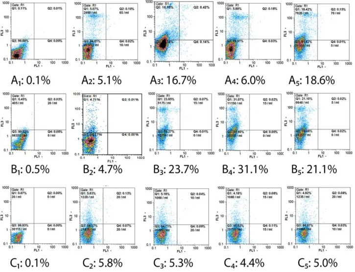
Antibody-mediated cell cytotoxicity, including apoptosis, in all treatment conditions, was studied by flow cytometry. As depicted in Figure 1 and Figure 2, the cytotoxicity rates of KG1a cell-mediated by anti-CD45Ra and/or anti-CD123 monoclonal antibodies
were about 16.7% with anti-CD45Ra mAb, 6% with anti-CD123 mAb, and 18.6% with anti-CD45Ra and CD123 mAbs. In the case of the Nalm6 cell line, under the same condition, the antibody-mediated cytotoxicity of 23.7%, 31.1%, and 21.1% was observed, respectively. Cytotoxicity of about 5% was observed for negative control cells, HUVEC cells, in the presence of complement.
Figure 2. Summarized data of cytotoxicity assessment (Figure 1) of anti-CD45Ra and/or anti-CD123 antibody treatments on KG1a, Nalm6, and HUVEC cells. Each value represents the mean ± S.D. (P ≤ 0.05) from triplicate experiments. Treatment with anti-CD45 Ra/anti-CD123 antibody alone or in combination had more influence on the Nalm6 cell line in comparison to the KG1a cell line. In both cell lines (Nalm6 and KG1a) in comparison to control cell line (HUVEC) cell cytotoxicity increased in all treatments.
Apoptosis Gene Profiling
To investigate alterations in the apoptosis-related genes caused by anti-CD45Ra and/or anti-CD123 mAbs treatments, the expression levels of PIM1, ID1, Survivin, cMyc, and MCL1 genes relative to eEF1A1 reference gene in KG1a, Nalm6, and HUVEC cells were evaluated by real-time PCR (Figure 3). The relative expression levels of the PIM1 gene did not change in KG1a cells in all antibody treatment conditions. In contrast, a significant increase was measured for this gene in Nalm6 cells after CD45Ra (2.59), CD123 (2.95), and anti-CD45Ra+anti-CD123 (1.61) mAbs treatments in comparison with the reference gene.1 Survivin transcript was decreased significantly after anti-CD45Ra (0.68) and anti-anti-CD45Ra+anti-CD123 (0.68) mAbs treatments for KG1a cell line, and
after anti-CD45Ra+anti-CD123 (0.71) mAbs treatment for Nalm6 cells. The c-Myc mRNA increased relatively after treatment with anti-CD123 (1.85) mAb in the Nalm6 cell line; while no changes were observed in KG1 cells in all treatment conditions. A significant decreased was detected for Mcl-1 mRNA expression level in KG1a cells after anti-CD45Ra+anti-CD123 (0.75) mAbs treatment. Moreover, no change was detected for the Nalm6 cell line for this gene. Finally, a decrease in the mRNA level of the Id1 gene was measured in anti-CD123 treated Nalm6 cells (0.51). The other treatments had no significant effects on this gene in none of the cell lines.
DISCUSSION
In order to show the effects of targeting CD123 and CD45Ra cell surface markers, we used two monoclonal antibodies and leukemic cell lines that were treated with these antibodies in different ways.
Analysis of the data demonstrated that treatment with anti-CD123 and/or anti-CD45Ra monoclonal antibodies in KG1a cells (AML cell line) and Nalm6 cells (ALL cell line) affects cell survival. The results of the cytotoxicity assay showed that treatment with anti-CD45Ra antibody in combination with anti-CD123 antibody or alone had more influence on KG1a cell line survival in comparison to treatment with anti-CD123 antibody alone. Accordingly, the antibody-mediated cytotoxicity rates of 18.6% and 16.7% were observed, respectively. Whereas in the Nalm6 cell line, treatment with anti-CD123 alone showed more cytotoxicity (31.1%) rather than treatment with anti-CD45 Ra alone (23.7%) or combination treatment (21.1%). In HUVEC cells as a negative control cell, cytotoxicity of about 5% was achieved through the treatments.
In spite of limited clinical data that is present to examine the influence of treating AML patients with CD123 antibodies, there are strong pre-clinical data for unconjugated anti-CD123. It was shown that h7007; an unconjugated humanized anti-CD123 monoclonal antibody, decreases LSCs and enhances survival in xenograft models.16,17 Interleukin-3 (IL-3) that is bound to CD123 is involved in cell proliferation and differentiation. Therefore, blocking CD123; using monoclonal antibodies reduces cell survival. Furthermore, outcomes of phase 1 studies with h7007 in AML patients showed that this agent induces ADCC in patients although has no remarkable clinical activity in relapsed/refractory active AML patients.18
In our study, the treatment of Nalm6 cells with anti-CD123 showed significant cell cytotoxicity rate (31.5%) but it did not exert any remarkable influences on KG 1a cells. However, anti-CD45 Ra treatment had more effects on KG 1a cells and demonstrated 16% cell cytotoxicity. CD45 is expressed on more than 85% of leukemic samples with about 200,000 molecules per cell copy number.19 There are limited studies on targeting CD45 in leukemic patients and more studies are needed to be done and show effects of immunotherapy through targeting this marker in leukemia.
In addition, the alterations induced by
anti-CD45Ra and/or anti-CD123 mAbs treatments in expression levels of the apoptosis-related genes were investigated in our study. Analysis of the collected data revealed that the relative expression level of the PIM1 gene in KG1a was not affected cells in all antibody treatment conditions. Nevertheless, a significant increase was detected for this gene in Nalm6 cells after CD123 (2.95), CD45Ra (2.59), and anti-CD45Ra+anti-CD123 (1.61) mAbs treatments in comparison with the reference gene.
The PIM-1 kinase modulates cell survival, proliferation, and differentiation is over-expressed in many malignancies, including leukemia and skin cancer. Recently, a novel Cinnamon-related natural product with Pim-1 inhibitory activity showed hopeful anti-cancer outcomes.20 Inhibition of PIM kinase paves a new therapeutic way to cancer treatment.21
In our study, treatment of KG1a and Nalm6 cells with anti-CD123 and CD45Ra did not reduce the gene expression level of PIM kinase, while treatment with these antibodies increased this kinase expression in Nalm6 cells.
In the present study Survivin gene expression level was significantly decreased after anti-CD45Ra (0.68) and anti-CD45Ra+anti-CD123 (0.68) mAbs treatments for KG1a cell line, and after anti-CD45Ra+anti-CD123 (0.71) mAbs treatment for Nalm6 cells.
There are several studies suggesting that Survivin expression in cancer is associated with drug resistance.22,23 Because of its deviated expression in cancer, its role in the inhibition of apoptosis and drug resistance as well as enhancement of cancer cell proliferation, Survivin is considered to be a potent therapeutic target in cancer.24
Regarding the c-Myc gene, it was found that in KG1 cells no change was detected after all antibody treatments but in the Nalm6 cell line, the gene expression of c-Myc increased relatively after treatment with anti-CD123 (1.85) mAb.
Furthermore, a significant decrease was detected for Mcl-1 mRNA expression level in KG1a cells after anti-CD45Ra+anti-CD123 (0.75) mAbs treatment and no change was detected in Nalm6 cell line for this gene. It is believed that the overexpression of Mcl-1 is the reason for resistance to some chemotherapeutic drugs.25
none of the cell lines. ID1 (inhibitor of DNA binding 1) transcription factor is crucial for the proliferation and progression of many cancers, including leukemia. However, this protein has not yet been targeted in leukemia.26
In this study, CD45Ra and CD123 antigens were targeted as two considered surface markers of the leukemic blasts in AML and ALL; using specific monoclonal antibodies. The results showed that anti-CD45Ra and anti-CD123 monoclonal antibodies had significant cytotoxic effects on the cancerous cells and altered gene expression of some important genes involved in the cancer emergence such as surviving and MCL-1. This suggests that targeting AML and ALL cells; using anti-CD45Ra and CD123 monoclonal antibodies may open a new window in the treatment of patients with leukemia; especially childhood leukemia and support the development of clinical studies; targeting CD123 and CD45 Ra in these patients.
ACKNOWLEDGEMENTS
This work was funded by the Iranian National Science Foundation (INSF) with project No. INSF-91004810.
REFERENCES
1. Hao T, Li-Talley M, Buck A, Chen W. An emerging trend of rapid increase of leukemia but not all cancers in the aging population in the United States. Scientific reports. 2019;9(1):1-13.
2. Leszczyniecka M, Roberts T, Dent P, Grant S, Fisher PB. Differentiation therapy of human cancer: basic science and clinical applications. Pharmacol Ther 2001; 90(2-3):105-56.
3. Nahar R, Muschen M. Pre-B cell receptor signaling in acute lymphoblastic leukemia. Cell cycle 2009; 8(23):3874-7.
4. Zafir-Lavie I, Michaeli Y, Reiter Y. Pre-B cell receptor signaling in acute lymphoblastic leukemia Oncogene 2007; 26(25):3714-33.
5. Jordan C, Upchurch D, Szilvassy S, Guzman M, Howard D, Pettigrew A, et al. The interleukin-3 receptor alpha chain is a unique marker for human acute myelogenous leukemia stem cells. Leukemia 2000; 14(10):1777-84. 6. He SZ, Busfield S, Ritchie DS, Hertzberg MS, Durrant S,
Lewis ID, et al. A Phase 1 study of the safety, pharmacokinetics and leukemic activity of the
anti-CD123 monoclonal antibody CSL360 in relapsed, refractory or high-risk acute myeloid leukemia. Leuk Lymphoma 2015; 56(5):1406-15.
7. Uckun FM, Gesner TG, Song CW, Myers DE, Mufson A. Leukemic B-cell precursors express functional receptors for human interleukin-3. Blood 1989; 73(2):533-42. 8. Djokic M, Bjorklund E, Blennow E, Mazur J, Soderhall
S, Porwit A. Overexpression of CD123 correlates with the hyperdiploid genotype in acute lymphoblastic leukemia. Haematologica 2009; 94(7):1016-9.
9. Goardon N, Marchi E, Atzberger A, et al. Coexistence of LMPP-like and GMP-like leukemia stem cells in acute myeloid leukemia. Cancer cell 2011; 19(1):138-52. 10. Ruella M, Barrett DM, Kenderian SS, Shestova O,
Hofmann TJ, Perazzelli J, et al. Dual CD19 and CD123 targeting prevents antigen-loss relapses after CD19-directed immunotherapies. J Clin Invest 2016; 126(10):3814-26.
11. Aguirre E, Renner O, Narlik-Grassow M, Blanco-Aparicio C. Genetic Modeling of PIM Proteins in Cancer: Proviral Tagging and Cooperation with Oncogenes, Tumor Suppressor Genes, and Carcinogens. Front Oncol 2014; 4:109.
12. Belmar J, Fesik SW. Small molecule Mcl-1 inhibitors for the treatment of cancer. Pharmacol Ther 2015; 145:76-84. 13. Ling F, Kang B, Sun XH. Id proteins: small molecules, mighty regulators. Curr Top Dev Biol 2014; 110:189-216. 14. Krishna A, Singh S, Kumar V, Pal US. Molecular concept in human oral cancer. Natl J Maxillofac Surg 2015; 6(1):9-15.
15. Soleimanpour E, Babaei E. Survivin as a Potential Target for Cancer Therapy. Asian Pac J Cancer Prev 2015; 16(15):6187-91.
16. Busfield S, Biondo M, Wong M, Ramshaw H, Lee E, Ghosh S, et al. Targeting of acute myeloid leukemia in vitro and in vivo with an anti-CD123 mAb engineered for optimal ADCC. Leukemia 2014; 28(11):2213-21. 17. Jin L, Lee EM, Ramshaw HS, Busfield SJ, et al.
Monoclonal antibody-mediated targeting of CD123, IL-3 receptor α chain, eliminates human acute myeloid leukemic stem cells. Cell stem cell 2009; 5(1):31-42. 18. Smith BD, Roboz GJ, Walter RB, Altman JK, Ferguson
A, Curcio TJ, et al. First-in man, phase 1 study of CSL362 (anti-IL3Rα/anti-CD123 monoclonal antibody) in patients with CD123+ acute myeloid leukemia (AML) in CR at high risk for early relapse. Blood 2014; 124(21):120.
with 90 Y but Not 177 Lu Is Effective Treatment in a Syngeneic Murine Leukemia Model. PloS one 2014; 9(12):e113601.
20. Kim J-E, Son JE, Jeong H, Kim DJ, Seo SG, Lee E, et al. A novel cinnamon-related natural product with Pim-1 inhibitory activity inhibits leukemia and skin cancer. Cancer Res 2015; 75(13):2716-28.
21. Keeton EK, McEachern K, Dillman KS, Palakurthi S, Cao Y, Grondine MR, et al. AZD1208, a potent and selective pan-Pim kinase inhibitor, demonstrates efficacy in preclinical models of acute myeloid leukemia. Blood 2014; 123(6):905-13.
22. Chandele A, Prasad V, Jagtap JC, Shukla R, Shastry PR. Upregulation of survivin in G2/M cells and inhibition of caspase 9 activity enhances resistance in staurosporine-induced apoptosis. Neoplasia 2004; 6(1):29-40.
23. Tirrò E, Consoli ML, Massimino M, Manzella L, Frasca F, Sciacca L, et al. Altered expression of c-IAP1, survivin, and Smac contributes to chemotherapy resistance in thyroid cancer cells. Cancer Res 2006; 66(8):4263-72.
24. Feng W, Yoshida A, Ueda T. YM155 induces caspase-8 dependent apoptosis through downregulation of survivin and Mcl-1 in human leukemia cells. Biochem Biophys Res Commun 2013; 435(1):52-7.