Open Access
Research
Effects of cigarette smoke on endothelial function of pulmonary
arteries in the guinea pig
Elisabet Ferrer
1, Víctor Ivo Peinado
1,2, Marta Díez
1, Josep Lluís Carrasco
3,
Melina Mara Musri
1, Anna Martínez
1, Robert Rodríguez-Roisin
1,2and
Joan Albert Barberà*
1,2Address: 1Department of Pulmonary Medicine, Hospital Clínic-IDIBAPS, Barcelona, Spain, 2Ciber de Enfermedades Respiratorias, Barcelona, Spain and 3Biostatistic Unit, Department of Public Health, Universitat de Barcelona, Barcelona, Spain
Email: Elisabet Ferrer - [email protected]; Víctor Ivo Peinado - [email protected]; Marta Díez - [email protected];
Josep Lluís Carrasco - [email protected]; Melina Mara Musri - [email protected]; Anna Martínez - [email protected]; Robert Rodríguez-Roisin - [email protected]; Joan Albert Barberà* - [email protected]
* Corresponding author
Abstract
Background: Cigarette smoking may contribute to pulmonary hypertension in chronic obstructive pulmonary disease by altering the structure and function of pulmonary vessels at early disease stages. The objectives of this study were to evaluate the effects of long-term exposure to cigarette smoke on endothelial function and smooth muscle-cell proliferation in pulmonary arteries of guinea pigs.
Methods: 19 male Hartley guinea pigs were exposed to the smoke of 7 cigarettes/day, 5 days/ week, for 3 and 6 months. 17 control guinea pigs were sham-exposed for the same periods. Endothelial function was evaluated in rings of pulmonary artery and aorta as the relaxation induced by ADP. The proliferation of smooth muscle cells and their phenotype in small pulmonary vessels were evaluated by immunohistochemical expression of α-actin and desmin. Vessel wall thickness, arteriolar muscularization and emphysema were assessed morphometrically. The expression of endothelial nitric oxide synthase (eNOS) was evaluated by Real Time-PCR.
Results: Exposure to cigarette smoke reduced endothelium-dependent vasodilatation in pulmonary arteries (ANOVA p < 0.05) but not in the aorta. Endothelial dysfunction was apparent at 3 months of exposure and did not increase further after 6 months of exposure. Smoke-exposed animals showed proliferation of poorly differentiated smooth muscle cells in small vessels (p < 0.05) after 3 months of exposure. Prolonged exposure resulted in full muscularization of small pulmonary vessels (p < 0.05), wall thickening (p < 0.01) and increased contractility of the main pulmonary artery (p < 0.05), and enlargement of the alveolar spaces. Lung expression of eNOS was decreased in animals exposed to cigarette smoke.
Conclusion: In the guinea pig, exposure to cigarette smoke induces selective endothelial dysfunction in pulmonary arteries, smooth muscle cell proliferation in small pulmonary vessels and reduced lung expression of eNOS. These changes appear after 3 months of exposure and precede the development of pulmonary emphysema.
Published: 14 August 2009
Respiratory Research 2009, 10:76 doi:10.1186/1465-9921-10-76
Received: 25 June 2009 Accepted: 14 August 2009
This article is available from: http://respiratory-research.com/content/10/1/76 © 2009 Ferrer et al; licensee BioMed Central Ltd.
Introduction
Patients with chronic obstructive pulmonary disease (COPD) show intimal hyperplasia in pulmonary muscu-lar arteries, which results from the proliferation of smooth muscle cells (SMCs), and an increased proportion of mus-cularized arterioles. In addition, pulmonary arteries of COPD patients show abnormal endothelium-dependent vascular reactivity [1,2]. Studies conducted in healthy smokers have also revealed intimal hyperplasia in pulmo-nary muscular arteries, which does not differ from that in patients with mild COPD [3]. Furthermore, endothelial function of pulmonary arteries in healthy smokers lies between that in non-smokers and COPD patients, thereby indicating that endothelial dysfunction is present at the origin of the disease [2]. The impairment of endothelial function results from changes in the expression and release of vasoactive mediators that also regulate cell growth [4]. Overall, these initial alterations may lead to persistent changes in the vascular structure and function that underlie the development of pulmonary hyperten-sion in COPD [5].
Studies performed in animal models have attempted to reproduce some of the pulmonary alterations that occur in COPD [6,7]. Among these, the model of airflow obstruction resulting from exposure to cigarette smoke (CS) is probably the most satisfactory approach. Chronic exposure of the guinea pig to CS is a widely recognized model of COPD [6,8]. In this model, Wright et al. [9-12] have shown muscularization of small pulmonary vessels, which precedes the development of emphysema, as well as changes in the expression of vascular mediators. In a recent study performed in guinea pig precision-cut lung slices, short-term exposure to CS induced a delayed response to vasoactive agents in intracinar arteries [13]. Whether long-term exposure to CS in this animal model produces endothelial dysfunction in pulmonary arteries has not been addressed using the organ-bath methodol-ogy, which is the conventional method to assess endothe-lial function of pulmonary arteries in humans [2,14] and in animal models [15-18]. Furthermore, the extent to which changes in endothelial function are related to ves-sel remodeling and/or expression of endothelium-derived mediators remains to be established.
We hypothesized that in the guinea pig, long-term expo-sure to CS alters endothelial function, induces the prolif-eration of poorly differentiated SMCs in pulmonary vessels, and reduces the expression of endothelium-derived vasodilators, in a similar way as in humans [2-4]. Accordingly, the present study was addressed to investi-gate in the guinea pig the effects of chronic exposure to CS on the endothelial function of pulmonary arteries and the lung expression of endothelial nitric oxide synthase
(eNOS), and to determine whether CS induces muscular-ization in small pulmonary vessels. We also studied the chronological sequence of the functional and morpholog-ical changes induced by CS on pulmonary vessels.
Methods
Animals and cigarette smoke exposure
Thirty-six male Hartley guinea pigs (Harlam Ibérica, Spain), each weighing 300 g, were given a diet of standard chow and water supplemented with vitamin C (1 g/L; Roche Pharma, Madrid, Spain) ad libitum. A group of 19 animals was exposed to the smoke of 7 research cigarettes (1R3F; Kentucky University Research; Lexington, KY, USA) per day, 5 days a week, using a nose-only system [6] (Protowerx Design Inc; Langley, British Columbia, Can-ada) for a period of 3 and 6 months (n = 6 and n = 13, respectively). Controls (n = 17) were sham-exposed dur-ing the same periods of time (n = 9 for 3 months, n = 8 for 6 months). Animals that died during the study were excluded from the sample size. All procedures involving animals and their care were approved by the Ethics Com-mittee of the University of Barcelona and were conducted following institutional guidelines that comply with national (Generalitat de Catalunya decree 214/1997, DOGC 2450) and international (Guide for the Care and Use of Laboratory Animals, National Institutes of Health, 85-23, 1985) laws and policies.
Endothelial function
M, Sigma Aldrich. St Louis, USA) 30 min before the exper-iments in order to inhibit the synthesis of cyclo-oxygenase products. Indomethacin was kept in the solution through-out the experiment. The rings were then contracted with norepinephrine (NE; 1 × 10⁻⁷ to 0.2 × 10⁻⁶ M, Sigma Aldrich.) to obtain a stable plateau of tension. Endothelial function was evaluated by adding adenosine-5'-diphos-phate (ADP, Boehringer GmbH, Mannheim, Germany), an endothelial nitric oxide (NO)-dependent vasodilator, to the organ bath. One of the rings of the aorta and the left branch of the pulmonary artery were tested to cumulative concentrations of ADP (10-9 to 10-5M). Response to cumulative concentrations of the exogenous NO donor, sodium nitroprusside (SNP; 10-10 to 10-5 M, Sigma Aldrich.), was also tested in the other two rings. To corrob-orate the endothelial function assay performed with ADP, all the procedures were repeated in the presence of NG -monomethyl-L-arginine (L-NAME; 10-1 M, Sigma Aldrich.), an inhibitor of eNOS. Endothelium-dependent vasodilator responses were assessed by the maximal relax-ation induced by ADP, the dose that caused 50% relaxa-tion (EC50), and the area under the curve (AUC) [19] (Sigmaplot 10.0, Systat Software Inc, San José, CA, USA). Whereas EC50 is a single-point estimated value, the AUC is a summary measure obtained from all experimental points in the dose-response curve, providing a complete profile of vessel responsiveness. Each curve was evaluated by an observer without knowledge of the smoke exposure status.
Histological Assessment
Explanted lungs were inflated with 4% formaldehyde at a constant pressure of 25 cm H2O during 24 h, and then embedded in paraffin. Histological examination was per-formed in 4-μm sagital sections stained with hematoxy-lin-eosin. The presence of emphysema was evaluated by measuring the mean linear intercept of alveolar septa in 20 randomly selected fields per slice using an image anal-ysis system (Leica Qwin, Leica Microsystems Image Solu-tions Ltd, Cambridge, UK). Pulmonary vessels were analyzed in lung tissue sections stained with orcein. To assess the number of muscularized arterioles, all vessels with an external diameter <50 μm and with double elastic laminas were counted.
After the organ bath studies, all artery rings were fixed in 4% formaldehyde and cryo-embedded in optimal cutting temperature (O.C.T). Morphometric studies were per-formed in 4-μm slices of aorta and main pulmonary artery sections stained with elastin-Van Gienson. The external and internal elastic laminas were outlined and both total and lumen areas were computed using an image analysis system [2] (Leica Qwin). The area of the arterial wall was estimated as the difference between the total and luminal
areas. Wall thickness was calculated by dividing the arte-rial wall area by the external perimeter of the artery [20].
Immunohistochemical studies
The expression of desmin and α-actin in pulmonary ves-sels (< 50 μm) was assessed by immunohistochemistry using anti-α-actin and anti-desmin antibodies (Dako, Glostrup, Denmark). An avidin-biotin reaction was per-formed to amplify the signal. The immunoreactions to α -actin and desmin were quantified as the number of posi-tive vessels per mm2. The intensity and extension of immunoreaction to desmin were also semi-quantitatively evaluated in a scale from 1 to 3 (for intensity, 1: low, 2: medium, 3: high; and for extension, 1: 0–25% of the ves-sel wall, 2: 26–75%, 3: > 75%).
Real Time-PCR
Total RNA was extracted from lung tissue using TRIzol (Invitrogen, Paisley, Scotland, UK). For reverse transcrip-tion, 2.0 mg of total RNA was retrotranscribed using a high-capacity cDNA Archive kit (Applied Biosystems). Quantification of eNOS was done with real-time PCR using SYBR Green I chemistry (SensiMix (dT) DNA Kit, Quantance Ltd, Ballards Lane, London). Normalization of gene expression levels was performed by using β-actin as endogenous housekeeping gene. To generate a standard curve, 7-fold serial dilutions of each purified PCR product were used for templates. Primers were designed based on guinea pig (eNOS) sequence from GeneBank using spe-cific software (Primer Express, Applied Biosystems, Foster City, CA.). Amplification was performed on Chromo4 thermocycler (MJ Research, BioRad, Hercules, CA), and each sample was run in duplicate. The identities of the amplified products were examined using 12% poly-acry-lamide gel electrophoresis and melt curve analysis. The primer sequences for eNOS were 3'-AGCCAACGCGGT-GAAGATC-5' and 5'-TTAGCCATCACCGTGCCC-3' and for β-actin 3'-ATATCGCTGCGCTCGTTGTC-5' and 5'-AACGATGCCGTGCTCAATG-3'.
Statistics
Mann Whitney test. A p-value lower than 0.05 was consid-ered significant.
Results
Five of the 24 guinea pigs exposed to CS died during the study while no deaths occurred in the control group. The animals that died during the experiment were excluded from the analysis.
At the end of the study, animals showed normal behavior and activity. CS-exposed guinea pigs were smaller and had lower body weights than the controls (data not shown). The anesthesia was deep in all cases and anesthetic dos-ages were identical between controls and CS-exposed ani-mals. The anatomical examination revealed no signs of respiratory infection or other major abnormalities in lung tissue.
Vascular contractility
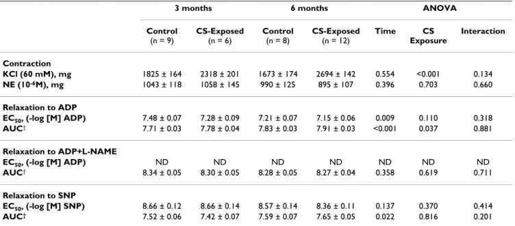
In pulmonary arteries, maximal contraction to KCl was greater in animals exposed to CS at 3 and 6 months than in controls (Table 1). Maximal contraction to NE was sim-ilar in all groups at all times of exposure.
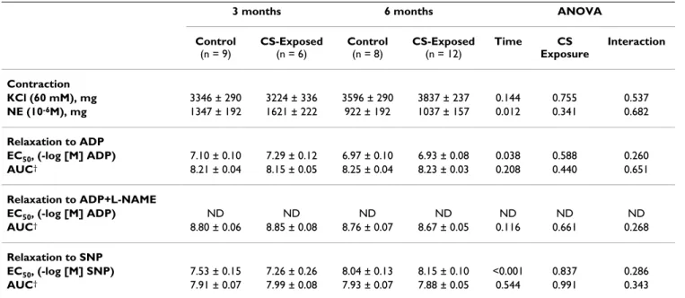
In the aorta, there were no differences in the maximal con-traction to KCl between CS-exposed animals and controls at any time of exposure. The ANOVA revealed a time effect in the contractile response to NE (Table 2).
Endothelial function
Guinea pigs exposed to CS showed a shift to the right in the dose-response curve of pulmonary arteries to ADP, as shown by a greater AUC and higher (less diluted) EC50, compared with control guinea pigs (Table 1, Figure 1). The ANOVA failed to show any interaction between CS exposure and time (Table 1), thereby indicating that reduced reactivity of pulmonary arteries in CS-exposed animals was independent of the length of exposure.
The ANOVA also revealed a marked effect of time on endothelium-dependent relaxation. Irrespective of whether the animals were exposed to CS, the endothe-lium-dependent vasodilatation of pulmonary arteries was lower at 6 months than at 3 months.
The maximal relaxation induced by ADP was close to 100% in all groups. When pulmonary artery rings were exposed to the competitive eNOS inhibitor, L-NAME, the relaxation induced by ADP was almost completely abol-ished (Table 1), indicating that ADP operated through the L-arginine-NO pathway. In all cases, pulmonary arteries reached maximal relaxation when they were assessed with SNP, an exogenous NO donor.
In rings of the aorta, no effect of CS exposure was found in any of the relaxation responses to the pharmacological agents that were tested (Table 2). Yet, the ANOVA revealed
Table 1: Vascular response of pulmonary artery
3 months 6 months ANOVA Control
(n = 9)
CS-Exposed
(n = 6)
Control
(n = 8)
CS-Exposed
(n = 12)
Time CS Exposure
Interaction
Contraction
KCl (60 mM), mg 1825 ± 164 2318 ± 201 1673 ± 174 2694 ± 142 0.554 <0.001 0.134
NE (10-6M), mg 1043 ± 118 1058 ± 145 990 ± 125 895 ± 107 0.396 0.703 0.660
Relaxation to ADP
EC50, (-log [M] ADP) 7.48 ± 0.07 7.28 ± 0.09 7.21 ± 0.07 7.15 ± 0.06 0.009 0.110 0.318
AUC† 7.71 ± 0.03 7.78 ± 0.04 7.83 ± 0.03 7.91 ± 0.03 <0.001 0.037 0.881
Relaxation to ADP+L-NAME
EC50, (-log [M] ADP) ND ND ND ND ND ND ND
AUC† 8.34 ± 0.05 8.30 ± 0.05 8.28 ± 0.05 8.27 ± 0.04 0.358 0.619 0.711
Relaxation to SNP
EC50, (-log [M] SNP) 8.66 ± 0.12 8.66 ± 0.14 8.57 ± 0.14 8.36 ± 0.11 0.137 0.370 0.414
AUC† 7.52 ± 0.06 7.42 ± 0.07 7.59 ± 0.07 7.65 ± 0.05 0.022 0.816 0.201
Definition of abbreviation: NE: Norepinephrine; ADP: Adenosine diphosphate; SNP: Sodium nitroprusside; EC50: dose that causes 50% of relaxation; AUC: area under the curve; L-NAME: NG-monomethyl-L-arginine
Values are mean ± SEM
†log-transformed
a time effect in both the endothelium-dependent and – independent relaxation responses.
Morphological evaluation
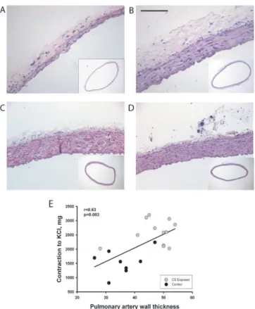
The thickness of the walls of the right and left main pul-monary arteries was enlarged in CS-exposed animals (Fig-ure 2A and 2B). Wall thickening of pulmonary arteries was due to both smooth muscle cell proliferation and elastic fiber deposition (data not shown). In contrast, the thickness of the aorta was not affected at either 3 or 6 months (Figure 2C and 2D, Table 3).
The percentage of intra-pulmonary vessels with double elastic laminas increased 2-fold after 6 months of CS exposure compared to controls (12.3 ± 4.8 vs. 6.3 ± 5.7 respectively). This effect was not observed at 3 months (6.1 ± 3.9 vs. 7.4 ± 8.1 respectively) (Figure 3). There were no differences in the percentage of muscularized arterioles at 3 months of exposure.
Immunohistochemical evaluation
Immunohistochemical evaluation was performed in ves-sels with a diameter <50 μm. The number of vessels that were positive to smooth muscle α-actin was significantly greater in CS-exposed animals, at both 3 and 6 months of exposure (Figures 4A, B and 4C). In contrast, no changes were observed in the number of desmin-positive vessels at 3 or 6 months (Figure 4D), nor in the intensity or exten-sion of protein expresexten-sion (data not shown).
Alveolar space size
An increase in the mean distance between alveolar septa was observed in animals exposed to CS for 6 months (control vs. exposed: 52 ± 8 vs. 59 ± 7 μm, p < 0.05). This finding is consistent with the presence of pulmonary emphysema. No differences between control animals and those exposed to CS for 3 months were found.
Gene expression of eNOS
Gene expression of eNOS was evaluated by Real-Time PCR in whole lung homogenates and normalized by the expression of β-actin. Compared with control animals, eNOS expression was decreased at 3 and 6 months of exposure to CS (Figure 5).
Correlation
The wall thickness of pulmonary arteries correlated signif-icantly with the contraction to KCl (r = 0.63, p = 0.003) (Figure 2E). This correlation was not observed in aortas. There was no correlation between the endothelial func-tion of the main pulmonary artery and the number of α -actin-positive intrapulmonary vessels or muscularized arterioles.
Discussion
Our results show that guinea pigs chronically exposed to CS developed endothelial dysfunction in the pulmonary artery, which was already apparent after 3 months of exposure. In this period of time, animals exposed to CS
Table 2: Vascular response of aorta
3 months 6 months ANOVA Control
(n = 9)
CS-Exposed
(n = 6)
Control
(n = 8)
CS-Exposed
(n = 12)
Time CS Exposure
Interaction
Contraction
KCl (60 mM), mg 3346 ± 290 3224 ± 336 3596 ± 290 3837 ± 237 0.144 0.755 0.537
NE (10-6M), mg 1347 ± 192 1621 ± 222 922 ± 192 1037 ± 157 0.012 0.341 0.682
Relaxation to ADP
EC50, (-log [M] ADP) 7.10 ± 0.10 7.29 ± 0.12 6.97 ± 0.10 6.93 ± 0.08 0.038 0.588 0.260
AUC† 8.21 ± 0.04 8.15 ± 0.05 8.25 ± 0.04 8.23 ± 0.03 0.208 0.440 0.651
Relaxation to ADP+L-NAME
EC50, (-log [M] ADP) ND ND ND ND ND ND ND
AUC† 8.80 ± 0.06 8.85 ± 0.08 8.76 ± 0.07 8.67 ± 0.05 0.116 0.661 0.268
Relaxation to SNP
EC50, (-log [M] SNP) 7.53 ± 0.15 7.26 ± 0.26 8.04 ± 0.13 8.15 ± 0.10 <0.001 0.837 0.286
AUC† 7.91 ± 0.07 7.99 ± 0.08 7.93 ± 0.07 7.88 ± 0.05 0.544 0.991 0.343
Definition of abbreviation: NE: Norepinephrine; ADP: Adenosine diphosphate; SNP: Sodium nitroprusside; EC50: dose that causes 50% of
relaxation; AUC: area under the curve; L-NAME: NG-monomethyl-L-arginine
Values are mean ± SEM
†log-transformed
showed reduced expression of eNOS in lung tissue and developed SMC proliferation in small intrapulmonary arteries. Longer exposure resulted in complete musculari-zation of small pulmonary vessels, as well as emphysema-tous changes in the alveolar spaces.
Endothelial dysfunction in pulmonary arteries was shown by a diminished response to the endothelium-dependent vasodilator ADP, which was abolished by the eNOS inhibitor L-NAME. Contrasting with this observation, no differences between groups were found in the
endothe-Endothelial function of pulmonary and aorta arteries
Figure 1
lium-dependent relaxation of the aorta, thereby suggest-ing that CS exposure exerted a direct effect on pulmonary circulation. These results are in agreement with previous studies showing diminished endothelium-dependent relaxation in pulmonary arteries of smokers [2,4] and in guinea pigs after a short period of CS exposure [13]. More-over, in the present study endothelial dysfunction pre-ceded the complete muscularization of small intrapulmonary vessels (vessels with double elastic lam-ina), thereby supporting the hypothesis that in COPD endothelial dysfunction is an early event that antecedes pulmonary vascular remodeling [22]. The mechanisms by which CS impairs pulmonary endothelium remain to be established. Our results show a decrease in eNOS gene expression in the lungs of animals exposed to CS. This finding is in agreement with those of Su and co-workers [23], who demonstrated that eNOS is down-regulated in endothelial cell cultures exposed to cumulative doses of CS extract. The expression of eNOS is also reduced in pul-monary arteries of smokers and in patients with different degrees of COPD severity [4,24]. Accordingly, it is con-ceivable that CS might alter endothelium-dependent relaxation by down-regulating eNOS expression in pul-monary arteries of guinea pigs.
We also observed a marked effect of time on the endothe-lial function of pulmonary arteries. The vascular relaxa-tion induced by ADP was lower at 6 months than at 3 months, irrespective of whether the animals were exposed to CS or not. The effect of time on vascular reactivity was also apparent in the aorta. Since the guinea pigs used in our study were in their growing period (mean weight increased by 231% at 3 months, and by 404% at 6 months), we consider that growth (or maturation) might affect vascular reactivity, in particular the endothelial function of pulmonary arteries, by mechanisms that have
Table 3: Morphometric measurements in main pulmonary artery and aorta
3 months 6 months Control
(n = 8)
CS-Exposed
(n = 5)
Control
(n = 8)
CS-Exposed
(n = 13)
Pulmonary artery Diameter (mm) 2.26 (2.10–2.49) 2.37 (2.11–2.45) 2.26 (2.13–2.39) 2.19 (2.15–2.40)
Wall area (mm2) 0.26 (0.24–0.33) 0.38 (0.30–0.42) 0.27 (0.24–0.31) 0.35* (0.32–0.41)
Wall thickness (μm) 38 (36–43) 50 (43–57) 35 (33–40) 51‡ (45–53)
Aorta Diameter (mm) 2.21 (2.14–2.30) 2.34 (2.17–3.80) 2.22 (2.07–2.32) 2.28 (2.24–2.36)
Wall area (mm2) 0.58 (0.51–0.65) 0.77 (0.59–0.92) 0.50 (0.46–0.65) 0.63 (0.57–0.74)
Wall thickness (μm) 82 (74–92) 96 (86–117) 77 (70–87) 88 (80–97) Wall thickness was calculated as total area/external perimeter
Values are median and interquartile range * p < 0.05
‡ p ≤ 0.01
Morphometry of pulmonary artery
Figure 2
not been fully elucidated. Indeed, it has been observed that hormonal changes associated with sexual maturity may affect posttranscriptional and/or translational regula-tion of eNOS protein and result in lower plasma NO lev-els in adult male pigs, thereby exerting an effect on vascular function [25]. On the other hand, maturation also induces increased production of reactive oxygen spe-cies (ROS) in vessels, which, in turn, may impair vessel function as a result of decreased NO bioavailability [26-28].
We characterized the phenotype of the SMC responsible for vascular remodeling in guinea pigs exposed to CS by evaluating the expression of the intermediate filaments smooth muscle α-actin and desmin in small pulmonary vessels. Animals exposed to CS for 3 months showed an increase in the number of α-actin-positive vessels, which persisted after 6 months of exposure. In contrast, there were no differences in the number of vessels positive for the contractile filament desmin, either at 3 or at 6 months. Accordingly, CS-exposed guinea pigs showed prolifera-tion of α-actin+/desmin- SMC, which represent a subpop-ulation of less differentiated SMCs with synthetic capacity [5]. These results are consistent with those obtained in COPD showing that vascular remodeling is produced by the intimal proliferation of poorly differentiated SMCs [3]. The structural alterations in the pulmonary circula-tion of CS-exposed guinea pigs might be a consequence of changes in the synthesis and release of vasoactive and cell proliferative mediators, since endothelin and VEGF expression are increased in the arterial wall of remodeled vessels in animals exposed to CS [12]. After 6 months of exposure, we found an increased percentage of vessels with double elastic laminas. In the same experimental model, Wright et al. [9] also reported that the greater number of vessels with double elastic laminas was accom-panied by an increase in pulmonary artery pressure (PAP). This observation suggests that muscularization of small vessels induced by CS exposure is associated with pulmo-nary hypertension. In keeping with this, we observed a wall enlargement in main pulmonary arteries after 6 months of CS exposure, which correlated with the con-tractility to KCl.
Smooth muscle cell proliferation in small vessels was already present at 3 months of exposure and preceded the development of emphysema. These findings confirm pre-vious observations made by Yamato et al. [29] and are in agreement with studies performed in healthy smokers, who showed pulmonary vascular remodeling and endothelial dysfunction [2]. Although hypoxemia, which is associated with emphysema, is one of the strongest agents producing vasoconstriction and vessel remodeling,
Double elastic lamina presence in small intrapulmonary arter-ies
Figure 3
our results corroborate that CS exposure may have a sim-ilar effect on pulmonary vessels [30]. Nevertheless, the presence of hypoxemia in patients with COPD might exert a synergistic effect on the pathogenesis of pulmonary hypertension. Further studies are required to elucidate the potential synergism between cigarette smoke and hypoxia in this experimental model.
It is interesting to note that endothelial dysfunction and vessel remodeling associated with CS exposure affected selectively pulmonary arteries while the aorta remained unaltered. We consider this could be due to the fact that pulmonary vessels are exposed to greater concentrations of CS products, whereas the effects on the aorta might be eventually caused by products diffusing to the blood. We
do not disregard that longer exposure to CS might exert an effect on the aorta or systemic vessels. Although, it is con-ceivable that longer exposure would also result in greater structural and functional damage of pulmonary vessels. Yet, it is noteworthy that in this experimental model pul-monary vessels develop changes after a short period of CS exposure that antecede changes in lung structure or sys-temic vascular involvement.
In conclusion, the guinea pig chronically exposed to CS develops endothelial dysfunction selectively in pulmo-nary arteries, without presenting changes in systemic arteries. This endothelial dysfunction is accompanied by reduced lung expression of eNOS and proliferation of poorly differentiated SMCs in small pulmonary vessels.
Smooth muscle cell proliferation in small intrapulmonary arteries
Figure 4
Smooth muscle cell proliferation in small intrapulmonary arteries. Immunohistochemistry for α-actin in small vessels of a control guinea pig (A) and an animal exposed to cigarette smoke (CS) (B). The vessel of the exposed animal shows a thicker wall with a strong immunoreactivity to α-actin. Scale bar, 50 μm. Bar graphs show the number of vessels/mm2 with
These changes are already apparent after a relatively short period of CS exposure and precede the full musculariza-tion of small pulmonary vessels and the development of emphysema. Results in this experimental model confirm that CS has direct and deleterious effects on the structure and function of pulmonary vessels that might contribute to the development of pulmonary hypertension in COPD.
Competing interests
The authors declare that they have no competing interests.
Authors' contributions
EF carried out the experimental work, participated in the analysis of the data and in the preparation of the manu-script. VIP was involved in the conception of the study, participated in its design and coordination, the analysis of the data and in the elaboration of the manuscript. MD provided support in the experimental work and data col-lection. JLC performed the statistical analysis and contrib-uted to the analysis of the data. MMM carried out the RT-PCR experiments. AM contributed in the implementation of the study and in the immunohistochemical studies. RRR provided funding support and contributed in the analysis of the data. JAB conceived the study, raised fund-ing support, participated in the design and implementa-tion of the study, and in the revision of the manuscript for important intellectual content. All authors read and approved the final manuscript.
Acknowledgements
We thank Blanca Reyes, Ma Dolores Cano, Montserrat Cerrillo and Belén
González for technical assistance. We also thank Lluís de Jover and Josep Ramírez for their valuable contributions.
Funded by grants from the Fondo de Investigación Sanitaria (02/0026 and 04/1424), the Sociedad Española de Neumología y Cirugía Torácica (SEPAR-2001), and the European Commission (6th Framework Pro-gramme, LSHM-CT-2005-018725, PULMOTENSION).
References
1. Dinh-Xuan AT, Higenbottam TW, Clelland CA, Pepke-Zaba J, Cre-mona G, Butt AY, Large SR, Wells FC, Wallwork J: Impairment of endothelium-dependent pulmonary-artery relaxation in chronic obstructive lung disease. N Engl J Med 1991,
324:1539-1547.
2. Peinado VI, Barbera JA, Ramirez J, Gomez FP, Roca J, Jover L, Gimfer-rer JM, Rodriguez-Roisin R: Endothelial dysfunction in pulmo-nary arteries of patients with mild COPD. Am J Physiol 1998,
274:L908-L913.
3. Santos S, Peinado VI, Ramirez J, Melgosa T, Roca J, Rodriguez-Roisin R, Barbera JA: Characterization of pulmonary vascular remod-elling in smokers and patients with mild COPD. Eur Respir J
2002, 19:632-638.
4. Barbera JA, Peinado VI, Santos S, Ramirez J, Roca J, Rodriguez-Roisin R: Reduced expression of endothelial nitric oxide synthase in pulmonary arteries of smokers. Am J Respir Crit Care Med 2001,
164:709-713.
5. Peinado VI, Pizarro S, Barbera JA: Pulmonary vascular involve-ment in COPD. Chest 2008, 134:808-814.
6. Wright JL, Churg A: A model of tobacco smoke-induced airflow obstruction in the guinea pig. Chest 2002, 121:188S-191S. 7. Brusselle GG, Bracke KR, Maes T, D'hulst AI, Moerloose KB, Joos GF,
Pauwels RA: Murine models of COPD. Pulm Pharmacol Ther 2006,
19:155-165.
8. Simani AS, Inoue S, Hogg JC: Penetration of the respiratory epi-thelium of guinea pigs following exposure to cigarette smoke. Lab Invest 1974, 31:75-81.
9. Wright JL, Churg A: Effect of long-term cigarette smoke expo-sure on pulmonary vascular structure and function in the guinea pig. Exp Lung Res 1991, 17:997-1009.
10. Wright JL, Sun JP: Dissociation of chronic vascular cell prolifer-ation and vascular contractility after chronic cigarette smoke exposure. Eur Respir J 1999, 14:832-838.
11. Wright JL, Tai H, Dai J, Churg A: Cigarette smoke induces rapid changes in gene expression in pulmonary arteries. Lab Invest
2002, 82:1391-1398.
12. Wright JL, Tai H, Churg A: Cigarette smoke induces persisting increases of vasoactive mediators in pulmonary arteries. Am J Respir Cell Mol Biol 2004, 31:501-509.
13. Wright JL, Churg A: Short-term exposure to cigarette smoke induces endothelial dysfunction in small intrapulmonary arteries: analysis using guinea pig precision cut lung slices. J Appl Physiol 2008, 104:1462-1469.
14. Furchgott RF, Zawadzki JV: The obligatory role of endothelial cells in the relaxation of arterial smooth muscle by acetyl-choline. Nature 1980, 288:373-376.
15. Tasatargil A, Sadan G, Karasu E: Homocysteine-induced changes in vascular reactivity of guinea-pig pulmonary arteries: role of the oxidative stress and poly (ADP-ribose) polymerase activation. Pulm Pharmacol Ther 2007, 20:265-272.
16. Jin N, Packer CS, Rhoades RA: Pulmonary arterial hypoxic con-traction: signal transduction. Am J Physiol 1992, 263:L73-L78. 17. Johns RA, Linden JM, Peach MJ: Endothelium-dependent
relaxa-tion and cyclic GMP accumularelaxa-tion in rabbit pulmonary artery are selectively impaired by moderate hypoxia. Circ Res
1989, 65:1508-1515.
18. Carville C, Raffestin B, Eddahibi S, Blouquit Y, Adnot S: Loss of endothelium-dependent relaxation in proximal pulmonary arteries from rats exposed to chronic hypoxia: effects of in vivo and in vitro supplementation with L-arginine. J Cardiovasc Pharmacol 1993, 22:889-896.
Gene expression of eNOS in whole lungs evaluated by real time-PCR
Figure 5
Publish with BioMed Central and every scientist can read your work free of charge "BioMed Central will be the most significant development for disseminating the results of biomedical researc h in our lifetime."
Sir Paul Nurse, Cancer Research UK
Your research papers will be:
available free of charge to the entire biomedical community
peer reviewed and published immediately upon acceptance
cited in PubMed and archived on PubMed Central
yours — you keep the copyright
Submit your manuscript here:
http://www.biomedcentral.com/info/publishing_adv.asp
BioMedcentral
19. Buikema H, Monnink SH, Tio RA, Crijns HJ, de Zeeuw D, van Gilst WH: Comparison of zofenopril and lisinopril to study the role of the sulfhydryl-group in improvement of endothelial dys-function with ACE-inhibitors in experimental heart failure.
Br J Pharmacol 2000, 130:1999-2007.
20. James AL, Hogg JC, Dunn LA, Pare PD: The use of the internal perimeter to compare airway size and to calculate smooth muscle shortening. Am Rev Respir Dis 1988, 138:136-139. 21. Motulsky H, Christopoulos A: Fitting Models to Biological Data Using
Lin-ear and NonlinLin-ear Regression: a practical guide to curve fitting Oxford: Oxford Univesity Press; 2004.
22. Barbera JA, Peinado VI, Santos S: Pulmonary hypertension in chronic obstructive pulmonary disease. Eur Respir J 2003,
21:892-905.
23. Su Y, Han W, Giraldo C, De Li Y, Block ER: Effect of cigarette smoke extract on nitric oxide synthase in pulmonary artery endothelial cells. Am J Respir Cell Mol Biol 1998, 19:819-825. 24. Melgosa M, Peinado VI, Santos S, Morales J, Ramirez J, Roca J,
Rod-riguez-Roisin R, Barbera JA: Expression of endothelial nitric oxide synthase (eNOS) and endothelin-1 (ET-1) in pulmo-nary arteries of patients with severe COPD. Eur Respir J 2003,
22:20s [http://www.ers-education.org/pages/ default.aspx?id=335&idBrowse=20419&det=1].
25. Chatrath R, Ronningen KL, Severson SR, LaBreche P, Jayachandran M, Bracamonte MP, Miller VM: Endothelium-dependent responses in coronary arteries are changed with puberty in male pigs.
Am J Physiol Heart Circ Physiol 2003, 285:H1168-H1176.
26. Barton M, Cosentino F, Brandes RP, Moreau P, Shaw S, Luscher TF:
Anatomic heterogeneity of vascular aging: role of nitric oxide and endothelin. Hypertension 1997, 30:817-824.
27. Tschudi MR, Barton M, Bersinger NA, Moreau P, Cosentino F, Noll G, Malinski T, Lüscher TF: Effect of age on kinetics of nitric oxide release in rat aorta and pulmonary artery. J Clin Invest 1996,
98:899-905.
28. van der Loo B, Labugger R, Skepper JN, Bachschmid M, Kilo J, Powell JM, Palacios-Callender M, Erusalimsky JD, Quaschning T, Malinski T, Gygi D, Ullrich V, Lüscher TF: Enhanced peroxynitrite formation is associated with vascular aging. J Exp Med 2000,
192:1731-1744.
29. Yamato H, Churg A, Wright JL: Guinea pig pulmonary hyperten-sion caused by cigarette smoke cannot be explained by cap-illary bed destruction. J Appl Physiol 1997, 82:1644-1653. 30. Thompson BT, Steigman DM, Spence CL, Janssens SP, Hales CA: