SYNTHESIS, STRUCTURE, AND MAGNETIC BEHAVIOR OF
[Cu
2(PYRAZINE)
3(CH
3O)
2](ClO
4)
2Benjamin L. Solomon
[a],
Christopher P. Landee
[b],
Jerry P. Jasinski
[c],
xxxxxxxxxxxxx
Mark M. Turnbull
[a]*Keywords: Pyrazine, Copper(II), Synthesis, X-ray Structure, Magnetism.
Reaction of copper perchlorate with sodium nitrite and pyrazine in methanol gives rise to the compound [Cu2(pyrazine)3(CH3O)2](ClO4)2
(1). This complex has been characterized through IR, combustion analysis, single crystal X-ray diffraction, and temperature dependent mag-netic susceptibility measurements. Compound 1 crystallizes in the triclinic space group P-1 with the copper coordination geometry being nearly square pyramidal. The Cu(II) ions are bridged by the methoxide ions to form a dimeric structure. Compound 1 exhibits an extended 3-D network with pyrazine rings forming sheets of the copper dimers, and bridging those sheets to form a distorted honeycomb. Magnetic susceptibility measurements show a strong antiferromagnetic exchange within the methoxy-bridged copper dimers with 2J ~ –880 K.
* Corresponding Authors Fax: +1-508-793-8861 E-Mail: [email protected]
[a] Carlson School of Chemistry and Biochemistry, Clark University, 950 Main Street, Worcester, Massachusetts 01610
[b] Department of Physics, Clark University, 950 Main Street, Worcester, Massachusetts 01610
[c] Department of Chemistry, Keene State College, 229 Main Street, Keene, New Hampshire 03435
INTRODUCTION
Transition metal ions form interesting and diverse coordination complexes that vary widely in structure with a change of ligand, or ion. To study this phenomenon many different families of Cu(II) complexes have been made to establish the effects of modification of the structure and electronic effects on the packing motif.1-4 Copper(II), with
its single unpaired electron and almost negligible internal magnetic field, as indicated by a g factor close to 2.00,5 is an
exemplary choice to study quantum magnetic interactions.
Pyrazine is an interesting ligand for research in the realm of copper(II) coordination chemistry due to the second donor nitrogen in the 4 position, making pyrazine a possible bidentate ligand.6,7 The bidenticity of pyrazine can also
create an efficient pathway for spin exchange and subsequent magnetic interactions.8,9 Pyrazine is a much
weaker base than the well-studied monodentate ligand pyridine10 (pyrazine
pKa= 1.1; pyridine pKa= 5.2).11 The
weaker basicity of pyrazine decreases the favorability of the ligand deprotonating another species in the solution, allowing for the synthesis of unique complexes compared to when pyridine is the ligand. Pyrazine complexes can also have varied dimensionalities depending on the stoichiometry of the starting materials.12
We are involved in a project to produce families of two-dimensional quantum Heisenberg antiferromagnets (2D-QHAF).13,14 A recent report of [M(pyrazine)
2NO2]ClO4,
where M is Co(II) or Cu(II), by Gao et al.15 is of interest to
our group due to the similarities in structure to some high temperature superconductors.16 Our attempts to grow single
crystals of the copper complex using the published synthesis proved ineffective. However, during those attempts, we
encountered a novel pyrazine/methoxide-bridged, three-dimensional coordination polymer. Here we present the synthesis, crystal structure, and magnetic properties of [Cu2(pyrazine)3(CH3O)2](ClO4)2(1).
EXPERIMENTAL
Pyrazine and copper perchlorate hexahydrate were purchased from Aldrich Chemical Company and used without further purification. Sodium nitrite was purchased from Fisher Scientific and used without further purification. IR spectra were recorded as KBr pellets on a Perkin-Elmer Spectrum 100. X-Ray powder diffraction was carried out on a Bruker AXS-D8 X-ray Powder Diffractometer. Elemental analysis was carried out by Marine Science Institute, University of California, Santa Barbara, CA 93106.
Synthesis of [Cu2(pyrazine)3(CH3O)2](ClO4)2 (1)
Copper perchlorate hexahydrate(3.625 g, 9.9 mmol), sodium nitrite (0.686 g, 9.94 mmol), and pyrazine (1.595 g, 19.9 mmol) were placed in three separate 30 mL beakers and dissolved in 5 mL of methanol, 5 mL of methanol, and 5 mL of water, respectively. All three beakers were placed in a 1000 mL beaker. The large beaker was slowly filled with methanol to a level of 0.8 cm over the rim of the smaller beakers and four drops of HClO4 were added to the solution.
The 1000 mL beaker was then covered with parafilm. After 15 days, black needles (0.268 g, 6.44 % yield) had formed on the top of the sodium nitrite-containing beaker, with a green powder (0.760 g, 18.27 % yield) at the bottom of the beaker. Black prisms (0.492 g, 11.83 % yield) had formed on the bottom of the 1000 mL beaker. The products were isolated by vacuum filtration, washed with cold methanol, and air-dried. The needles and prisms both became a green color upon powdering, meaning the crystals are a very dark green. All three proved to be 1, as indicated by identical IR spectra. Attempts to synthesize (1) by other methods using varying ratios of starting materials and differing orders of addition proved unsuccessful. IR (KBr, ν in cm–1): 3108 (w),
X-ray Structure Analysis
Data for 1 was collected on an Agilent Technologies Gemini Eos CCD-Xray Diffractometer using the CrysAlis Pro software package with MoKα radiation (λ = 0.71073 Å) via ω-scans at 173(2) K employing a graphite monochromator. Cell parameters were determined and refined using CrysAlisPro17 and absorption corrections were
made using SADABS.18 The structure was solved by direct
methods and refined via least-squares analysis using SHELXS97-2.19 All non-hydrogen atoms were refined
anisotropically. Hydrogen atoms were added in calculated positions and refined using a riding model with fixed isotropic thermal parameters. Crystallographic information and details of the data collection can be found in Table 1.
Table 1. X-ray data for 1.
Empirical Formula C14H18N6O10Cl2Cu2
Formula weight (g mol–1) T (K)
Wavelength (Ĺ) Crystal System Space Group a (Ĺ) b (Ĺ) c (Ĺ) α (°) β (°) γ (°) V (A3)
Z
Crystal Size (mm)
Absorption coefficient (mm–1) F (0,0,0)
θmin, θmax
Index Ranges
Reflections collected Independent reflections Restraints/parameters Final R index [I > 2σ(I)] R index (all data) Largest peak/hole (e/A3)
628.32 173(2) 0.71073 Triclinic P-1
7.7047(7) 9.0747(8) 9.1003(11) 60.124(11) 82.573(9) 85.277(7) 546.96(10) 1
0.30 x 0.25 x 0.20 2.254
316 3.50, 27.87 –10 < h < 9 –11 < k < 8 –11 < l < 11 5849 2993 0/155 0.0413 0.0427
1.95(near Cu1)/–0.51
Magnetic Susceptibility Data Collection
Magnetic data was collected using a Quantum Design MPMS-XL SQUID magnetometer. Finely ground samples of the crystals were packed in gelatin capsules. The moment was measured using magnetic fields from 0 to 50 kOe at 1.8 K. Several data points were collected as the field was brought back to 0 kOe to check for hysteresis; none was observed. Magnetization was then measured from 1.8 to 310 K in a 1 kOe field. The contributions from the sample holder were measured independently and subtracted from the data set. The data was also corrected for the temperature independent paramagnetism of the Cu(II) ion (60 × 10–6
emu/molOe) and for the diamagnetism of the constituent atoms (–226.5 × 10⁻⁶ emu/molOe) taken from Pascal’s constants.5
RESULTS AND DISCUSSION
Reaction of one equivalent of sodium nitrite, 1.4 equivalents of copper perchlorate, and two equivalents of pyrazine in methanol and water over the course of two weeks produced black crystals of [Cu2(pyrazine)3(CH3O)2
]-(ClO4)2 (1) in 11.83% yield.
Scheme 1. Preparation of compound 1.
Attempts to prepare the complex by a variety of other methods, varying concentrations, solvents, and omitting the NaNO2 proved unsuccessful. Although there is no nitrite
ion in the final product, it is clear that its presence is required for successful preparation of 1. We presume that the role of the nitrite ion is that of base/buffer to allow deprotonation of the methanol (most likely as a coordinated methanol molecule). Nitrite is a somewhat stronger base than pyrazine (pKa HNO2 = 3.3)20 and thus more suited to
that role.
Crystal Structure Analysis
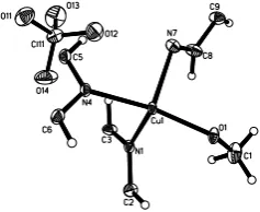
Compound 1 crystallizes in the triclinic space group P-1. The asymmetric unit is shown in Figure 1. A crystallog-raphic inversion center lies at (0.5, 0.5, 0.5) with respect to the Cu(II) ion, producing a methoxy-bridged dimer (Figure 2). Selected bond lengths and angles are given in Table 2.
Figure 1. Thermal ellipsoid plot of the asymmetric unit of 1
showing 50% probability ellipsoids. H atoms were placed in calculated positions and are not labeled.
Figure 2. Thermal ellipsoid plot showing the dimeric core of 1
(50% probability ellipsoids). Only the nitrogen atoms of the N7/N7A pyrazine rings are shown for clarity.
!
Table 2. Selected bond lengths (Å) and angles (°) for 1.
Cu1-O1 Cu1-N1 Cu1-N4 Cu1-N7 O1-C1
1.928(3) 2.015(3) 2.007(3) 2.255(3) 1.431(5)
Cu1-O1-Cu1A O1A-Cu1-O1 O1-Cu1-N4 O1A-Cu1-N4 O1-Cu1-N1 O1A-Cu1-N1 N4-Cu1-N1 O1-Cu1-N7 O1A-Cu1-N7 N4-Cu1-N7 N1-Cu1-N7
101.7(1) 78.33(1) 165.6(1) 94.9(1) 94.4(1) 162.8(1) 88.5(1) 94.7(1) 93.5(1) 98.5(1) 102.7(1)
The Cu ion has a nearly square pyramidal geometry as indicated by an Addison parameter (τ) of 0.05.21 The Cu(II)
ion lies 0.257 (2) Å above the N1-N4-O1-O1A mean plane. The three symmetry-independent pyrazine rings lie across crystallographic inversion centers located at (0.5, 0.5, 1) for the N1 ring, (0.5, 1, 0.5) for the N4 ring, and (1, 0.5, 0.5) for the N7 ring. The N1 and N4 pyrazine rings are unre-markable22,23 and connect the copper dimers together to
form a sheet parallel to the bc-plane (Figure 3). The N1-Cu1 vector lies 15.8(2)° below the copper-oxygen plane while N4-Cu1 vector is inclined 12.9(1)° in the same direction. The N7 pyrazine ring is on the opposite face and the Cu1-N7 bond is canted 5.4(1)° from the normal to the copper-oxygen plane (Figure 4). The N7-pyrazine ring occupies the axial site of the square pyramidal structure and the Cu1-N7 bond is 0.25 Å longer than the Cu1-N1 and Cu1-N4 bonds as expected.
Figure 3. Sheet formed by methoxy-bridged copper dimers. The N7 pyrazine rings are removed for clarity.
Figure 4. Links between sheets of dimers formed by the N7 pyrazine rings. The N1 and N4 pyrazine rings are removed for clarity.
This combination of the sheet formed by the N1 and N4 pyrazine rings, and the N7 pyrazine ring nearly normal to the Cu(II) ion gives rise to a 3D distorted honeycomb structure (Figures 5 and 6). The ClO4– ions occupy the holes
of the distorted honeycomb, balancing the charge of the Cu(II) ion, and are nearly tetrahedral and otherwise unremarkable.
Figure 5. Packing structure of 1 viewed parallel to the a axis. Perchlorate ions have been removed for clarity.
Figure 6. Packing structure of 1 viewed parallel to the bc-face diagonal. Perchlorate ions have been removed for clarity.
Similar covalently bonded networks of copper dimers bridged by alkoxy ligands have been synthesized. The compound [Cu2(μ-(6-oxyquinoline))(PPh3)2] forms
copper-oxygen dimers comparable to those found in 1, but bridge via the oxyquinoline ligands to give chains of dimers.24 A
2-D sheet is formed by the coordination polymer [Cu2(salicylate)2(pyrazine)(H2O)2].25 Two copper atoms and
two deprotonated phenolic oxygen atoms form copper-oxygen dimers that are linked by pyrazine units to yield a zigzag chain. Further dimensionality is added to the complex by the carboxyl groups of the salicylates, which bridge the zigzag chains to give a 2-D sheet. However, the Cu(II) ions have an octahedral geometry, unlike the square pyramidal geometry in 1.
The two short and one long Cu-N bonds found in 1 are present in the polymer [Cu2(monoethanolamine)2
-(pyrazine)2](CF3SO3)226 to give a 3-D structure akin to 1.
The two short Cu-N bonds belong to the pyrazine that forms the 2-D sheets of copper dimers and the amino group that resides in the monoethanolamine ligand. The long Cu-N
!
!
!
bond is to the axial pyrazine ring that links the 2-D sheets of copper dimers together to form a 3-D structure as in 1. As opposed to 1, the coordination of the 2-D sheet is built through one ligand instead of two, as the monoethanolamine bridges one half of the dimer to itself by the form-ation of a five-membered ring. Related compounds have been synthesized that yield analogous structures such as [Cu2(propanolamine)2(4,4’-bipyridine)2](ClO4)327 and
[Cu2(ethanolamine)2(bis(4-pyridyl)ethylene)2](ClO4)2.28
Magnetic Study
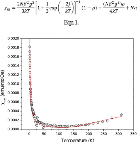
Magnetic susceptibility data were collected for compound 1 from 1.8 to 310 K in a 0.1 T field (Figure 7). Compound 1 is a strong antiferromagnet that has a maximum suscep-tibility well above room temperature. The data were fit to the Bleaney-Bowers equation (where represents a paramagnetic impurity, and Nα represents the temperature independent paramagnetism, see below).29 The best data fit
yields 2J = –880 (±170) K, g = 2.1 (± 1.7), and ρ = 0.009 (± 0.005). The large errors in these fitted values arise due to the limited data available for the magnetic contribution of the dimer since the maximum in chi occurs well above room temperature (and the limit of our instrument). Thus, these values are presented as rough approximations only.
Eqn.1.
Figure 7. χm vs. T plot for 1 in a 0.1 T field. The solid line
represents the best fit to a dimer model with a paramagnetic impurity term and was calculated from equation 1.
At room temperature, compound 1 exhibits a T product of 0.053 emuK/molOe, well below the expected value of above 0.375 emuK/molOe for an S = ½ ion with g = 2.00. This suggests very strong exchange. This was supported by the M vs. H data, which showed a saturation magnetization of 36.7 emu/mol at 5 T and 1.8 K. For a Cu(II) ion, a saturation magnetization near 6000 emu/mol is expected,5
implying that the response being measured at low temperature is not due to the bulk sample, but rather due to a paramagnetic impurity. The sharp increase in susceptibility
at lower temperatures also agrees with the presence of a trace paramagnetic impurity.
Copper dimers bridged by alkoxy,30,31 phenoxy,32,33 or
hydroxy34,35 groups are part of a family of antiferromagnetic
compounds with strong exchange. The value of the Cu-O-Cu angle ranges from 99.1(1)° ({[2-(1-(2-dimethylamino-ethylamino)ethyl]-phenoxy]Cu(NCCCN)}2; 2J = –530 K]32
to 103.9(2)° {[Cu(Me(6-R-2-pyridylmethyl)-(2-pyridyl)ben-zylamine)MeO]2(ClO4)2; 2J = –1840 K]31 in these
compo-unds, which all have maximum molar susceptibilities above room temperature. The magnitude of the 2J parameter has been related to the Cu-O-Cu angle,33,36 with larger angles
exhibiting a larger exchange.37,38 The Cu-O-Cu angle of 1 is
101.7(1)°, agreeing with a maximum molar susceptibility occurring at greater than room temperature. Exchange may also occur within and between the layers of copper dimers through the pyrazine rings, but the very strong exchange within the dimers prevents any observation of that exchange.
ACKNOWLEDGEMENTS
Financial assistance from the NSF (IMR-0314773) and the Kresge Foundation toward the purchase of the MPMS-XL SQUID magnetometer are greatly appreciated. The Bruker D8-Advance powder X-ray Diffractometer was purchased with the assistance of funds from the Kresge Foundation and PCISynthesis, Inc. BLS is grateful for the PCISynthesis, Inc. Summer Research Fellowship. JPJ acknowledges the NSF MRI program (grant No. CHE-1039027) for funds to purchase the X-ray diffractometer.
APPENDIX. Supplementary Data
CCDC (940687) contains the supplementary crystal-lographic data for 1. This data can be obtained free of charge via from
http://www.ccdc.cam.ac.uk/conts/retrie-ving.html, or from the Cambridge Crystallographic Data
Centre, 12 Union Road, Cambridge CB2 1EZ, UK; fax:
(+44) 1223-336-033; or e-mail: [email protected].
REFERENCES
1 Adhikary, C., Koner, S., Coord. Chem. Rev.2010, 254, 2933. 2 Herringer, S. N., Turnbull, M. M., Landee, C. P., Wikaira, J. L.,
J. Coord. Chem.2009, 62, 863.
3 Tremelling, G. W., Foxman, B. M., Landee, C. P., Turnbull, M.
M., Willett, R. D., Dalton Trans.2009, 47, 10518.
4 Zhao, J.P., Hu, B.W., Sanudo, E., Yang, Q., Zeng, Y.F., Bu,
X.H., Inorg. Chem.2009, 48, 2482.
5 Carlin, R. L., Magnetochemistry, Springer-Verlag, 1986. 6 Otieno, T., Rettig, S., Thompson, R., Trotter, J., Can. J. Chem.
1989, 67, 1964.
7 Otieno, T., Gipson, A. M., Parkin, S., J. Chem. Crystallog.2002,
32, 81.
8 Leznoff, D. B., Xue, B.-Y., Stevens, C. L., Storr, A., Thompson,
R. C., Patrick, B. O., Polyhedron2001, 20, 1247.
9 Manson, J. L., Huang, Q.Z., Lynn, J. W., Koo, H.J., Whangbo,
M.H., Bateman, R., Otsuka, T., Wada, N., Argyriou, D. N., Miller, J. S., J. Amer. Chem. Soc.2001, 123, 162.
10 Tomasik, P., Ratajewicz, Z., Newkome, G. R., Strekowski, L.,
Pyridine-metal complexes, John Wiley & Sons, 1985.
0 50 100 150 200 250 300 350
0.0000 0.0002 0.0004 0.0006 0.0008 0.0010 0.0012 0.0014 0.0016 0.0018 0.0020
cmo
l
(
e
m
u
/m
o
lO
e
)
11 Keyworth, D., The J. Organ. Chem.1959, 24, 1355.
12 Carlucci, L., Ciani, G., Proserpio, D. M., Sironi, A., J. Amer.
Chem. Soc.1995, 117, 4562.
13 Gale, A. J., Landee, C. P., Turnbull, M. M., Wikaira, J. L.,
Polyhedron2012, 52, 986.
14 Abdalrahman, M., Landee, C. P., Telfer, S. G., Turnbull, M. M.,
Wikaira, J. L., Inorg. Chim. Acta2012, 389, 66.
15 Liu, T., Chen, Y.H., Zhang, Y.J., Wang, Z.M., Gao, S., Inorg.
Chem.2006, 45, 9148.
16 Monthoux, P., Balatsky, A., Pines, D., Phys. Rev. B1992, 46,
14803.
17 CrysAlisPro Oxford Diffraction Ltd., Version 1.171.35.19
(release 27-10-2011 CrysAlis171.NET).
18 Sheldrick, G.M., SADABS v 2.01: An empirical absorption
correction program, Bruker AXS Inc., Madison, WI (1999).
19 Sheldrick, G. M., Acta Cryst. A2008, 64, 112.
20 Schwartz, S. E., White, W. H., Adv. Environ. Sci. Eng.1981, 4,
1.
21 Addison, A. W., Rao, T. N., Reedijk, J., van Rijn, J., Verschoor,
G. C., J. Chem. Soc., Dalton Trans.1984, 1349.
22 Ramírez, J., Stadler, A.M., Rogez, G., Drillon, M., Lehn, J.M.,
Inorg. Chem.2009, 48, 2456.
23 Albrecht, A. S., Landee, C. P., Slanic, Z., Turnbull, M. M., Mol.
Cryst. Liq. Cryst.1997, 305, 333.
24 Ponikiewski, L., Shi, W., Rothenberger, A., Z. Anorg. Allg.
Chem.2008, 634, 1770.
25 Long-Guan, Z., Kitagawa, S., J. Inorg. Organomet. Polym.2002,
12, 23.
26 Marin, G., Kravtsov, V., Simonov, Y. A., Tudor, V., Lipkowski,
J., Andruh, M., J. Mol. Struct.2006, 796, 123.
27 Tudor, V., Marin, G., Kravtsov, V., Simonov, Y. A., Lipkowski,
J., Brezeanu, M., Andruh, M., Inorg. Chim. Acta2003, 353, 35.
28 Marin, G., Tudor, V., Kravtsov, V. C., Schmidtmann, M.,
Simonov, Y. A., Müller, A., Andruh, M., Cryst. Growth Des.
2005, 5, 279.
29 Bleaney, B., Bowers, K., Proc. R. Soc. London, Ser. A. 1952,
214, 451.
30 Garcia, A. M., Manzur, J., Garland, M. T., Baggio, R., Gonzales,
O., Pena, O., Spodine, E., Inorg. Chim. Acta1996, 248, 247
31 Rojas, D., García, A. M., Vega, A., Moreno, Y.,
Venegas-Yazigi, D., Garland, M. T., Manzur, J., Inorg. Chem.2004,
43, 6324.
32 Biswas, A., Drew, M. G., Ribas, J., Diaz, C., Ghosh, A., Inorg.
Chim. Acta2011, 379, 28.
33 Thompson, L. K., Mandal, S. K., Tandon, S. S., Bridson, J. N.,
Park, M. K., Inorg. Chem.1996, 35, 3117.
34 Massoud, S. S., Louka, F. R., Xu, W., Perkins, R. S., Vicente,
R., Albering, J. H., Mautner, F. A., Eur. J. Inorg. Chem.
2011, 50, 3469.
35 Cvetkovic, M., Batten, S. R., Moubaraki, B., Murray, K. S.,
Spiccia, L., Inorg. Chim. Acta2001, 324, 131.
36 Crawford, V. H., Richardson, H. W., Wasson, J. R., Hodgson, D.
J., Hatfield, W. E., Inorg. Chem.1976, 15, 2107.
37 Merz, L., Haase, W., J. Chem. Soc., Dalton Trans.1980, 6, 875. 38 Melnik, M., Coord. Chem. Rev.1982, 42, 259.