REVIEW
Focal hand lesions: review and radiological approach
Chau Hung Lee&Ankit Tandon
Received: 9 December 2013 / Revised: 9 April 2014 / Accepted: 15 April 2014 / Published online: 17 May 2014 #The Author(s) 2014. This article is published with open access at Springerlink.com
Abstract Focal hand lesions are commonly encountered in
clinical practice and are often benign. Magnetic resonance (MR) imaging is the imaging modality of choice in evaluating these lesions as it can accurately determine the nature of the lesion, enhancement pattern and exact location in relation to surrounding tissues. However, while MR features of various soft tissue lesions in the hand have been well described, it is often still difficult to differentiate between benign and malig-nant lesions. We review the MR imaging features of a variety of focal hand lesions presenting at our institution and propose a classification into“benign”,“intermediate grade” (histolog-ically benign but locally aggressive with potential for recur-rence) and frankly“malignant”lesions based on MR findings. This aims to narrow down differential diagnoses and helps in further management of the lesion, preoperative planning and, in cases of primary malignancy, local staging.
Teaching Points
• Hand lesions are often benign and MR is essential as part of the workup.
• MR features of various hand lesions are well described but are often non-specific.
• Certain MR features may help for the diagnosis but histo-logical examination is usually required.
• We aim to classify hand lesions based on MR features such as margin, enhancement and bony involvement.
• Classifying these lesions can help narrow down differential diagnoses and aid management.
Keywords magnetic resonance imaging . hand . wrist . soft
tissue neoplasms . bone neoplasms
Introduction
Most soft tissue lesions in the hand are benign [1]. Imaging is often required to determine the nature of the lesion. Plain radiography has limited utility but is useful in demonstrating calcification. Ultrasound is a cheap and relatively quick meth-od for determining the cystic or solid nature of the lesion. Magnetic resonance (MR) is the imaging modality of choice as it can accurately determine the nature of the lesion, en-hancement pattern and exact location in relation to surround-ing tissues given its high contrast and spatial resolution. However, while MR features of various soft tissue lesions in the hand have been well described, preoperative diagnosis of these lesions is often difficult, and even distinguishing benign from malignant lesions remains challenging. We review the MR imaging features of a variety of hand lesions presenting at our institution and propose a classification into “benign”, “intermediate grade”(histologically benign but locally aggres-sive with potential for recurrence) and frankly “malignant” lesions based on MR findings. This aims not just to narrow down differential diagnoses but also to help in further man-agement of the lesion in terms of preoperative planning, and for cases of primary malignancy, local staging and prognosis.
Imaging technique
At our institute, MR imaging is performed on a 1.5- or 3-T scanner. Several technical factors need to be considered to get the best images of the hand and wrist. Important technical factors to consider are patient positioning, choice of coil and sequences. Based on the lesion location and extent of C. H. Lee (*)
:
A. TandonTan Tock Seng Hospital, 11 Jalan Tan Tock Seng, Singapore 308433, Singapore
e-mail: powerlee_1999@yahoo.com
A. Tandon
the region of interest to isocenter. When the region of interest is close to the periphery of the coil, auto shimming is used. Skin markers are used to localise small lumps.
The routinely used sequences at our institute include T1-weighted (T1w) sequences in the axial and coronal planes, T2-weighted fat-saturated (T2w-FS) or short tau inversion recov-ery (STIR) sequences in the axial plane and T2-weighted (T2w) sequences without fat saturation in either the sagittal or coronal plane. A gradient-echo (GRE) sequence is also acquired, which is particularly useful if a vascular lesion or giant-cell tumour of the tendon sheath is suspected. Post-contrast T1w-FS sequences with intravenous gadolinium compounds are acquired in all patients unless contraindicated. Routine FOV of 16–20 cm, slice thickness of 4.0 mm and matrix of at least 512×256 is used, although when higher resolution imaging is critical, a smaller FOV of 8–12 cm and thinner slice thickness of 1.5–3.0 mm are preferred.
Benign lesions
Ganglion cyst
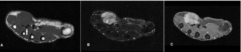
Ganglion cysts are the most common lumps encountered in the hand and wrist region [2]. They tend to occur in young adults and are three times more frequent in females. They are thought to represent degeneration of connective tissue caused by chronic irritation [3]. The most common location is in the dorsum of the wrist where they usually arise from the scapholunate joint. Less typical sites include the volar aspect of the wrist from the radio-scaphoid or scapho-trapezial joint, at the metacarpophalangeal joint in relation to flexor tendons and distal interphalangeal joints [4]. MR shows a well-circumscribed unilocular or multilocular lesion of fluid signal, although the signal may vary depending on the amount of proteinaceous contents (Fig.1). Mild rim enhancement of the capsule may be seen, but there is usually no enhancement of internal contents. Differential diagnoses include synovial cysts and other cystic lesions such as epidermal cysts.
Epidermal cyst
Epidermal cysts are common hand lesion resulting from pro-liferation of epidermal cells within a confined space in the dermis. They can be congenital, a result of occlusion of the
key to distinguish epidermal cysts from more sinister lesions such as neurogenic tumours or sarcomas, particularly if het-erogeneous signal is present because of internal debris. They may also be mistaken for ganglion cysts if located close to the joint or tendon sheath.
Fibroma of the tendon sheath
Fibromas of the tendon sheath (FTS) are uncommon lesions thought to be a reactive fibrosis. It has a peak incidence at 20– 50 years old, is three times more common in males, and 82 % of them occur in the hand and wrist region [7,8]. The low signal on all MR pulse sequences expected of a fibrous lesion is not always seen. FTS are generally well defined and of low signal on T1w sequence. Signal intensity on T2w sequence and contrast enhancement is more varied and heterogeneous (Fig.3), likely reflecting varying proportions of fibrous and cellular tissue [9]. Differential diagnoses include more sinister lesions such as soft tissue sarcomas and giant cell tumours of the tendon sheath (GCTTS). FTS and GCTTS are believed to represent two end points of a single clinicopathological entity based on histological studies, the former with a predominance of myofibrolastic markers while the latter with a predomi-nance of macrophage-related components [10]. They can be differentiated based on certain imaging features. On GRE sequence, GCTTS usually demonstrates susceptibility arte-facts, which are absent in FTS. Bony scalloping is also often seen in GCTTS but rare in FTS, and while both are most commonly located in the hand, GCTTS is more likely to be found in the feet compared to FTS [9].
Focal nodular synovitis
projections into the joint. On MR, focal nodular synovitis is usually well defined, isointense or slightly hyperintense to muscle on T1w sequence and of low signal on T2w sequence because of collageneous stroma (Fig. 4). In larger lesions, variable signal on T2w sequence and heterogeneous enhance-ment may be seen because of proliferating capillaries [13].
Nodular fasciitis
Nodular fasciitis is a benign reactive fibroblastic lesion usu-ally in young adults, thought to be associated with trauma, most commonly seen in the upper extremities [14]. On MRI, it shows low signal on T1w sequence and heterogeneous, mixed high signal on T2w sequence [15], depending on the distribu-tion of myxoid and fibrous components (Fig.5). Lesions with predominantly cellular content or myxoid degeneration ap-pear hyperintense on T2w sequence while those with highly collagenous contents are hypointense. Contrast enhancement pattern is most commonly diffuse but it may also be peripheral in lesions with cystic degeneration [16]. Nodular fasciitis can be easily confused with a malignant lesion given the clinical presentation of a rapidly growing lump and imaging findings of inhomogeneous signal characteristics and enhancement,
lobulated borders and crossing of compartments. Differential diagnoses include neurogenic tumour, soft tissue sarcoma or early stage of myositis ossificans especially when it occurs in an intramuscular location [17]. Complete local excision is usually curative and hence careful histological analysis is necessary to avoid misdiagnosis and unnecessary radical surgery [18].
Lipoma
Lipomas are the most common tumour in the human body usually developing in later adult life. A rather suggestive sonographic feature is an encapsulated hyperechoic lesion with fine linear internal echoes [19,20]. Characteristic MR features of a well-circumscribed lesion that is hyperintense on both T1w and T2w sequences with homogeneous signal loss on the STIR or fat-saturation sequences allow confident diag-nosis (Fig.6). It can be difficult to distinguish among benign lipomas, atypical lipomatous lesions or well-differentiated liposarcomas on imaging. Findings of thick enhancing septation or a nodular component should raise suspicion of the latter [21]. Interestingly, findings of infiltrating or insinu-ating margins tend to suggest benign lipoma rather than liposarcoma [22].
Fig. 2 Epidermal cyst in a 42-year-old male presenting with a lump over
the palm of about 10-year duration. (a) T1w sequence shows a low-signal, smooth, well-circumscribed subcutaneous lesion superficial to the thenar musculature. (b) The lesion is hyperintense on T2w-FS sequence
with faint low-signal foci within indicated squames (arrow). A thin hypointense capsule is seen. (c) Faint rim enhancement is seen with no significant internal enhancement
Fig. 1 Ganglion cyst in a 24-year-old female who presented with a
slowly growing, firm, painless hand lump for about 6 months. (a) T1w sequence shows a smooth, well-circumscribed lesion within the thenar musculature of homogeneous low signal. (b) The lesion is hyperintense
Lipofibromatous hamartoma
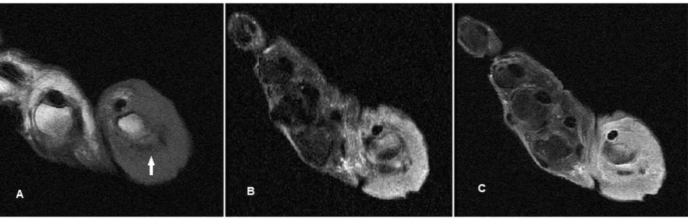
Lipofibromatous hamartomas are rare benign tumours usually involving the median nerve at the wrist [23]. It commonly presents as a progressively enlarging lump or symptoms of compressive neuropathy, most occurring in the first 3 decades of life. Excessive fibroadipose tissue proliferates along the perineurium, surrounding the nerve bundles within the nerve sheath. This gives the lesion its pathognomic appearance on MR imaging. MR typically shows a fusiform swelling representing the enlarged nerve with serpinginous, “spaghetti” or “ ca-ble”-like appearance, representing the low-signal axons s u r r o u n d e d b y f a t t y t i s s u e [2 4, 2 5] ( F i g . 7) . Management is usually conservative as surgical resec-tion is almost always accompanied by neurologic mor-bidity [26].
Haemangioma
Haemangiomas are the fourth most common hand tumour, usually occuring in the younger age group, with a slight female predominance [27]. On MR, they are typically very hyperintense on T2w sequence and show lobulations, septations or low-signal foci much more frequently than other soft tissue masses [28]. Marked hyperintensity of the lesions on T2w sequence is due to increased fluid content secondary to stagnant blood flow in vessels. Signal on T1w sequence is usually iso- to hyperintense compared to muscle because of the presence of fat and blood products (Fig.8). Lesions that are typically larger than 2 cm contain various amounts of fat, smooth muscle, myxoid stroma, thrombi and haemosiderin. Susceptibility artefacts on GRE sequence may be due to phleboliths or blood products, and these can be distinguished on plain radiograph. Larger lesions may also demonstrate
Fig. 4 Focal nodular synovitis in a 62-year-old male presenting with a
painless lump over the dorsal aspect of the right middle finger for a few months. (a) T1w sequence shows a lobulated, homogeneously low-signal subcutaneous nodule over t he dorsal a spect o f the t hird
metacarpophalangeal joint abutting the extensor tendon (arrow). (b) The lesion is also of low signal on T2w-FS sequences. No bony destruction is seen. (c) There is no significant contrast enhancement
Fig. 3 Fibroma of the tendon sheath in a 51-year-old female presenting
with a 6-month history of a lump in the left hand over the thenar eminence near the first metacarpal base. (a) T1w sequence shows a lobulated mass deep to the thenar musculature. It is slightly hyperintense to muscle, which is unusual. (b) The lesion is heterogeneously hyperintense on
fluid-fluid levels [29]. Enhancement on post-contrast imaging can be variable from minimal to heterogeneous to more ho-mogeneous. Superficial haemangiomas are easy to diagnose because of the presence of skin discolouration and MRI may only be needed to assess their extent for surgical planning.
Aneurysm/pseudoaneurysm
Aneurysms/pseudoaneurysms are rare lesions, usually sec-ondary to trauma-related intimal injury, or iatrogenic from arterial punctures or arteriovenous shunts for dialysis [30].
True aneurysms can rarely occur secondary to vasculitis. MR features are variable depending on presence of thrombus or turbulent flow. They are generally slightly hyperintense on T1w and T2w sequences with areas of signal void reflecting high flow, and susceptibility artefacts may be seen on GRE sequence if there is thrombosis (Fig. 9). Either MR or conventional angiography can demonstrate continuity with a parent artery, essentially excluding other differen-tial diagnoses [31]. Imaging features are highly suggestive even in the presence of a thrombus and allow avoiding catastrophic biopsies.
Fig. 6 Lipoma in a 60-year-old female presenting with a soft swelling
over the left thenar eminence of 1-year duration. (a) T1w sequence shows a well-circumscribed hyperintense lobulated subcutaneous lesion in the volar aspect of the hand involving thenar and mid-palmar spaces,
insinuating between the flexor tendons (arrow). (b) There is homoge-neous signal loss on T2w-FS sequence. (C) Faint enhancement of thin internal septation is noted (arrow), with no suspicious nodular component
Fig. 5 Nodular fasciitis in a
Schwannoma
Schwannomas are the most common benign tumour of the peripheral nerve sheath, usually occuring in the 4th to 6th decades. On MR schwannomas are generally well circumscribed, show low-to-intermediate signal on T1w se-quence, high signal on T2w sequence and homogeneous con-trast enhancement (Fig.10). It is often difficult to distinguish schwannomas from neurofibromas, vascular lesions or even soft tissue sarcomas. Its close relation to an expected course of a major nerve may suggest the diagnosis [32]. Larger tumours may demonstrate a dural tail,“split-fat sign”(peripheral rim of perineural fat compressed by the tumour), “fascicular sign” (central small ring-like structures representing nerve fibres) or
“target sign”(central low signal with surrounding high signal on T2-weighted sequence) [33]. Schwannomas can undergo cystic or fatty degeneration. Malignant change is very rare.
Intermediate-grade lesions
Neurofibroma
Neurofibromas are common benign peripheral nerve sheath tumours, occurring in isolation or in relation to neurofibroma-tosis type 1. On MR imaging, superficial neurofibromas tend to be asymmetric, lack fascicular morphology and target-like signal intensity, and are likely to involve skin [34], features
Fig. 8 Haemangioma in a 32-year-old female presenting with a soft lump
along the medial aspect of the volar aspect of right hand, occasionally painful. (a) T1w sequence shows a multilobulated subcutaneous lesion superficial to the hypothenar muscles slightly hyperintense to muscle, insinuating between adjacent flexor tendons (arrow). There is suggestion of a small intramuscular component (arrowhead). (b) The lesion is
extremely hyperintense on T2w-FS sequence with areas of low signal (arrow) and suggestion of fluid-fluid level (arrowhead). (c) Susceptibility artefacts are seen on GRE sequence indicating phleboliths or blood products (arrow). (d) There is avid and near-homogeneous enhancement (arrow)
Fig. 7 Fibrolipomatous hamartoma in a 45-year-old male presenting
with a lump of 1-year duration in the first web space of the left hand. (a) T1w sequence shows a well-circumscribed hyperintense lobulated subcutaneous lesion within the first web space, with curvilinear low-signal structures (arrow), similar to a“spaghetti-like”appearance. (b) On T2w-FS sequence the lesion shows homogeneous signal loss
that are evident in our case (Fig. 11). They are hypo- to isointense to muscle on T1w sequence and heterogeneously hyperintense on T2w sequence [35, 36]. Neurofibromas have a propensity for recurrence as complete resection, which would require sacrificing the whole nerve, is usually not possible [34].
Desmoplastic fibroblastoma (collagenous fibroma)
Desmoplastic fibroblastomas are rare benign myofibroblastic tumours arising in the subcutaneous tissue or skeletal muscle. There is a male predominance usually around the 5th decade. On MR these are usually of low-to-very-low signal intensity on T1w and T2w sequences and show only minimal contrast
enhancement, reflecting their collagenous nature and low vascularity. Small intermixed areas of T2 hyperintensity may be seen depending upon the amount of cellular components [37]. They are only minimally infiltrative, and the presence of calcifications and cystic changes are unusual. One specific MR characteristic reported in the literature that was also seen in our case is the rim enhancement of the capsule [38,39] (Fig. 12). Collagenous fibromas are often confused with desmoid tumours on imaging. However, desmoid tumours are often painful and more infiltrative at presentation. Moreover, desmoid tumours show prominent areas of high signal on T2w sequence and internal enhancement because of their cellular nature. Preoperative differentiation is important to avoid overtreatment and unnecessary extensive procedures
Fig. 10 Schwannoma in a 40-year-old male with a 15-year history of
non-enlarging swelling over the right palm hypothenar eminence, asso-ciated with occasional sharp pains and paresthesia. (a) T1w sequence shows a smooth well-circumscribed nodule within the hypothenar
musculature iso-intense to adjacent muscle. (b) On T2w-FS sequence the lesion shows a typical target-sign appearance with a hypointense centre (arrow) and peripheral hyperintense rim (arrowhead). (c) Fairly homogeneous enhancement is seen
Fig. 9 Aneurysm/pseudoaneurysm in a 32-year-old female presenting
with a lump over the palmar aspect of the right hand over 4 years. There was no prior invasive medical procedure although a vague history of trauma was obtained. (a) T1w sequence shows a well-circumscribed subcutaneous lesion with a central, relatively high signal (arrow) located superficial to the flexor tendons. (b) On T2w-FS sequence the lesion is
performed for desmoid tumours. Other differential diagnoses include FTS, calcifying fibrous tumours, leiomyoma and GCTTS.
Giant cell tumour of tendon sheath
GCTTSs are common tumours, usually presenting as pain-less masses at 30–50 years, with a slight female predilec-tion. They are typically found in the hands or feet asso-ciated with degenerative joints and thought to be reactive lesions to adjacent inflammation rather than true neo-plasms [40]. GCTTSs are histologically benign but pres-sure changes in adjacent bone can be seen on plain radiographs in 10-20 % of cases [41] (Fig. 13a). MR shows a lesion in close relation to joints and tendons, predominantly of low signal on T1w sequence and of intermediate to slightly high signal on T2w sequence (Fig. 13b-e). Susceptibility artefacts on GRE sequence are typical because of haemosiderin deposition and this is a helpful feature [42]. Strong enhancement is seen because of the presence of numerous proliferative capil-laries in the collagenous stroma. Differential diagnoses
include focal nodular synovitis, which also contains haemosiderin while a more heterogeneous signal and en-hancement can result in confusion with soft tissue sarcomas.
Glomus tumour
Glomus tumours are benign disordered proliferation of the neuromyoarterial apparatus that serves to regulate skin circulation [43]. They usually occur in the 4th to 5th decade and are three times more frequent in females. Patients with glomus tumour seek medical attention early, but the mass is frequently too small to be identified on physical examination. The classic triad of moderate pain, temperature sensitivity and point tenderness is inconsis-tently present [44]. Useful distinguishing features on MR are its characteristic location, pressure erosion of under-lying bone, very high and homogeneous signal on T2w se-quence, low signal on T1w sequence and intense enhance-ment [45] (Fig.14). Although these MR signal characteristics can be associated with any vascular tumour, the typical subungual location and its small size should lead one to suspect glomus tumour in most cases. In particular, T2w-FS
Fig. 11 Neurofibroma in a 53-year-old male with a painless left thumb
nailbed swelling of 4–5-year duration. The patient had no personal or family history of neurofibromatosis or any other clinical manifestations, and this was likely an isolated lesion. (a) T1w sequence shows a lobulated
subcutaneous lesion at the tip of the thumb, isointense to muscle involv-ing the skin, with mild pressure erosion on the underlyinvolv-ing distal phalanx (arrow). (b) The nodule is heterogeneously hyperintense on T2w-FS sequence. (c) Fairly homogeneous enhancement is seen
Fig. 12 Desmoplastic fibroblastoma in a 40-year-old female who
pre-sented with a swelling of 3-month duration over the dorsum of the first web space of the right hand. (a) T1w sequence shows a lobulated lesion of very low signal within the first dorsal interosseous muscle with loss of
and post-contrast sequences are very helpful in delineating small tumours. The lesion is typically painful, and surgery is
the treatment of choice for symptom relief and histological confirmation.
Fig. 13 GCTTS in a 23-year-old female presenting with a 1-year history
of right thumb swelling and mild pain. (a) Plain radiograph shows well-defined lucency at the head of the first metacarpal (arrow). (b) T1w sequence shows a lobulated subcutaneous lesion of intermediate signal encasing the flexor pollicis longus tendon (arrow). (c) The lesion shows
heterogeneous high signal on T2w-FS sequence. There is pressure ero-sion on the underlying bone. (d) Foci of susceptibility are demonstrated on the GRE sequence (arrow). (e) There is avid and fairly homogeneous enhancement
Fig. 14 Glomus tumour in a 30-year-old female with a painful lump at
the tip of the left ring finger. (a) Plain radiograph shows well-defined scalloping along the ulnar aspect of the left ring finger distal phalanx (arrow). (b) T1w sequence revealed a smooth, well-circumscribed
occurs in the soft tissues of the retroperitoneum and proximal extremities [46]. Occurrence in the hand is rare. MR is the imaging modality of choice to stage and characterise the tumour. It is typically intermediate to low signal on T1w sequence and heterogeneously high signal on T2w sequence; however, appear-ance may vary depending on the presence of calcification, fi-brous tissue, haemorrhage or necrosis (Fig.15). The tumour is fairly well defined despite its malignant nature because of a pseudocapsule, but may exert a mass effect or encase neurovascular bundles. Other less common subtypes of pleomor-phic sarcoma have been identified based on the proportion of myxoid, fibrous or cellular components [47].
Primary squamous cell carcinoma of the skin
Primary squamous cell carcinoma (SCC) of the skin usually occurs in the older age group commonly over the sun-exposed back of hands [48]. Diagnosis is usually suspected clinically and easily confirmed on bed-side punch biopsy. Imaging is utilised for local staging. On MR, the tumour has an infiltrative appearance and can be diffuse (Fig.16) or focal (Fig.17). It is usually of low to intermediate signal on T1w sequence and heterogeneously hyperintense on T2w sequence with variable enhancement. Its epicentre in the cutaneous layer, irregular and infiltrative appearance should raise suspicion for a primary skin malignancy.
Malignant bony lesions
Osseous metastases to the hand (acrometastases) is uncommon. The most common primary sites of malignancies are the lung,
of a benign chondroid lesion such as an enchondroma. Plain radiograph shows a typical “ring-and-arc” matrix with endosteal scalloping and cortical thinning, but in higher grade subtypes there is often bony destruction and irregular margins [50] (Fig. 19a). MR is usually used to for local staging (Fig. 19b-d). Differential diagnosis for a lesion in the distal phalanx is a glomus tumour, but the plain radiograph showing a primarily expansile bony lesion effectively excludes it.
Fibrosarcoma of the tendon sheath
Fibrosarcoma is a rare malignancy of mesenchymal origin com-posed of fibroblasts and collageneous matrix. The primary adult form is slightly more common in males in the 35–55-year age group. They usually arise from the joint capsule or bones around the knee and pelvis, less commonly from soft tissue such as muscle or the tendon sheath [51]. Secondary forms occur in a setting of prior irradiation or other malignant transformation from benign bony disorders such as Paget’s disease. On MR, the lesion is usually of low signal on both T1w and T2w sequences reflecting a fibrous matrix but may be interspersed with high-signal areas on T2w sequence from increased cellularity or necrosis (Fig.20). Post-contrast there is usually intense enhance-ment, and a “spoke-wheel” pattern of enhancement has been described [52]. Necrosis and haemorrhage commonly seen in high-grade fibrosarcomas is uncommon in the low-grade variety. Differentiating low-grade fibrosarcomas from fibromatosis and its variants may be difficult. Histological analysis is required for the definite diagnosis.
Fig. 15 Undifferentiated pleomorphic sarcoma in a 50-year-old female
presenting with a 3-month history of a firm, fixed, enlarging lump over the hypothenar eminence of the right hand. (a) T1w sequence shows a hypointense, predominantly subcutaneous lesion with irregular margins.
Pseudo-masses
Inflammatory pseudotumour
Inflammatory pseudotumours are thought to be an abnormal inflammatory response to trauma or an infectious agent [53]. They occur over a wide age range with equal male to female distribution. Usually seen in the orbits, head and neck, lung and various sites in the abdomen, their occurrence in the hand is rare [54]. MR features are non-specific, usually those of a well-circumscribed lesion of low signal on T1w sequence and variable signal on T2w sequence with variable enhancement
(Fig. 21). Although this lesion is considered benign, recur-rence and malignant change have been reported and complete surgical resection is required. Histologically, this entity en-compasses a spectrum from early inflammatory lesions to chronic calcifying/sclerotic ones [55].
Gout
Gouty tophi can present as focal masses. Plain radiographs usually suggest the diagnosis. Juxta-articular erosions with overhanging edges and associated calcified soft tissue masses are typical findings (Fig.22a). MR findings of gouty tophi are
Fig. 17 SCC in a 76-year-old female presenting with a focal ulcerating
skin lump along the radial aspect of the right thumb base. (a) T1w sequence shows an irregular, ill-defined exophytic skin lesion at the base
of thumb, of intermediate signal. (b) The lesion is heterogeneously hyperintense on STIR sequence. (c) Heterogeneous enhancement is seen with the lesion extending to the subdermis (arrow)
Fig. 16 SCC in an 84-year-old female presenting with diffuse
circum-ferential swelling over the base of the right thumb associated with ulcerations. (a) T1w sequence shows infiltrative circumferential skin and subcutaneous soft tissue thickening around the thumb metacarpophalangeal joint of intermediate signal. There is involvement
also rather characteristic. The lesions are of low to intermedi-ate signal on all MR sequences mainly because of the presence of calcification and can show peripheral enhancement [56] (Fig.22b-d).
Tendon abnormalities
Tenosynovitis refers to inflammation of the tendon and tendon sheath. Localised inflammation can appear mass-like clinically.
Fig. 19 Low-grade chondrosarcoma in a 47-year-old female presenting
with a slowly enlarging firm swelling over the distal phalanx of the left little finger for several months with nail deformity. (a) Plain radiograph shows an expansile lytic bony lesion in the distal phalanx of the little finger with marked endosteal scalloping and thinning with disruption of the dorsal cortex. Ring-and-arc densities are suggestive of chondroid
matrix (arrows). (b) T1w sequence shows an expansile bony lesion in the little finger distal phalanx of intermediate signal, with a soft tissue component destroying and breaking through the dorsal cortex to involve the skin and nailbed. (c) The lesion is very hyperintense on T2w-FS sequence with hypointense areas (arrow), typical of a chondroid matrix. (d) Post-contrast there is heterogeneous enhancement
of the fifth metacarpal with infiltrative soft tissue,
MRI findings of tenosynovitis include increased fluid signal within the tendon sheath, tendon sheath distension, synovial proliferation and enhancement (Fig.23). According to one study, flexor tenosynovitis of the hand diagnosed by MRI is a strong predictor of early rheumatoid arthritis [57].
In unusual cases of chronic tendinopathy, chronic inflamma-tion and swelling of a tendon presented as focal swelling resulting in bony scalloping and a striated pattern of calcification (Fig.24). Main differential diagnosis in this case is a GCTTS but MR effectively excludes an underlying mass lesion.
Discussion
MR characteristics of various soft tissue lesions in the hand are well described; however, preoperative diagnosis is difficult
as imaging features are usually non-specific and features of different lesions may overlap. Certain features on MR can suggest the nature of the lesion. For example, vascular lesions tend to have slightly increased signal on T1w sequence, fatty lesions are hyperintense on both T1w and T1w sequences, while fibrous lesions tend to demonstrate low signal on all pulse sequences. The location of a lesion based on its relation to the carpi, metacarpals or phalanges can also help limit differential diagnoses [58]. Several studies have attempted to distinguish benign from malignant lesions on imaging. For example, benign lesions tend to have homogeneous signal and well-defined margins while malignant soft tissue lesions tend to demonstrate less well-defined margins, lobulation, fascial oedema, haemorrhage and necrosis (hence heterogeneous sig-nal and enhancement) [59, 60]. Larger and deep-seated tu-mours have been shown to be associated with increased signal
Fig. 20 Fibrosarcoma of the
tendon sheath in a 47-year-old female presenting with focal swelling along the ulna aspect of the right wrist, gradually increasing in size over a few months. (a) Ultrasound shows a well-defined hypoechoeic lesion along the ulnar side of the wrist abutting the extensor carpi ulnaris (arrow). (b) T1w sequence shows a hypointense well-circumscribed subcutaneous lesion abutting the extensor carpi ulnaris (arrow). (c) The lesion is predominantly of low signal on T2w sequence, with small areas of high signal (arrow) indicating a more cellular/necrotic component. (d) Heterogeneous enhancement is seen, in contrast to a benign FTS (Fig.3), which shows only minimal enhancement
Fig. 21 Inflammatory pseudotumour in a 50-year-old female presenting
with a painless mobile lump over the medial aspect of the palm for a few months. (a) Ultrasound shows a solid heterogeneous lesion in the palm over the fourth web space close to the flexor tendon of the ring finger (arrow). (b)
heterogeneity and hence likelihood of malignancy [61]. However, this has been countered by Chung et al., who found that 43 % of histologically proven benign soft tissue tumours were >5 cm in size and 57 % were deeply located beneath the superficial fascia [62]. Despite these findings in the literature, differentiating benign from malignant soft tissue lesions pre-operatively remains difficult as most soft tissue lesions,
benign or malignant, often demonstrate smooth borders and homogeneous MR signal [63]. A classification based on MR imaging features would be more helpful for management as well as prognosis in cases of malignant soft tissue lesions. Established staging systems for extremity soft tissue sarcomas involve three parameters of size, depth and histological grade, and it is found that the staging system that incorporates all (arrow). (c) The lesion was also
hypointense on T2w-FS sequence. (d) Mild peripheral enhancement is seen, and there were also erosions of the underlying ulna styloid (arrow)
Fig. 23 Tenosynovitis in a
three parameters (Memorial Sloan-Kettering Cancer Centre Staging System) was the best predictor of relapse [64]. In addition, the Surgical Staging System (SSS) classifies soft tissue sarcomas based on compartmental status [65]. Although the hand is thought to be poorly compartmentalised, the subfascial spaces can be divided into five main compart-ments: the mid-palmar, thenar and hypothenar spaces ventral-ly, the dorsal subaponeurotic space deep to the extensor ten-dons and superficial to the metacarpals, and the space of Parona around the wrist, superficial to the pronator quadratus and interosseus membrane and deep to the flexor digitorum profundus and superficialis tendons [66]. SSS utilises a more practical approach to compartmental status. In the hand, intra-compartmental lesions lie close to or involve the bony struc-tures, digital soft tissues, extensor and flexor tendons and intrinsic musculature, while extra-compartmental lesions in-volve the nerves, vasculature and subcutaneous soft tissues. For malignant lesions extracompartmental involvement indi-cates at least SSS stage IB, IIB or IIIB disease [67] and this classification enables stage-appropriate management.
We propose a classification of hand lesions in our review (excluding the pseudo-masses) into“benign”,“intermediate grade”(histologically benign but locally aggressive with po-tential for recurrence) and“malignant”lesions. This is based on specific MR features in terms of signal, enhancement, lesion margins, presence of bony destruction and compart-mental involvement (Table1).
The most consistent observation from our review is that lesions classified as benign often do not show significant internal enhancement. Those that enhance (such as haemangiomas, schwannomas) tend to be homogeneous. They also usually demonstrate smooth margins and
homogeneous signal on T2w sequence while intermediate-grade and malignant lesions tend to show more heterogeneous signal. Larger benign lesions may show lobulated margins, insinuate around adjacent structures or cross compartments (such as large lipomas), resembling intermediate-grade lesions in this respect. However, the surrounding fat planes are pre-served, while intermediate-grade lesions (such as desmoplastic fibromas) may show loss of surrounding fat planes. Bony scalloping with no overt bony destruction is suggestive of an intermediate-grade lesion (GCTTS, glomus tumours, neurofibromas). Frank bony destruction and irregu-lar, infiltrative margins certainly suggest a malignant lesion. One exception is benign lipomas, which are more likely to demonstrate insinuating margins and crossing of compart-ments than liposarcomas [24]. We postulate that benign lipo-mas tend to be softer and hence insinuate more easily than their malignant counterpart. Another exception is that of soft tissue sarcomas. Despite their malignant nature, they may show fairly well-defined or lobulated margins and can be confused with the intermediate-grade lesions radiologically. In these situations, the pattern of enhancement can be a useful distinguishing feature. Malignant lesions tend to show hetero-geneous enhancement while intermediate-grade lesions tend to enhance homogeneously. Another notable exception is that of nodular fasciitis. While this is a benign entity and almost never recurs after excision, the heterogeneous signal and enhancement on MR resembles that of a malignant lesion, such as the case of pleomorphic sarcoma. Only histological examination confirms the benign nature of this lesion and avoids more extensive surgery.
Our classification of“benign”and “malignant”lesions is also consistent with the WHO classification of soft tissue and
Fig. 24 Chronic calcific tendinopathy in a patient presenting with
10-year history of focal swelling along the volar aspect of the left middle finger.(a) Plain radiograph shows well-defined scalloping along the volar aspect of the middle finger proximal phalanx with an overlying striated pattern of calcification (arrow). (b) T1w sequence shows focal swelling of the flexor digitorum longus tendon (arrow) causing scalloping of the
bone tumours [68]. Although two lesions in our review clas-sified as “intermediate-grade” lesions (GCTTS and desmoplastic fibroblastoma) are deemed “benign” in the WHO classification, we attribute this difference to the fact that our classification is based on preoperative imaging criteria, while the WHO classification is based on known histology and biological behavior.
Conclusion
Imaging, particularly MR, plays an important role in charac-terisation of hand lesions. We propose a classification of these lesions into benign, intermediate-grade and malignant lesions based on MR features. Together with established classifica-tions based on lesion depth, size and compartment status, we believe our classification will help further management of focal hand lesions in terms surgical planning and, in cases of primary malignancy, local staging and prognosis. A formal prospective study or systematic review would be useful to validate our proposed classification.
Open AccessThis article is distributed under the terms of the Creative
Commons Attribution License which permits any use, distribution, and reproduction in any medium, provided the original author(s) and the source are credited.
References
1. Johnson J, Kilgore E, Newmeyer W (1985) Tumorous lesions of the hand. J Hand Surg [Am] 10:284–286
2. Thornburg LE (1999) Ganglions of the hand and wrist. J Am Acad Orthop Surg 7:231–238
3. Soren A (1966) Pathogenesis and treatment of ganglion. Clin Orthop Relat Res 48:173–179
4. Blam O, Bindra R, Middleton W, Gelberman R (1998) The occult dorsal carpal ganglion: usefulness of magnetic resonance imaging and ultrasound in diagnosis. Am J Orthop (Belle Mead NJ) 27:107– 110
5. Hong SH, Chung HW, Choi JY et al (2006) MRI findings of subcu-taneous epidermal cysts: emphasis on the presence of rupture. AJR Am J Roentgenol 186:961–966
6. Baek HJ, Lee SJ, Cho KH et al (2010) Subungual tumors: clinico-pathologic correlation with US and MR imaging findings. Radiographics 30:1621–1636
7. Ciatti R, Mariani PP (2009) Fibroma of tendon sheath located within the ankle joint capsule. J Orthop Traumatol 10:147–150
8. Chung EB, Enzinger FM (1979) Fibroma of tendon sheath. Cancer 44:1945–1954
9. Fox MG, Kransdorf MJ, Bancroft LW, Peterson JJ, Flemming DJ (2003) MR imaging of fibroma of the tendon sheath. AJR Am J Roentgenol 180:1449–1453
10. Maluf HM, DeYoung BR, Swanson PE, Wick MR (1995) Fibroma and giant cell tumor of tendon sheath: a comparative histological and immunohistological study. Mod Pathol 8:155–159
11. Huang GS, Lee CH, Chan WP, Chen CY, Yu JS, Resnick D (2003) Localized nodular synovitis of the knee: MR imaging
Cancer 49:1668–1678
15. Coyle J, White LM, Dickson B, Ferguson P, Wunder J, Naraghi A (2013) MRI characteristics of nodular fasciitis of the musculoskeletal system. Skeletal Radiol 42:975–982
16. Kim ST, Kim HJ, Park SW, Baek CH, Byun HS, Kim YM (2005) Nodular fasciitis in the head and neck: CT and MR imaging findings. AJNR Am J Neuroradiol 26:2617–2623
17. Dinauer PA, Brixey CJ, Moncur JT, Fanburg-Smith JC, Murphey MD (2007) Pathologic and MR imaging features of benign fibrous soft-tissue tumors in adults. Radiographics 27:173–187
18. Kijima H, Okada K, Ito H, Shimada Y, Nanjo H, Itoi E (2005) Nodular fasciitis of the finger. Skeletal Radiol 34:121–123 19. Inampudi P, Jacobson JA, Fessell DP et al (2004) Soft-tissue lipomas:
accuracy of sonography in diagnosis with pathologic correlation. Radiology 233:763–767
20. Bhawan KP, James FG, Darshana DR, Chow LTC, Kumta SM, Ahuja A (2010) Ultrasound features of deep-seated lipomas. Insights Imaging 1:149–153
21. Gaskin CM, Helms CA (2004) Lipomas, lipoma variants and well-differentiated liposarcomas (atypical lipomas): results of MRI evalu-ations of 126 consecutive fatty masses. AJR Am J Roentgenol 182: 733–739
22. Ohguri T, Aoki T, Hisaoka M et al (2003) Differential diagnosis of benign peripheral lipoma from well-differentiated liposarcoma on MR imaging: is comparison of margins and internal characteristics useful? AJR Am J Roentgenol 180:1689–1694
23. Nardella D, Sohawon S, Carlier A (2009) Lipofibromatous hamartoma of the median nerve. Three case reports J Plast Reconstr Aesthet Surg 62:314–317
24. Toms AP, Anastakis D, Bleakney RR, Marshall TJ (2006) Lipofibromatous harmatoma of the upper extremity: a review of the radiologic findings for 15 patients. AJR Am J Roentgenol 186:805–811 25. Amadio PC, Reiman HM, Dobyns JH (1988) Lipofibromatous
hamartoma of nerve. J Hand Surg [Am] 13:67–75
26. Murphey MD, Carroll JF, Flemming DJ, Pope TL, Gannon FH, Kransdorf MJ (2004) From the archives of the AFIP: benign musculoskeletal lipomatous lesions. Radiographics 24: 1433–1466
27. Palmieri TJ (1983) Subcutaneous hemangiomas of the hand. J Hand Surg [Am] 8:201–204
28. Teo ELHJ, Strouse PJ, Hernandez RJ (2000) MR Imaging differen-tiation of soft-tissue hemangiomas from malignant soft-tissue masses. AJR Am J Roentgenol 174:1623–1628
29. Theumann NH, Bittoun J, Goettmann S, Le Viet D, Chevrot A, Drapé JL (2001) Hemangiomas of the fingers: MR imaging evaluation. Radiology 218:841–847
30. Millender LH, Nalebuff EA, Kasdon E (1972) Aneurysms and thromboses of the ulnar artery in the hand. Arch Surg 105:686–690 31. Anderson SE, De Monaco D, Buechler U et al (2003) Imaging features of pseudoaneurysms of the hand in children and adults. AJR Am J Roentgenol 180:659–664
32. Beaman FD, Kransdorf MJ, Menke DM (2004) Schwannoma: radiologic-pathologic correlation. Radiographics 24:1477–1481 33. Koga H, Matsumoto S, Manabe J, Tanizawa T, Kawaguchi N (2007)
Definition of the target sign and its use for the diagnosis of schwannomas. Clin Orthop Relat Res 464:224–229
Roentgenol 183:629–633
37. Singh NG, Mannan AR, Kahvic M (2011) Desmoplastic fibroblastoma (collagenous fibroma): report of a case. Indian J Pathol Microbiol 54:206–207
38. Yamamoto A, Abe S, Imamura T et al (2013) Three cases of collag-enous fibroma with rim enhancement on postcontrast T1-weighted images with fat suppression. Skeletal Radiol 42:141–146
39. Shuto R, Kiyosue H, Hori Y, Miyake H, Kawano K, Mori H (2002) CT and MR imaging of desmoplastic fibroblastoma. Eur Radiol 12: 2474–2476
40. Jaffe HL, Lichtenstein HL, Elsutro CJ (1941) Pigmented villonodular synovitis, bursitis, and tenosynovitis. Arch Pathol 31:731–765 41. Peh WC, Shek TW, Ip WY (2001) Growing wrist mass. Ann Rheum
Dis 60:550–553
42. Sherry CS, Harms SE (1989) MR evaluation of giant cell tumors of the tendon sheath. Magn Reson Imaging 7:195–201
43. Carroll RE, Berman AT (1972) Glomus tumors of the hand: review of the literature and report on twenty-eight cases. J Bone Joint Surg Am 54:691–703
44. Van Geertruyden J, Lorea P, Goldschmidt D et al (1996) Glomus tumours of the hand. a retrospective study of 51 cases. J Hand Surg (Br) 21:257–260
45. Drape JL, Idy-Peretti I, Goettmann S et al (1995) Subungual glomus tumors: evaluation with MR imaging. Radiology 195:507–515 46. Gazziola C, Cordani N, Wasserman B, Carta S, Colombatti A, Perris
R (2003) Malignant fibrous histiocytoma: a proposed cellular origin and identification of its characterizing gene transcripts. Int J Oncol 23:343–351
47. Matushansky I, Charytonowicz E, Mills J et al (2009) MFH classifi-cation: differentiating undifferentiated pleomorphic sarcoma in the 21st Century. Expert Rev Anticancer Ther 9:1135–1144
48. Johnson TM, Rowe DE, Nelson BR, Swanson NA (1992) Squamous cell carcinoma of the skin (excluding lip and oral mucosa). J Am Acad Dermatol 26:467–484
49. Flynn CJ, Danjoux C, Wong J et al (2008) Two cases of acrometastasis to the hands and review of the literature. Curr Oncol 15:51–58
50. Ollivier L, Vanel D, Leclère J (2004) Imaging of chondrosarcomas. Cancer Imaging 4:36–38
51. Baciu C, Stanciulescu P, Zgarbura I (1962) Fibrosarcoma of the synovial sheath of the tendon. Acta Orthop Belg 28:280–282 52. Laffan EE, Ngan BY, Navarro OM (2009) Pediatric soft-tissue
tu-mors and pseudotutu-mors: MR imaging features with pathologic cor-relation: part 2. Tumors of fibroblastic/myofibroblastic, so-called fibrohistiocytic, muscular, lymphomatous, neurogenic, hair matrix, and uncertain origin. Radiographics 29:e36
53. Frey JL, Huerter CJ, Shehan JM (2007) Inflammatory pseudotumor of the skin: a case report and review of the literature. Internet Journal of Dermatology Volume 6 Number 1
54. Son SB, Heo YS, Shin WW, Oh TS, Song HJ, Oh CH (2010) A case of cutaneous inflammatory myofibroblastic tumor. Ann Dermatol 22: 91–95
56. Chen CK, Yeh LR, Pan HB et al (1999) Intra-articular gouty tophi of the knee: CT and MR imaging in 12 patients. Skeletal Radiol 28:75– 80
57. Eshed I, Feist E, Althoff CE et al (2009) Tenosynovitis of the flexor tendons of the hand detected by MRI: an early indicator of rheuma-toid arthritis. Rheumatology (Oxford) 48:887–891
58. Teh J, Whiteley G (2007) MRI of soft tissue masses of the hand and wrist. Br J Radiol 80:47–63
59. Calleja M, Dimigen M, Saifuddin A (2012) MRI of superficial soft tissue masses: analysis of features useful in distinguishing between benign and malignant lesions. Skeletal Radiol 41:1517–1524 60. Binkovitz LA, Berquist TH, McLeod RA (1990) Masses of the hand
and wrist: detection and characterization with MR imaging. AJR Am J Roentgenol 154:323–332
61. Datir A, James SL, Ali K, Lee J, Ahmad M, Saifuddin A (2008) MRI of soft-tissue masses: the relationship between lesion size, depth, and diagnosis. Clin Radiol 63:373–380
62. Chung WJ, Chung HW, Shin MJ (2012) MRI to differentiate benign from malignant soft-tissue tumours of the extremities: a simplified
systematic imaging approach using depth, size and heterogeneity of signal intensity. Br J Radiol 85:831–836
63. Crim JR, Seeger LL, Yao L, Chandnani V, Eckardt JJ (1992) Diagnosis of soft-tissue masses with MR imaging: can benign masses be differentiated from malignant ones? Radiology 185:581–586 64. Wunder JS, Healey JH, Davis AM, Brennan MF (2000) A
compar-ison of staging systems for localized extremity soft tissue sarcoma. Cancer 88:2721–2730
65. World Health Organization (2002) Pathology and genetics of tu-mours of soft tissue and bone. World Health Organization, Geneva. Available viahttp://www.iarc.fr/en/publications/pdfs-online/pat-gen/ bb5/BB5.pdf
66. Enneking WF, Spanier SS, Goodman MA (1980) A system for the surgical staging of musculoskeletal sarcoma. Clin Orthop 153:106–120 67. Jebson PJ (1998) Deep subfascial space infections. Hand Clin 14:
557–566


![[μ 2,3,5,6 Tetrakis(2 pyridyl)pyrazine]bis[chloroplatinum(II)] bis[trichloro(dimethyl sulfoxide κS)platinate(II)]](data:image/gif;base64,R0lGODlhAQABAIAAAP///wAAACH5BAEAAAAALAAAAAABAAEAAAICRAEAOw==)