Address for correspondence Dr. Betty Ekawati Suryanigsih Department of Dermatovenereology,
Faculty of Medicine Universitas, Gadjah Mada, Yogyakarta, Indonesia
Email: [email protected]
Original Article
Characteristics of facial melasma on Javanese women
in Yogyakarta, Indonesia
Introduction
Melasma is a hyperpigmentation disorder found symmetrically distributed primarily on the face and most commonly on women of reproductive age with skin types III-V.1,2 The actual prevalence of melasma in many countries remains poorly understood. Prevalence reportedly varies between 1.5% and 33.3% depending on the location of the population. In pregnant women, its prevalence is 50% to 70%.3 In Southeast Asia, its prevalence is approximately 0.25% to 4% among dermatology clinic patients.1 Its distribution determines its type: the centrofacial type appears on the forehead, chin, above the lips, and on the cheeks
and nose (65%); the maxillary type appears on the nose and cheeks (20%); and the mandibular type appears on the ramus of the mandible (15%).3,4,5
Melasma is a heterogeneous multifactorial disorder involving interactions between genetics and environmental, hormonal, or inflammatory factors. Sun exposure and family history are the most dominant factors playing roles in the development of melisma (Guinot et al., 2010; Sonthalia and Sarkar, 2015; Trivedi et al., 2017).3,6,7 Clinically, melasma and its severity differ depending on the geographic location of the person. In this study, we aimed to understand the characteristics of melasma on Javanese women in Yogyakarta.
Methods
This cross-sectional study was conducted between May 2016 and March 2017, enrolling Betty Ekawati Suryanigsih
Department of Dermatovenereology, Faculty of Medicine Universitas, Gadjah Mada, Yogyakarta
Abstract
Objective To understand the clinical characteristics of melasma in Javanese women.Methods We used a cross-sectional study design, conducting a clinical examination and collecting questionnaires from93 Javanese women with melasma.
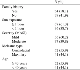
Results Among 93 patients, centrofacial melasma was more prevalent than maxillary melasma (55.9% vs. 44.1%). We found no mandibular melasma. Most cases were categorized as mild rather than moderate degree using the Melasma Area Severity Index (60.2%:39.8%), and most of these were found in women aged at least 40 years (55.9%). Family history (58.1%) and sun exposure exceeding 1 hour (61.3%) were among the factors contributing to melasma development.
Conclusion Characteristics of melasma in Javanese women are centrofacial type, mild severity and most common in those who are at least 40 year old with sun exposure and have a family history.
Key words
93 women with melasma. Subjects were recruited from the Dermato-Venereology Clinic of Dr. Sardjito General Hospital and the Be Queen Skin Care Clinic in Yogyakarta, Indonesia. This study was approved by the Medical and Health Research Ethic Committee of Faculty of Medicine Universitas Gadjah Mada and Sardjito Hospital with approval number (KE/FK/462/EC/2016). Subjects were interviewed using a standardized questionnaire that explored UV exposure and family history. Study participants were adult Javanese women aged 18 to 60 years (mean 40.6 ± 6.45 years) in Yogyakarta who had melasma. Exclusion criteria were use of hormonal contraceptives, pregnancy, and use of a whitening cream during the prior two weeks. We examined the skin
using a Bombtech Skin Diagnosis A-ONE®
(Korea) to identify the skin’s condition and the
areas with melasma. Mexameter® MX18
Courage-Khazaka (Germany) was used to measure skin pigmentation and calculate the Melasma Area Severity Index (MASI) score. This instrument utilizes inspections of facial skin to assess three factors: the involved area, hyperpigmentation, and homogeneity. The face was divided into four areas: the forehead, right malar region, left malar region, and chin, representing 30%; 30%; 30%, and 10% of the face, respectively. Each involved area was rated from 0 to 6 (0, unaffected; 1, less than 10%; 2, 10%–29%; 3, 30%–49%; 4, 50%–69%; 5, 70%– 89%; and 6, 90%–100%). Hyperpigmentation and homogeneity were each assessed from 0 to 4 (0, none; 1, thin; 2, medium; 3, clear; and 4, very clear). The final MASI result was the sum of hyperpigmentation and homogeneity multiplied by the involved facial area, rendering a value from 0 to 48.8,9,10
Results
All patients had skin type IV, and they were aged with the youngest age 29-year and the
oldest 58-year; then the age categorization was divided into below year-old and above
40-year-old (Table 1). Of 93 participants, 54
subjects had family history of melasma and 39 subjects did not (58.1% vs 41.9%). Melasma was found most frequently in subjects with accumulated sun exposure for more than 1 hour are 57 subject (61.3%) and for less than 1 hour are 36 (38.7%). Using the MASI score, severity of melasma was classified as mild in 56 (60.2%)
patients and moderate in 37 (39.8%).
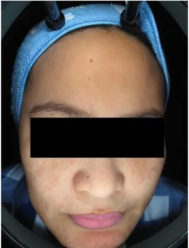
Centrofacial type of melasma was the most common type seen in this study compared to
maxillaris (55.9%: 44.1%) patients (Figure 1
and 2). No mandibular melisma was found.
Patients’ characteristics are presented in Table 1.
Discussion
In this study, all patients had skin type IV. Therefore, all had similar responses to sun exposure. People with skin types IV and V have acidic melanocyte dendrites because melanosomes are present in greater quantities compared to those with skin types I and II. This causes the corneal layer of skin to be acidic (pH 4.6). An acidic corneal layer increases the skin’s integrity and its healing process after
Table 1 Patients’ characteristics (n=93). N (%) Family history
Yes 54 (58.1)
No 39 (41.9)
Sun exposure
≥ 1 hour 57 (61.3)
< 1 hour 36 (38.7)
Severity (MASI)
Mild 56 (60.2)
Moderate 37 (39.8)
Melasma type
Centrofacial 52 (55.9)
Maxillary 41 (44.1)
Age
≥ 40 years 52 (55.9)
Figure 1 Centrofacial type of melasma (printed after permission of subject).
Figure 2 Maxillary type of melasma (printed after permission of subject).
photodamage. However, in those with melasma, a disorder in the integrity of the corneal layer creates a slower healing process.11,12,13 A study by Ortonne et al. (2009)14 using 324 women with melasma showed that 83% had skin types III-VI; 33% had skin type IV.A study in Tunisia showed that 45% of melisma patients had skin type IV.11 In addition, a Brazilian study found that 302 (38.4%) melasma patients had skin type IV.5 We found a similar result. These might be
reasons for the high of melasma in people with skin type IV, because the healing process is slow after UV exposure. However, these conditions depended on the duration and intensity of sun exposure and geographic location.
We found that most patients (54, 58.1%) had family histories of melasma. Several studies show that genetic factors have important roles in melasma development. A study of 324 women with melasma showed that 48% had family histories, and 90% had dark skin.14 A Brazilian study showed that 56.3% of 302 participants with melasma had family histories.5 A study in the sub-Himalayan showed that 75% of 100 melasma patients had family history of disease.15 We found that a family history or genetic factors seem to be related to the development of melasma.
sun exposure is a risk factor for melasma in addition to a family history of melasma.
People with skin type IV are primarily brown-skinned.19 Skin type IV has more eumelanin and has greater photoprotectant abilities.20 Three factors play role during sun exposure: melanin, amino acid, and light reflection by melanin and amino acid. Increased melanin and a thicker epidermis protects the DNA and reduces inflammation caused by photons.21,22 The amount and type of melanin determine skin color and its protectiveness against sun exposure. Differences in the amount of melanin can have a 100-fold difference in the response to UV exposure. Melanin is a polymer, and its content remains poorly understood. The ratio between melanin eumelanin and pheomelanin differs depending on the population and the geographic location where the individual originated.22 Eumelanin can act as a photoprotector by absorbing visible and nonvisible UV rays, especially between the wavelengths of 720 nm and 620 nm. It has an exponential effect between 300 nm and 600 nm. An ultrastructure study showed that the epidermal eumelanosome was intact on dark skin after sun exposure. On the other hand, the eumelanosome was not intact on white skin after sun exposure.20,22 A study in Tunisia enrolling participants with skin type III, brown hair, and sun-exposed skin showed moderate melasma based on MASI score (16-32.9). Mild melasma was commonly found in people with skin type IV. Severe melasma was commonly found in patients with skin type V who followed a regimen of oral contraceptives.6 We found mild melasma in 56 patients (95% CI, 13.34-16.53). This could be because all our patients had skin type IV and a high eumelanin-to-pheomelanin ratio. Therefore, the skin was more photoprotective. Our results are comparable to those of the study in Tunisia enrolling participants with mild or moderate melasma. Differences lie in our lack of patients with
severe melasma, a result of excluding those experiencing hormonal factors (contraception use and pregnancy) from our study.
The centrofacial type of melasma is most common (65%), followed by maxillary melasma (20%) and then mandibular melasma (15%).4,5 We classified melasma according to its distribution on the face and found results similar to those found by Tamega et al. (2012).5 Studies in India and Brazil showed that centrofacial melasma was the most common type (69.2%) followed by malar melasma (43.4%). Mandibular melasma was not found in the Brazilian study because most mandibular melasma (68.8%) spread to the parotid area; melasma purely on the ramus of mandible was found in only 2 patients (3.7%).5,23 On the other hand, in Singapore, malar melasma was most common (89%) followed by centrofacial melasma (8%) and mandibular melasma (3%).16 Similar results were found in Tunisia, where centrofacial melasma was the most common (76.1%) among 197 patients, and malar melasma and mandibular melasma were found in 22.9% and 1%, respectively.6
Conclusion
The characteristic features of melasma in Javanese women are centrofacial type, and for severity of most sufferers is mild. It is seen most frequently in women aged above 40 year old, in those who received at least 1 hour of sun exposure, and in those with family histories of melasma. Sun exposure and family history are the most influencing factors in the characteristics of melasma.
References
1. Achar A, Rathi SK. Melasma: A clinicoepidemiological study of 312 cases. Indian J Dermatol. 2011;56:380-2.
2. Jiang J, Akinseye O, Tovar-Garza A, Pandya AG. The effect of melasma on self-esteem: A pilot study. Int J Womens Dermatol. 2017;4:38-42.
3. Sarkar R, Arora P, Garg VK, Sonthalia S, Gokhale N. Melasma update. Indian Dermatol Online J. 2014;5:426-35.
4. Damevska K. New Aspects of Melasma/ Novi aspektimelazme. Serbian J Dermatol. Venereol. 2014;6:5-18.
5. Tamega Ade A, Miot LD, Bonfietti C, Gige TC, Marques ME, Miot HA. Clinical patterns and epidemiological characteristics of facial melasma in Brazilian women: Clinical patterns and epidemiology of melasma. J Eur Acad Dermatol Venereol. 2013;27:151-6.
6. Guinot C, Cheffai S, Latreille J, Dhaoui M, Youssef S, Jaber K et al. Aggravating factors for melasma: a prospective study in 197 Tunisian patients. J Eur Acad Dermatol Venereol. 2010;24:1060-9.
7. Trivedi MK, Yang FC, Cho BK. A review of laser and light therapy in melasma. Int J Womens Dermatol. 2017;3:11-20.
8. Majid I, Haq I, Imran S, Keen A, Aziz K, Arif T. Proposing melasma severity index: A new, more practical, office-based scoring system for assessing the severity of melasma. Indian J Dermatol. 2016;61 :39-44.
9. Pandya A, Berneburg M, Ortonne J-P, Picardo M. Guidelines for clinical trials in melasma. Pigmentation Disorders Academy. Br J Dermatol. 2006;156 Suppl 1:21-8.
10. Tay EY, Gan EY, Tan VW, Lin Z, Liang Y, Lin F et al. Pilot study of an automated method to determine Melasma Area and Severity Index. Br J Dermatol. 2015;172:1535-40.
11. Gunathilake R, Schurer NY, Shoo BA, Celli A, Hachem J-P, Crumrine D et al. pH-Regulated Mechanisms Account for Pigment-Type Differences in Epidermal Barrier Function. J Invest Dermatol. 2009;129:1719-29.
12. Lee A-Y. Recent progress in melasma pathogenesis. Pigment Cell Melanoma Res. 2015;28:648-60.
13. Lee A-Y. An updated review of melasma pathogenesis. Dermatol Sin. 2014;32:233-9. 14. Ortonne J, Arellano I, Berneburg M, Cestari
T, Chan H, Grimes P et al. A global survey of the role of ultraviolet radiation and hormonal influences in the development of melasma. J Eur Acad Dermatol Venereol. 2009;23:1254-62.
15. Halder S, Halder A, Nag SC, Rajesh D, Sarkar P. Melasma in the people of Sub-Himalayan region of Eastern India. J Pak Assoc Dermatol. 2016;23:139-42.
16. Goh CL, Dlova CN. A retrospective study on the clinical presentation and treatment outcome of melasma in a tertiary dermatological referral centre in Singapore. Singapore Med J. 1999;40:455-8.
17. Moin A, Jabery Z, Fallah N. Prevalence and awareness of melasma during pregnancy. Int J Dermatol. 2016;45:285-8.
18. Al-Hamdi KI, Hasony HJ, Jareh HI. Melasma in Basrah : A Clinical and Epidemiological study. Med J Basrah Univ. 2008;26(1):1-5.
19. Shah SK, Alexis AF, editors. Cosmetic Dermatology for Skin of Color. Philadelphia: McGraw-Hill; 2009. p.1-11. 20. Rouzaud F, Kadekaro AL, Abdel-Malek ZA,
Hearing VJ. MC1R and the response of melanocytes to ultraviolet radiation. Mutat Res Mol Mech Mutagen. 2005;571:133-52. 21. Alaluf S, Barrett K, Blount M, Carter N.
Ethnic variation in tyrosinase and TYRP1 expression in photoexposed and photoprotected human skin. Pigment Cell Res. 2003;16:35-42.
22. Rees JL. The genetics of sun sensitivity in humans. Am J Hum Genet. 2004;75:739-51. 23. Katsambas A, Antoniou CH. Melasma.