R E V I E W
Open Access
Organoid technology in female
reproductive biomedicine
Heidar Heidari-Khoei
1†, Fereshteh Esfandiari
1†, Mohammad Amin Hajari
1, Zeynab Ghorbaninejad
1,
Abbas Piryaei
2,3*and Hossein Baharvand
1,4*Abstract
Recent developments in organoid technology are revolutionizing our knowledge about the biology, physiology, and function of various organs. Female reproductive biology and medicine also benefit from this technology. Organoids recapitulate features of different reproductive organs including the uterus, fallopian tubes, and ovaries, as well as trophoblasts. The genetic stability of organoids and long-lasting commitment to their tissue of origin during long-term culture makes them attractive substitutes for animal and in vitro models. Despite current limitations, organoids offer a promising platform to address fundamental questions regarding the reproductive system’s physiology and pathology. They provide a human source to harness stem cells for regenerative medicine, heal damaged epithelia in specific diseases, and study biological processes in healthy and pathological conditions. The combination of male and female reproductive organoids with other technologies, such as microfluidics technology, would enable scientists to create a multi-organoid-on-a-chip platform for the next step to human-on-a-chip platforms for clinical applications, drug discovery, and toxicology studies. The present review discusses recent advances in producing organoid models of reproductive organs and highlights their applications, as well as technical challenges and future directions.
Keywords:Organoids, Reproductive organs, Reproductive medicine, Organoid-on-a-chip
Background
The female reproductive system is of utmost importance to a woman’s quality of life; it produces sex hormones and oocytes, provides the site for fertilization, and supports the fetal development [1, 2]. Diseases and disorders of the female reproductive system are not adequately studied, especially in the areas of endometriosis, gynecological cancers, sexually transmitted diseases (STDs), and preg-nancy disorders (including intrauterine growth restriction,
miscarriage, and recurrent miscarriage), in addition to medications that have deleterious effects on the reproduct-ive system [2–5]. Limited access to reproductive material, especially those for maternal-embryo interactions, and unavailable reliable experimental models are serious chal-lenges for studies in this field [6].
Conventional in vitro systems that include two-dimensional (2D) and three-two-dimensional (3D) cell cultures have serious pitfalls that make them unsuitable for studying the female reproductive system. The 2D culture of immor-talized cell lines or those derived from tumors in reproduct-ive organs have led to numerous insights about the biology and physiology of the reproductive system; their advantages include reproducibility and ease of access. However, they lack many complex features of in vivo microenvironments, including a cell-cell/cell-extracellular matrix (ECM), the display of a relatively homogenous phenotype, and are not
© The Author(s). 2020Open AccessThis article is licensed under a Creative Commons Attribution 4.0 International License, which permits use, sharing, adaptation, distribution and reproduction in any medium or format, as long as you give appropriate credit to the original author(s) and the source, provide a link to the Creative Commons licence, and indicate if changes were made. The images or other third party material in this article are included in the article's Creative Commons licence, unless indicated otherwise in a credit line to the material. If material is not included in the article's Creative Commons licence and your intended use is not permitted by statutory regulation or exceeds the permitted use, you will need to obtain permission directly from the copyright holder. To view a copy of this licence, visithttp://creativecommons.org/licenses/by/4.0/. The Creative Commons Public Domain Dedication waiver (http://creativecommons.org/publicdomain/zero/1.0/) applies to the data made available in this article, unless otherwise stated in a credit line to the data.
* Correspondence:piryae@sbmu.ac.ir;Baharvand@royaninstitute.org †Heidar Heidari-Khoei and Fereshteh Esfandiari contributed equally to this work.
2Urogenital Stem Cell Research Center, Shahid Beheshti University of Medical Sciences, Tehran, Iran
1Department of Stem Cells and Developmental Biology, Cell Science Research Center, Royan Institute for Stem Cell Biology and Technology, ACECR, P.O. Box: 16635-148, Tehran 1665659911, Iran
representative of in vivo cellular diversity. Because of genetic alterations in cell lines, they fail to recapitulate key features of native tissues as well as different active cell signaling pathways and function from those in the native cells [7]. Moreover, rapid loss of phenotype and tissue-related functions occur following 2D culture of primary cells [8]. Therefore, 2D culture systems do not adequately show the natural 3D environment of cells; in turn, they fail to mimic in vivo cellular functions and signaling pathways, and may provide misleading and non-predictive data for in vivo responses. The 3D cell aggregates exhibit improved function, but lack cellular polarity and the 3D organization that is present in vivo. Other 3D culture systems include spheroids that often lack the capacity for self-renewal and differentiation due to the absence of relevant progenitor or stem cells [9, 10]. The organ explant or organotypic slice cultures that recapitulate the complex 3D architecture, cellular heterogeneity, and function of the native organ are very useful for the study of development and physiology, but they are limited by their inability to proliferate, their short-term nature, and cell phenotype drift due to improper orientation in culture media [11,12].
The recently developed organoid technology provides new in vitro models to serve as both tissue and organ proxies to bridge the gap between in vitro and in vivo [13]. Organoids are self-organizing structures that recap-itulate a numerous of biological and pathological features of organs such as spatially restricted lineage commitment, specific functions of the organ, multiple organ-specific cell types, and cell-cell and cell-matrix interactions. They rep-resent genetic stability and maintain commitment to their tissue of origin during the long-term culture that allows for access to an unlimited source of material to support research in this field. Moreover, they can serve as substi-tutes for animal and in vitro models and provide the opportunity for high throughput studies, personalized medicine, drug and toxicity testing, disease modeling, and present a promising way for autologous transplantation. Organoids recapitulate features of different reproductive organs. In this regard, the uterus, fallopian tubes, and ovaries as well as trophoblast organoids have been created in the laboratory. In this review, we will discuss recent advances in producing organoid models of reproductive organs and highlight their applications, in addition to discussing technical challenges and future directions.
Main text Ovarian organoids
Ovarian cancer represents the fifth leading cause of can-cer deaths in women in the US [14]. Access to reliable experimental models that address clinical challenges, such as early detection, tumor recurrence, and acquired chemotherapy resistance, is a high priority in ovarian cancer research. Recently, successful generation ovarian
cancer organoids was reported by two separate groups [15, 16]. These organoids recapitulate histological and genomic features of the lesions from which they were derived and represent intra- and interpatient heterogen-eity [15,16]. Moreover, ovarian cancer organoids display somatic mutations and amplifications/deletions [15] and recapitulated the parent tumor’s marker expression and mutational landscape [16]. Moreover, the organoids showed tumor-specific sensitivity to chemotherapeutic drugs and therefore provide a reliable preclinical tools for drug screening and discovery [15, 16]. Moreover, xenografting these organoids can provide an in vivo platform for drug screening [15]. Maru et al. have previ-ously reported that ovarian organoids recapitulated mutation profile and intra-tumor heterogeneity [17].
Developing organoids from other ovarian disorders such as premature ovarian failure and polycystic ovary would provide a platform to study the mechanisms that underlie these diseases and enable the development of new drugs and treatments.
Fallopian tube organoids
The fallopian tube (oviduct or uterine tube) is a central organ of the female reproductive system that plays an essential role in oocyte maturation and selection, gamete and embryo transportation, sperm reservoir, control of polyspermy, fertilization and early embryonic develop-ment [18]. It is believed that the fallopian tube is the origin site of several clinically important diseases such as high-grade serous ovarian cancer (the deadliest form of gynecological cancer), pelvic inflammatory disease (PID), and infertility [19, 20]. Direct examination and study of fallopian tubes in alive patients is extremely difficult and somehow impossible due to its intra-abdominal location and structure. Therefore, an in vitro organoid model that recapitulates the in vivo structure and function of fallopian tubes is advantageous for supporting studies in this field.
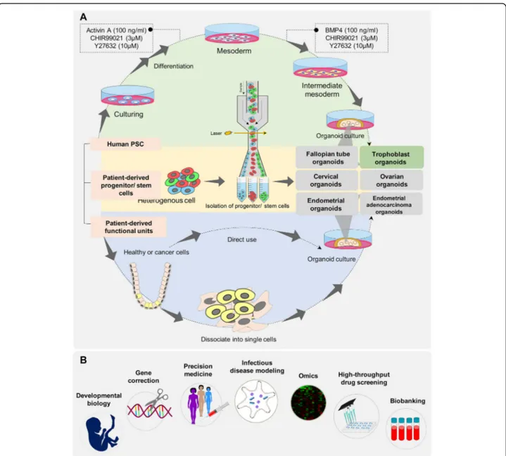
After the differentiation period, the cells secreted an oviduct fluid surrogate that could support embryonic development up to the blastocyst stage without the addition of embryo culture medium [21]. Despite the polarized structure and in vivo-like function, this culture system differs from the current organoid concept. This culture system lacks the tubular folded architecture and inserts, and does not permit live imaging or perfusion studies that limits its use to study gamete interactions and early embryo development in detail [8, 21]. In self-organizing organoids, the fallopian tube was generated based on the modified intestinal organoid proto-col. Isolated fallopian epithelial cells were seeded in a 2D culture, followed by culture in 3D Matrigel matrix supple-mented with growth factors (epidermal growth factor [EGF], fibroblast growth factor [FGF], and TGF-β), niche specific factors (Wnt3a, R-spondin-1 [RSPO1], ALK4/5, and Nog-gin), and an inhibitor of anoikis (ROCK inhibitor) (Fig. 1, Table 1) [25]. In contrast to other organoid models, this study showed that the addition of Wnt3a and RSPO1 main-tained the stem cell subpopulations for an extended period of time and also allowed full differentiation. The presence of EGF doubled the number of organoids, addition of RSPO1 increased their size, and addition of ALK4/5 was crucial for quasi-indefinite expansion (Table 2). Monoclonal cystic organoids that contained ciliated and secretory cells have been successfully generated from a single EpCAM+ cell. Fallopian tube organoids faithfully recapitulate the structure of native tissue, show highly polarized columnar cells, fully developed inter-cellar junctions, fully assembled cilia, active secretion, and an orientation of the apical pole to the luminal side. These organoids are responsive to hormonal stimulation, show robust growth and can be maintained long-term in culture. Successful generation of monoclonal organoids from different donors has confirmed the presence of stem cells in the generated organoids, as well as fallopian tube epithelium (FTE).
Scientists reported a human iPSC reprogramming method for generating FTE organoids. In this study, different WNT and BMP signaling were modulated to successful direct differentiation of human pluripotent stem cells into Müllerian cells and subsequent pro-Müllerian growth factors were used to develop FTE precur-sors. Then, FTE precursors were cultured in Matrigel with phenol red where they formed an organoid structure. How-ever, when cultured in Matrigel without phenol red, they became branched and formed an unorganized matrix [22]. Phenol red is widely used in cell culture as a pH indicator; it bears structural similarity to nonsteroidal estrogens, exhibits estrogen-like bioactivity, and promotes prolifera-tion in estrogen-sensitive cells such as fallopian tube cells [29,30]. Therefore, their results have shown that estrogen effects FTE differentiation and maturation [22]. Human iPSC-derived FTE organoids were grown in 3D Matrigel with estrogen and progesterone supplemented media for an
extended period. Immunocytochemistry results showed that FTE organoids formed secretory (PAX8+) and ciliated (TUBB4A+) cells. Expression of a mature epithelial cell marker (CDH1) in the organoid was comparable to fresh human fallopian tube tissue. In addition, the proper differ-entiation of iPSC-derived organoids into fallopian tube cells was confirmed using heat map analysis [22].
The described fallopian tube organoid models closely mimic normal physiology and architecture of the human FTE. Therefore, they provide promising models to study the biology and pathology of fallopian tubes with regards to screening technologies, cancer biology, and repro-ductive medicine [25]. However, this system has limita-tions for gamete or embryo interaction studies due to its small size and inaccessible luminal compartment that re-quire labor-intensive approaches, such as microinjection.
Endometrial organoids
The human endometrium is a dynamic tissue that under-goes cyclic changes in response to steroid hormones as well as paracrine and autocrine factors to be prepared for embryo implantation. Embryo implantation is a highly complex process that requires a receptive endometrium, a competent blastocyst, and a synchronized maternal-embryo dialogue [31]. The endometrium is also involved in many gynecologic conditions, including infertility, dysmenorrhea, endometrial polyps, endometriosis, and endometrial cancer which is the most common cancer of the female reproduct-ive organs [32].
breaching and invasion of isolated cells into the surrounding Matrigel. However, the tumor derived-organoids were positive for glandular markers MUC1 and SOX17, which confirmed their glandular origin. Investigation of chromosomal stability of endometrial
organoids using a comparative genomic hybridization (CGH) array demonstrated that the established orga-noids preserved their genetic integrity over several months in culture. These genetically stable endomet-rial organoids could be expanded and frozen without
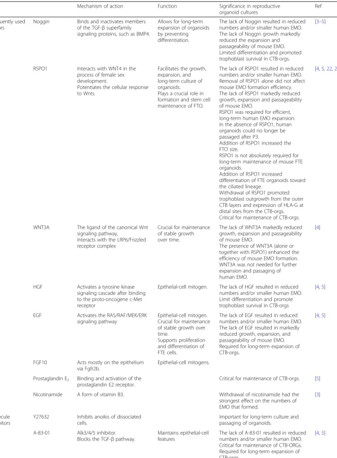
Table 1Frequently used growth media constituents, their working mechanisms, effects and applications
Mechanism of action Function Significance in reproductive organoid cultures
Ref
Frequently used factors
Noggin Binds and inactivates members of the TGF-βsuperfamily signaling proteins, such as BMP4.
Allows for long-term expansion of organoids by preventing differentiation.
The lack of Noggin resulted in reduced numbers and/or smaller human EMO. The lack of Noggin growth markedly reduced the expansion and passageability of mouse EMO. Limited differentiation and promoted trophoblast survival in CTB-orgs.
[3–5]
RSPO1 Interacts with WNT4 in the process of female sex development.
Potentiates the cellular response to Wnts.
Facilitates the growth, expansion, and long-term culture of organoids. Plays a crucial role in formation and stem cell maintenance of FTO.
The lack of RSPO1 resulted in reduced numbers and/or smaller human EMO. Removal of RSPO1 alone did not affect mouse EMO formation efficiency. The lack of RSPO1 markedly reduced growth, expansion and passageability of mouse EMO.
RSPO1 was required for efficient, long-term human EMO expansion. In the absence of RSPO1, human organoids could no longer be passaged after P3.
Addition of RSPO1 increased the FTO size.
RSPO1 is not absolutely required for long-term maintenance of mouse FTE organoids.
Addition of RSPO1 increased differentiation of FTE organoids toward the ciliated lineage.
Withdrawal of RSPO1 promoted trophoblast outgrowth from the outer CTB layers and expression of HLA-G at distal sites from the CTB-orgs. Critical for maintenance of CTB-orgs.
[4,5,22,23]
WNT3A The ligand of the canonical Wnt signaling pathway,
Interacts with the LRP6/Frizzled receptor complex
Crucial for maintenance of stable growth over time.
The lack of WNT3A markedly reduced growth, expansion and passageability of mouse EMO.
The presence of WNT3A (alone or together with RSPO1) enhanced the efficiency of mouse EMO formation. WNT3A was not needed for further expansion and passaging of human EMO.
[4]
HGF Activates a tyrosine kinase signaling cascade after binding to the proto-oncogene c-Met receptor
Epithelial-cell mitogen. The lack of HGF resulted in reduced numbers and/or smaller human EMO. Limit differentiation and promote trophoblast survival in CTB-orgs
[4,5]
EGF Activates the RAS/RAF/MEK/ERK signaling pathway
Epithelial-cell mitogen. Crucial for maintenance of stable growth over time.
Supports proliferation and differentiation of FTE cells.
The lack of EGF resulted in reduced numbers and/or smaller human EMO. The lack of EGF resulted in markedly reduced growth, expansion, and passageability of mouse EMO. Required for long-term expansion of CTB-orgs.
[4,5]
FGF10 Acts mostly on the epithelium via Fgfr2b.
Epithelial-cell mitogens.
Prostaglandin E2 Binding and activation of the prostaglandin E2 receptor.
Critical for maintenance of CTB-orgs. [5]
Nicotinamide A form of vitamin B3. Withdrawal of nicotinamide had the strongest effect on the numbers of EMO that formed.
[3]
Molecule inhibitors
Y27632 Inhibits anoikis of dissociated cells.
Important for long-term culture and passaging of organoids.
A-83-01 Alk3/4/5 inhibitor. Blocks the TGF-βpathway.
Maintains epithelial-cell features
The lack of A-83-01 resulted in reduced numbers and/or smaller human EMO. Critical for maintenance of CTB-ORGs. Required for long-term expansion of CTB-orgs.
loss of their proliferative ability after thawing to create a patient-specific biobank of endometrial tissues [3]. This culture system has the ability to expand the small quantity of starting material for a variety of high throughput assessments and could be a valuable plat-form for investigating implantation problems, the histotrophic nutrition period in early pregnancy, novel therapeutic strategies for gynecologic pathologies such as endometriosis and endometrial cancer, and generate an endometrial biobank. However, the current orga-noids lack stromal cells. It is well known that recipro-cal interaction between endometrial epithelial and stromal cells is responsible for physiological functions (proliferation, differentiation, and decidualization) and emergence of several pathologic conditions such as endometrial carcinoma [34–36]. Without a more complete complement of cell types, endometrial organoids will always lack the context they need to be actual mini-organs. Recently, a co-culture of intestinal organoids with stromal cells was developed by Stzepourginski et al. [37]. Co-culture of the present endometrial organoids with stromal cells could further complement the organoid model and provide a relevant model to study reciprocal epithelium stroma interactions that occur in vivo [36]. The inaccessible luminal compartment and apical aspect of the epithelium of organoids present a number of chal-lenges to physiologically relevant studies [38]. Unfolding the spherical organoid into a 2D planar tissue construct and monolayer culture of primary epithelial cells from organoids would provide an accessible luminal compart-ment for related studies, such as embryo cultures [38].
Trophoblast organoids
The placenta is an extraembryonic organ that is essential for survival and development of the mammalian embryo. During implantation in humans, the trophectoderm layer of the blastocyst attaches to the endometrial epi-thelium and continues to differentiate into trophoblast subtypes: the cytotrophoblast (CTB), extravillous CTB (EVT), and syncytiotrophoblast (STB). Undifferentiated
CTB cells grow through the STB to form cell columns and chorionic villi. The CTB cells at the tips of villi dif-ferentiate into EVT, which invade the deciduum. EVTs have two cell types: interstitial EVT cells that invade the decidualized endometrium and endovascular EVTs that invade and remodel the spiral arteries [39,40]. Multinu-cleated STB cells are responsible for nutrient exchange and synthesis. They secrete placental hormones such as placental lactogen, chorionic gonadotropin, and proges-terone in addition to numerous other proteins and steroids to maintain pregnancy. These cells are formed by fusion of interstitial CTB cells [41]. Placental dysfunc-tion and insufficiency results in major pregnancy-related disorders such as intrauterine growth restriction, pre-eclampsia, miscarriage, recurrent abortion, and preterm labor [42,43]. Our current knowledge about the human placenta is limited due to the lack of representative func-tional models [44]. Several experimental models, includ-ing animal models and in vitro cell culture models, have been used to study the placenta. However, distinct struc-tural and functional differences exist between human and other animal placentas; thus, data from animal models is not relevant to humans [45, 46]. In vitro models of human trophoblasts include placenta-derived cell lines, isolated primary placenta cells, and human placenta tissue explants. Several cell lines have been established from choriocarcinoma cells: JEG-3, BeWo, and JAR. Advantages of these cell lines include ease of use; unlimited supply of material; more pure cell source; less expensive to procure; ease of use for gene silencing approaches; and these sources bypass some of the ethical concerns associated with the use of animal or primary human tissues [47]. However, immortal cell lines have several drawbacks due to their malignant transforma-tions. This problem may be overcome by using immor-talized primary trophoblast cells, such as the HTR-8/ SVneo cell line derived by transfecting first trimester EVT cells with the gene that encodes for simian virus 40 large T antigen, or ACH-3P and AC1-M59 cell lines, which are choriocarcinoma cells fused with primary first Table 1Frequently used growth media constituents, their working mechanisms, effects and applications(Continued)
Mechanism of action Function Significance in reproductive organoid cultures
Ref
SB202190 p38 inhibitor. Decreasing concentrations of p38i were
beneficial for long-term expansion of endometrial cancer organoids.
[24]
SB431542 TGF-βR kinase inhibitor IV. Crucial for quasi-indefinite expansion of FTO.
Without TGF-β, RK inhibitor FTO had slower expansion and finally growth arrest by four to six passages (three to four months).
Important for formation and maintenance of large FTE organoids.
[23,25]
CHIR99021 Inhibitor of GSK3. Critical for maintenance of CTB-orgs. [5]
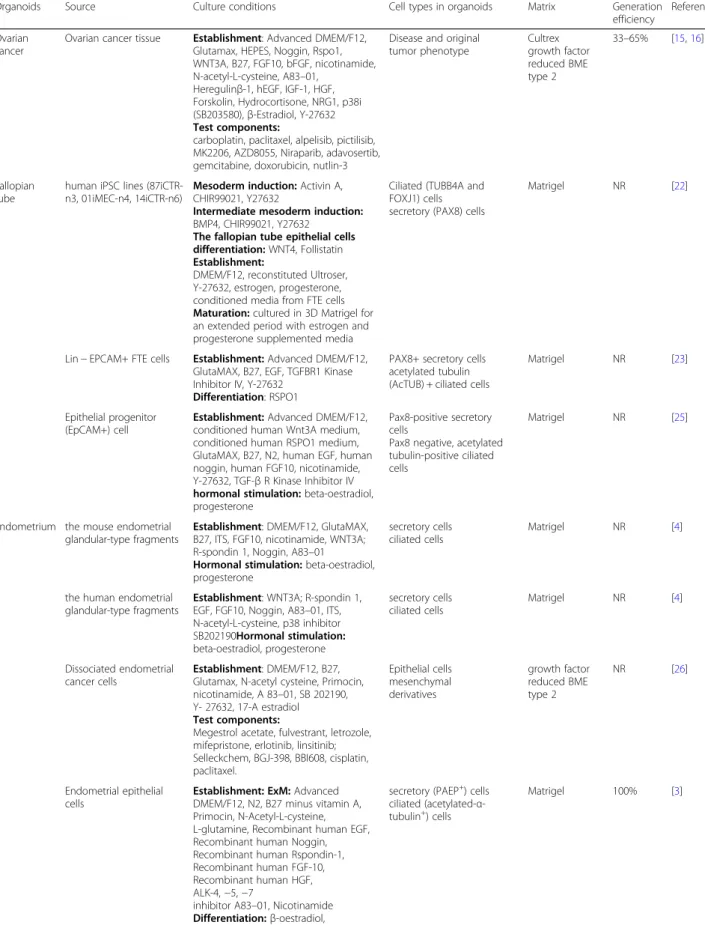
Table 2Summary of sources and culture conditions used in the development of various reproductive organoids
Organoids Source Culture conditions Cell types in organoids Matrix Generation efficiency
Reference
Ovarian cancer
Ovarian cancer tissue Establishment: Advanced DMEM/F12, Glutamax, HEPES, Noggin, Rspo1, WNT3A, B27, FGF10, bFGF, nicotinamide, N-acetyl-L-cysteine, A83–01,
Heregulinβ-1, hEGF, IGF-1, HGF, Forskolin, Hydrocortisone, NRG1, p38i (SB203580),β-Estradiol, Y-27632
Test components:
carboplatin, paclitaxel, alpelisib, pictilisib, MK2206, AZD8055, Niraparib, adavosertib, gemcitabine, doxorubicin, nutlin-3
Disease and original tumor phenotype
Cultrex growth factor reduced BME type 2
33–65% [15,16]
Fallopian tube
human iPSC lines (87iCTR-n3, 01iMEC-n4, 14iCTR-n6)
Mesoderm induction:Activin A, CHIR99021, Y27632
Intermediate mesoderm induction:
BMP4, CHIR99021, Y27632
The fallopian tube epithelial cells differentiation:WNT4, Follistatin
Establishment:
DMEM/F12, reconstituted Ultroser, Y-27632, estrogen, progesterone, conditioned media from FTE cells
Maturation:cultured in 3D Matrigel for an extended period with estrogen and progesterone supplemented media
Ciliated (TUBB4A and FOXJ1) cells secretory (PAX8) cells
Matrigel NR [22]
Lin−EPCAM+ FTE cells Establishment:Advanced DMEM/F12, GlutaMAX, B27, EGF, TGFBR1 Kinase Inhibitor IV, Y-27632
Differentiation: RSPO1
PAX8+ secretory cells acetylated tubulin (AcTUB) + ciliated cells
Matrigel NR [23]
Epithelial progenitor (EpCAM+) cell
Establishment:Advanced DMEM/F12, conditioned human Wnt3A medium, conditioned human RSPO1 medium, GlutaMAX, B27, N2, human EGF, human noggin, human FGF10, nicotinamide, Y-27632, TGF-βR Kinase Inhibitor IV
hormonal stimulation:beta-oestradiol, progesterone
Pax8-positive secretory cells
Pax8 negative, acetylated tubulin-positive ciliated cells
Matrigel NR [25]
Endometrium the mouse endometrial glandular-type fragments
Establishment: DMEM/F12, GlutaMAX, B27, ITS, FGF10, nicotinamide, WNT3A; R-spondin 1, Noggin, A83–01
Hormonal stimulation:beta-oestradiol, progesterone
secretory cells ciliated cells
Matrigel NR [4]
the human endometrial glandular-type fragments
Establishment: WNT3A; R-spondin 1, EGF, FGF10, Noggin, A83–01, ITS, N-acetyl-L-cysteine, p38 inhibitor SB202190Hormonal stimulation:
beta-oestradiol, progesterone
secretory cells ciliated cells
Matrigel NR [4]
Dissociated endometrial cancer cells
Establishment: DMEM/F12, B27, Glutamax, N-acetyl cysteine, Primocin, nicotinamide, A 83–01, SB 202190, Y- 27632, 17-A estradiol
Test components:
Megestrol acetate, fulvestrant, letrozole, mifepristone, erlotinib, linsitinib; Selleckchem, BGJ-398, BBI608, cisplatin, paclitaxel.
Epithelial cells mesenchymal derivatives
growth factor reduced BME type 2
NR [26]
Endometrial epithelial cells
Establishment: ExM:Advanced DMEM/F12, N2, B27 minus vitamin A, Primocin, N-Acetyl-L-cysteine, L-glutamine, Recombinant human EGF, Recombinant human Noggin, Recombinant human Rspondin-1, Recombinant human FGF-10, Recombinant human HGF, ALK-4,−5,−7
inhibitor A83–01, Nicotinamide
Differentiation:β-oestradiol, progesterone, cAMP, prolactin, human
secretory (PAEP+) cells ciliated (acetylated-α -tubulin+) cells
and third trimester trophoblast cells, respectively [48]. However, some of these cells do not meet the criteria for human trophoblast cells as proposed by Lee et al., and do not express some important protein markers (GATA3, KRT7, and TFAPC2), the human leukocyte antigen (HLA) class I profile, the chromosome 19 miRNA cluster (C19MC), and hypomethylation of the ELF5 promoter [49]. Alternative models, such as bone morphogenetic protein 4 (BMP4)-treated human embry-onic stem cells (hESCs), have also been established. Al-though these trophoblast-like cells are used as models
for understanding early trophoblast lineage segregation, their global gene expression profiles, trophoblast-specific markers, and HLA status considerably differ from pri-mary trophoblast populations [50].
Recently, two independent research groups developed organoid platforms for culturing first-trimester CTBs in vitro. Turcoet al.reported some growth cell clusters of
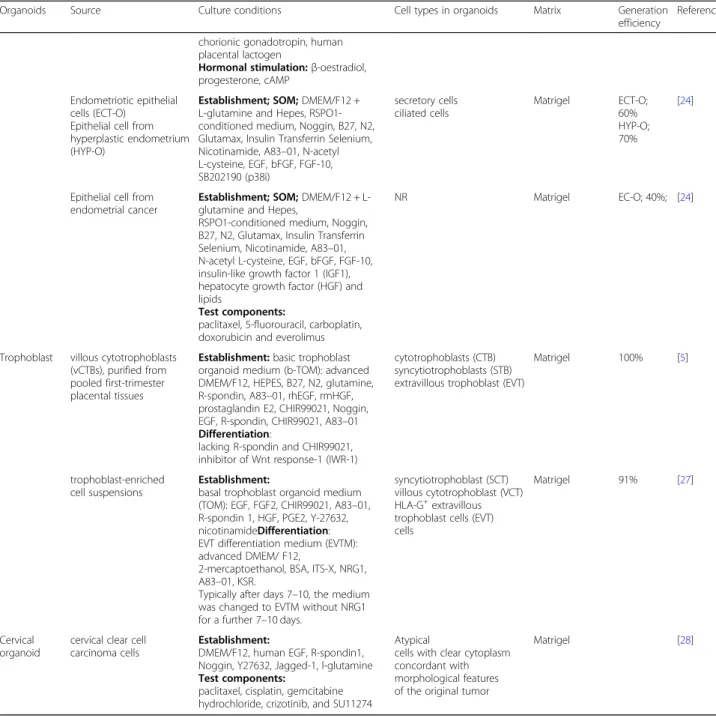
first-trimester placenta were seeded into Matrigel drops and grown in a basal trophoblast organoid medium (TOM) composed of EGF, FGF2, CHIR99021, A83–01, and RSPO1 (Table1, Fig.1). Addition of growth factors Table 2Summary of sources and culture conditions used in the development of various reproductive organoids(Continued)
Organoids Source Culture conditions Cell types in organoids Matrix Generation efficiency
Reference
chorionic gonadotropin, human placental lactogen
Hormonal stimulation:β-oestradiol, progesterone, cAMP
Endometriotic epithelial cells (ECT-O)
Epithelial cell from hyperplastic endometrium (HYP-O)
Establishment; SOM;DMEM/F12 + L-glutamine and Hepes, RSPO1-conditioned medium, Noggin, B27, N2, Glutamax, Insulin Transferrin Selenium, Nicotinamide, A83–01, N-acetyl L-cysteine, EGF, bFGF, FGF-10, SB202190 (p38i)
secretory cells ciliated cells
Matrigel ECT-O; 60% HYP-O; 70%
[24]
Epithelial cell from endometrial cancer
Establishment; SOM;DMEM/F12 + L-glutamine and Hepes,
RSPO1-conditioned medium, Noggin, B27, N2, Glutamax, Insulin Transferrin Selenium, Nicotinamide, A83–01, N-acetyl L-cysteine, EGF, bFGF, FGF-10, insulin-like growth factor 1 (IGF1), hepatocyte growth factor (HGF) and lipids
Test components:
paclitaxel, 5-fluorouracil, carboplatin, doxorubicin and everolimus
NR Matrigel EC-O; 40%; [24]
Trophoblast villous cytotrophoblasts (vCTBs), purified from pooled first-trimester placental tissues
Establishment:basic trophoblast organoid medium (b-TOM): advanced DMEM/F12, HEPES, B27, N2, glutamine, R-spondin, A83–01, rhEGF, rmHGF, prostaglandin E2, CHIR99021, Noggin, EGF, R-spondin, CHIR99021, A83–01
Differentiation:
lacking R-spondin and CHIR99021, inhibitor of Wnt response-1 (IWR-1)
cytotrophoblasts (CTB) syncytiotrophoblasts (STB) extravillous trophoblast (EVT)
Matrigel 100% [5]
trophoblast-enriched cell suspensions
Establishment:
basal trophoblast organoid medium (TOM): EGF, FGF2, CHIR99021, A83–01, R-spondin 1, HGF, PGE2, Y-27632, nicotinamideDifferentiation: EVT differentiation medium (EVTM): advanced DMEM/ F12,
2-mercaptoethanol, BSA, ITS-X, NRG1, A83–01, KSR.
Typically after days 7–10, the medium was changed to EVTM without NRG1 for a further 7–10 days.
syncytiotrophoblast (SCT) villous cytotrophoblast (VCT) HLA-G+extravillous trophoblast cells (EVT) cells
Matrigel 91% [27]
Cervical organoid
cervical clear cell carcinoma cells
Establishment:
DMEM/F12, human EGF, R-spondin1, Noggin, Y27632, Jagged-1, l-glutamine
Test components:
paclitaxel, cisplatin, gemcitabine hydrochloride, crizotinib, and SU11274
Atypical
cells with clear cytoplasm concordant with morphological features of the original tumor
Matrigel [28]
hepatocyte growth factor (HGF), PGE2, and Y-27632 increased cell viability and growth. In combination with TOM, the researchers observed rapid expansion of cells within a week (Fig. 2) [27]. Haider et al. showed that when villous CTB (vCTBs) were embedded in Matrigel that contained a defined cocktail of growth factors and signaling inhibitors, small cell clusters formed within several days of culture. These clusters rapidly grew and developed into organoids with irregular structures after 2–3 weeks [5]. Under these two well-defined conditions, organoid structures appeared and homogeneous tropho-blast organoids were established within 10–14 days (two passages). To confirm the fetal origin of trophoblast organoids, they used microsatellite analysis and HLA typing. The generated trophoblast organoids were genet-ically stable after consecutive passages for more than 6 months and had mitochondrial function after cryo-preservation. Finally, they provided evidence that the organoids were of clonal origin and that large organoids were formed from single cells within 3–4 weeks [5]. Identities of these trophoblast organoids were verified against trophoblast-specific criteria at similar or higher levels than the choriocarcinoma lines JEG-3 and JAR [27]. Principal component (PC) analysis of placental villi,
trophoblast organoids, placental stromal cells, and decid-ual organoids based on 12,673 probes showed that the trophoblast organoid clusters were more closely related to the placenta with enrichment for trophoblast-specific genes such as CGB3, GATA3, and PSG6 [27]. The trophoblast organoid structures closely recapitulated the organization of placental villi in vivo, including the pres-ence of a surrounding basement membrane beneath the VCT, SCT in the center of the organoids, syncytial masses which line the central cavity, abundant secretory organelles, and surface microvilli (confirmed by electron microscopy) [27]. The secretory function of trophoblast organoids was confirmed using proteomic analysis of the conditioned medium by liquid chromatography-tandem mass spectrometry (LC-MS/MS). Trophoblast organoids secrete a wide range of placental-specific peptides, hormones and enzymes, including pregnancy-specific glycoprotein (PSG), early placental insulin-like protein (INSL4 or EPIL), human chorionic gonadotropin (hCG), growth differentiation factor 15 (GDF15), kisspeptin (KISS1), chorionic somatomammotropin hormone 1 (CSH1), and aldose reductase [27]. HLA-G, a potent tol-erogenic molecule at the maternal-fetal interface, is highly expressed by both endovascular and interstitial
EVT and increases during trophoblast migration towards the spiral arteries [51]. In basic organoid medium, a few HLA-G+ cells were found in trophoblast organoids, but under an EVT differentiation protocol suggested by Okae et al. [52] and by using EVT differentiation medium (EVTM). HLA-G+cells that migrated out of the organoids appeared and invaded in the 3D culture, and adhered to the plastic dish. Hence, the organoids mim-icked the villous placenta in an anatomic, functional, metabolic, and endocrinologic manner [5,27].
Cumulatively, it is suggested that the trophoblast orga-noid model is a promising tool to study human placental development and investigate trophoblast dysfunction and insufficiency. Developing an advanced culture system that contains both endometrial and trophoblast organoids would allow researchers to study the mecha-nisms that underlie maternal-fetal interactions during pregnancy.
Female reproductive organoid applications Drug discovery and toxicology study
The discovery and development of a new drug is a com-plex, time-consuming, and costly process accompanied by a high rate of failure [53]. Despite substantial progress made in pharmaceutical research in recent years, only a single drug among thousands of laboratory tested com-pounds reach the marketplace [54]. The main reason for this high rate of failure is related to the lack of reliable disease and relevant human models, and inaccurate results from animal models [55–57]. The developmental and reproductive toxicity of a vast majority of about 75, 000–85,000 chemical substances in commerce has not been investigated [1]. Despite public concerns regarding the reproductive toxicity of chemicals, the field of repro-ductive toxicology (repro-toxicology) is in its infancy; therefore, more well-targeted research in this field is needed to better understand and prevent reproductive health risks. New approaches in medicine (precision medicine) focus on individual variations that lead to different patient responses for the same drug [58].
Although animal models are frequently used for drug screening and toxicity studies [59], they are not suitable. There is a need to confirm drug safety in both male and female humans [60] along with high-throughput screening of a wide range of drugs and compounds for different purposes, including development of novel contraceptive agents and vaccines, drug screening for infectious diseases, cancer drug development, gestational drug development and reproductive toxicity testing of drugs and compounds [61, 62]. Although traditional in vitro models such as the 2D monolayer cell culture provide valuable tools for drug screening and toxicity testing and have the potential to identify drug candidates, many challenges still remain. One of the main challenges is the change in cellular responses in
these model systems that contributes to their unnatural microenvironment [63]. Conventional models, including animal models and traditional culture systems, are unable to provide a platform for precision medicine. Limitations associated with these models have encouraged development and validation of new in vitro models that mimic the complex and dynamic biological features of human tissues, re-create the function and structure of these tissues, and recapitulate in vivo physiology. In this regard, organoids are 3D miniaturizations of human tissues that exhibit native tissue architecture [64] that carries out person-specific genomic and epigenetic information. Organoids provide several unique advantages for drug screening and toxicol-ogy studies. These structures are frequently derived from primary cells. They consist of multiple cell populations, possess stable genotypes, and their capacity for self-renewal facilitates their propagation and expansion for drug screen-ing and toxicity studies [65–67]. Since the use of organoids in reproductive medicine for toxicology studies, drug devel-opment and personalized medicine is still a naïve field, more studies are required to evaluate their potential as model systems.
organoid line was most sensitive to everolimus (an inhibitor of mammalian target of rapamycin [mTOR]), which sug-gested a strong dependence on the PI3K-AKT pathway and was in line with mutations in the pathway’s signaling medi-ators (PTEN, PIK3CA, AKT1) [24]. These evidences have shown that organoids are amenable to toxicology studies, drug development, and personalized medicine.
Organoid applications in studying reproductive infectious diseases
There are three types of human reproductive tract infections (RTIs) - STDs such as chlamydia, gonorrhea, syphilis, genital herpes, and human immunodeficiency virus (HIV); endogenous infections, which result from overgrowth of or-ganisms normally present in a healthy women’s genital tract; and iatrogenic infections, which are associated with medical procedures such as induced abortions, poor delivery prac-tices or intrauterine device (IUD) insertions [72, 73]. RTIs, generally seen as a ‘silent’ epidemic, are associated with complications of gynecologic and reproductive health, in-cluding endometritis, infertility, ectopic pregnancy, PID, chronic pelvic pain, miscarriage, and neonatal blindness. STIs/RTIs also increase the risk of HIV infection and can cause death [74–76]. The morbidity associated with RTIs af-fects economic productivity and the quality of life of many individuals and, ultimately, entire communities. Inaccessibil-ity of the internal organs for analysis of the long-term conse-quences of acute and chronic conseconse-quences of RTIs/STDs infections in vivo have encouraged the use of many experi-mental models such as in vitro, ex vivo, and animal models to study pathogenesis mechanisms, discover biomarkers, and perform drug candidate screenings [77–80]. Classically, because of the limited life span of primary cell cultures, the majority of studies have been conducted in immortalized cell lines, including HEC-1A cells [9], transformed endomet-rial line HEC-1B [81–83], Ishikawa cell line [83], uterine epi-thelial cell (UECs) line ECC-1 [84], OE-E6/E7 (OEC line) [85, 86], the transformed epithelial cervical line HeLa [87], and HEp-2 epithelial cell line [88, 89]. These cell lines are relatively easy to maintain and have provided important in-sights into understanding host-pathogen interactions. How-ever, the lack of complexity of these cell types, failure to recapitulate the architecture of in vivo tissues, and inability to produce some tissue-specific factors are main limitations for their use. Thus far, several 3D cell culture systems of hu-man reproductive tissues have been developed that have the potential for use in infectious disease research. These 3D cell culture systems include the rotating wall vessel (RWV) bioreactor [90–92], organoids [3,4,23,25], and organ-on-a-chip (OOAC) [21] models. A major challenge in infectious disease modeling is the recreation of a 3D microenviron-ment of native tissues to more accurately model the initi-ation and progression of an infection. Long et al. [93] and Nickerson et al. [94] have reported the first studies that
investigated viral and bacterial infections in 3D models, re-spectively. In recent years, 3D models have gained increasing interest in the field of infectious disease because of their po-tential to enhance understanding of disease pathogenesis and in drug screening.
Since organoids exhibit enhanced in vivo-like features, in-cluding apico-basal polarization, appropriate localization of cell adhesion molecules, cytokine production, responses to antimicrobials and microbial products, support of commen-sals, and/or susceptibility to infection, they are theoretically well-suited for infectious disease studies. Various pathogens that have been studied using organoids include Helicobac-ter pylori (stomach organoids) [95,96],Salmonella enterica
[97] and Clostridium difficile [98] in intestinal organoids, and Zika virus (ZIKV) in human neurospheres and brain organoids [99]. Kessler et al. used human fallopian tube organoids and genital Chlamydia trachomatis (C. tracho-matis)serovars D, K, and E for long-term in vitro infection analysis and investigated its effect on epithelial homeostasis [100]. The epithelial organoids responded to the infection with a fast, dynamic extrusion of intact Ctr inclusions and/ or infected cells into the lumen, and with compensatory cellular proliferation that demonstrated a role for epithelial cells in the defense against this pathogen. Furthermore, the acute infection led to activation of multiple paracrine growth factors (TGF-β1, EGF, FGF). LIF signaling is a key player in the maintenance of stemness in the organoids. The gradual decrease in the number of ciliated cells and increase in CD24 + Epcam+ and CD133 + cells suggests a sustained shift in the regulation of epithelial renewal during an infection. Infected organoids have a less differentiated phenotype with higher stemness potential. Moreover, their methylation data support a potential role for chronic C. trachomatis infection as an epigenetic modulator. C. trachomatis increases hypermethylation of DNA, which is an indicator of accelerated molecular aging [100]. This organoid approach could contribute to a better understand-ing of the long-term effects ofC. trachomatisinfections in the development of tubal pathologies, including the initi-ation of high-grade serous ovarian cancer.
Along with other infectious models, organoids also have limitations; the site of infection in most pathogens is the apical portion of the epithelium and delivery of the pathogen to the lumen of the cystic organoids is a challenge. To circumvent this limitation, microinjections have been used to deliver pathogens to the luminal side of the organoids.
Organoid platform for studying reproductive cancers
cancer (EOC) accounts for 85–90% of ovarian cancers and high-grade serous carcinoma (HGSC) represents nearly 70% of all EOCs, which is associated with a poorer prognosis [101]. The majority of HGSCs appear to arise from the secretory cells of the FTE rather than from the ovarian surface epithelium [102,103]. A better understanding of the HGSC biology, FTE transform-ation, and initiation and progression of HGSC using relevant in vitro human models can lead to new-targeted therapies and immunotherapeutic approaches. Recently, an iPSC- and patient-derived human FTE organoid in vitro model with the relevant cell types (ciliated and secretory) of the human fallopian tube and luminal architecture that closely mimic the tissue-specific structure has been established [22,25]. iPSC-derived organoids can help to understand physiological and pathological pro-cesses of fallopian tubes, provide a powerful platform for drug screening, and develop novel therapies. This in vitro model can provide an appropriate model to explore the fallopian tube origin of HGSC, investigate early processes in the initiation and progression of HGSC, and study germline mutations and genetic alterations involved in HGSC [25]. While iPSC-derived FTE organoids exhibit many features of in vivo tissue, their application in high-throughput screening remains difficult due to limited cul-ture scalability. Over the past decade, numerous literacul-tures in the field of ovarian cancer support an oncogenic role of Notch signaling in HGSC. Notch signaling may be in-volved in ovarian cancer initiation, progression, metas-tasis, resistance to chemotherapy, cancer stem cell activity, angiogenesis, and epithelial-to-mesenchymal transition (EMT) [104]. Kessler et al. have reported that Notch signaling is a crucial regulator of differentiation and stemness in fallopian tube organoids; inhibition of the Notch pathway by addition of DBZ (Notch γ -secretase inhibitor) changes the differentiation pattern and structure of these organoids. Their results showed that 78 of the 274 ‘stem cell signature genes’ were also significantly downregulated in the DBZ-treated orga-noids. This fallopian tube organoid along with the above findings enabled researchers to better understand tubal epithelium pathology, including its role in ovarian cancer because most models of tubal carcinogenesis postulate that secretory cell outgrowth is the initial step toward malignant transformation [25,105,106]. Potential therapeutic targeting of the molecular aberrations and cel-lular signaling pathways involved in initiation and progres-sion of ovarian cancer using organoid technology may provide novel treatment options for cancer patients [104]. The ability to efficiently introduce specific genetic alter-ations to epithelial precursor cells or human iPSCs and generate genetically engineered organoids using the CRISPR/Cas9 system will enable researchers to gain further insight in cancer research.
Uterine malignancies represent the most common di-agnosed gynecologic malignancy worldwide and the fourth most common malignancy in women [107]. About 95% of these malignancies are endometrial car-cinomas, the origin of which belongs to the endometrial glandular epithelium. The mesenchymal component such as endometrial stromal sarcoma or mixed epithelial and stromal tumors comprise the remainder [108]. The common treatment strategy for endometrial cancers includes surgery (hysterectomy with bilateral salpingo-oophorectomy) followed by chemotherapy and some-times radiation therapy. If fertility is desired, hormonal therapy may be administered. However, in two meta-analyses that reviewed trials of recurrent or advanced endometrial cancer and trials on progestin in the adju-vant setting, found no evidence of substantial benefits from these drugs [109,110]. During the past decade, our knowledge of the genetic basis for endometrial cancers has increased exponentially. Data from the Cancer Gen-ome Atlas (TCGA) project showed that 373 patients with endometrial carcinomas had frequent mutations in PTEN, CTNNB1, PIK3CA, ARID1A, PPP2R1A, KRAS, MYC, ERBB2, CTNNB1, CCNE1, FGFR3,S OX17, TP53, PTEN, ARID5B, PIK3R1, FBXW7, and POLE [111]. Today, treatment of endometrial cancers is not based on patients’genetic characteristics because of differences in mutations between patients. Therefore, personalized medicine that integrates data from whole genome sequen-cing (WGS) and whole exome sequensequen-cing (WES) to iden-tify patients with specific cancer-related mutations with the drug screening patient-derived organoid (PDO) models can provide a platform for developing an effective thera-peutic strategy for cancer patients. Successful treatment of endometrial carcinoma will require individualization of therapies based on the molecular and/or genetic make-up of the endometrial carcinoma cells. PDO cultures hold promise as an in vitro model for accurate recapitulation of a wide variety of normal and oncogenic in vivo cellular behaviors. To date, 3D organoids have been generated from biopsies and/or surgical resections of cancers of the colon [112], pancreas [113], lung [114], and prostate [115]. After the establishment of human endometrium organoid culture protocols, Turco et al. [3], Girda et al. [26], Pauli et al. [116], Dasari et al. [117], and Boretto et al. [24] have reported that it is feasible to grow organoids from primary endometrial cancer.
organoid model generated from tumor cells of type 1 endometrial carcinoma patient tissue [117]. VP-treated organoids had less expression of YAP and phospho-YAP, and higher expression of cleaved caspase-3. This finding suggested that VP induces apoptosis and more inactive YAP in the cells of organoids [117]. A high throughput drug screen that used a comprehensive library of up-to-date targeted agents was performed on uterine carcinosar-coma and endometrial adenocarcinoma organoids derived from patients with similar driver mutations in PIK3CA and PTEN. The results showed an association between genotype and drug response profiles. For the endometrial adenocarcinoma organoids, combined treatment with buparlisib (PI3K inhibitor) and olaparib (PARP inhibitor) was optimal when compared with other combination strategies. In contrast, for the endometrial adenocarcin-oma organoid, the combination of buparlisib and vorino-stat (HDAC inhibitor) was among the most effective treatments [116]. Results of a recent study of organoids from endometrial diseases indicated that the organoids were created from different grades and progression stages of endometrial cancer with lower efficiency compared to other endometrial conditions (20% for endometrial cancer organoids versus 100% for eutopic endometrial organoids and 70% for hyperplastic endo-metrial [HYP-O] organoids). An optimized culture con-dition by reducing the p38i concentration and adding insulin-like growth factor 1 (IGF1), HGF, and lipids en-hanced the efficiency of organoids generated from endometrial cancers. Endometrial cancer organoids have morphological heterogeneity. Those derived from low-grade/stage cancers exhibit glandular-like morph-ology with a defined lumen. However, endometrial can-cer organoids derived from high-grade/stage cancan-cers commonly appear to be dense and lack a visible lumen [24]. Microsatellite instability (MSI) involved in the pathogenesis of about 30% of endometrial cancer cases [118] was also observed in the endometrial cancer orga-noids [24]. CGH array or low-coverage WGS revealed that the large majority of no somatic copy number alteration (SCNA) in primary tumors were retained in the corresponding cancer organoids, and the majority of the genetic alterations in the primary tumors were retained in the organoids after long-term expansion. Interestingly, a considerable number of new substitu-tions were retrieved in cancer organoids after long-term expansion [24]. Endometrial cancer organoids recapitulated the disease phenotype in vivo. Subcutane-ously injected cancer organoids generated a cell mass that recapitulated histological and molecular features of the primary tumor. Orthotopic engraftment of high-grade cancer organoids into the uterine horn generated a large, invasive, and highly proliferative mass that had the potential for peritoneal metastasis [24]. These
endometrial cancer organoids offer researchers a way to probe cellular pathways involved in tumorigenesis and provide a unique alternative model for endocrine pro-file studies, drug sensitivity, and for correlating data with the genetic landscape of individual tumors prior to treatment in humans. Clinical studies are needed to correlate organoid assay results with patient outcomes.
Organoids have been generated from young women with cervical clear cell carcinoma (cCCC), an extremely rare subtype of cervical cancer. The optimized protocol for organoid generation from gynecological tumors was applied to produce cCCC organoids. cCCC organoids were expanded in the laboratory setting by a modified Matrigel bilayer organoid culture. These organoids were successfully cryopreserved and recovered after thawing. A few mutations were identified in cCCC organoids and CCC component following genomic analysis. Two of these mutations were detected both in cCCC organoids and CCC component. Moreover, development of a xeno-graft was confirmed following cCCC organoid transplant-ation into nude mice. Spheroids derived from cCCC organoids showed drug sensitivity and proliferative cap-acity upon exposure to the anti-cancer drugs commonly used for gynecological cancer (paclitaxel, cisplatin, and gemcitabine) [28].
However, additional studies are needed to clearly emphasize genomic heterogeneity and transcriptome pat-terns between organoids and their tumors of origin before replacing the exciting models with organoids, implement cancer-derived organoids for high-throughput preclinical screenings, and design targeted and personalized therapies.
Organoids to study gynecological diseases
Endometriosis is a gynecological disorder that affects 10–15% of women worldwide and 30–50% of women with infertility. The disease is characterized by the growth of ectopic endometrial tissue outside the uterus [119]. Recently, the organoids from endometriosis patients in various disease stages (I, II, III, and IV) from both eutopic and ectopic origins have been developed [24]. These organoids displayed genomic stability during long-term expansion in culture and showed the same hormonal receptor expression as the original tissue. Endometriosis organoids injected into the peritoneal cavity generated implants that expressed endometriosis markers [24]. However, the decreased efficiency of orga-noid generation from endometriosis samples compared to healthy endometrial biopsies is a challenge. Moreover, the ability of endometriosis organoids to respond to hormones has yet to be addressed.
Future therapeutic applications of endometrial organoids
tuberculosis, repeated implantation failure (RIF), and endo-metrial atrophy (EA) resistant to hormonal treatment are candidates for endometrial reconstruction [120]. AS is an uncommon gynecological disorder associated with infertil-ity, amenorrhea, hypomenorrhea, recurrent pregnancy loss, and abnormal placentation [121]. The current therapeutic approaches for AS are limited to surgical restoration and hormonal therapy [122] Surgical complications, serious side effects of hormonal therapy, and poor pregnancy outcomes in untreated AS cases indicate the need for new, safe, and effective treatment options [123,124]. Stem cell therapy is a possible solution that offers a promising AS and EA treat-ment with the ultimate goal of replenishing the cellular compartments of the endometrium [121, 124, 125]. The successful use of stem cells to improve endometrial func-tion and structure has been reported by several studies [126–129]. For successful stem cell therapy, transplanted cells must undergo several critical steps -proliferation, dif-ferentiation to tissue specific cell types, migration, distribu-tion into an accurate locadistribu-tion, and integradistribu-tion into the target tissue. In this process, a large number of stem cells may be eliminated from the chain of distribution, leaving only a few that survive and remain active. Expansion of stem cells without differentiation and genetic alteration is one of the main limitations of stem cell therapy [130]. The true value of a cultured cell as a candidate for cell-based therapy depends on its fidelity and expansion capacity, as well as its ability to maintain a normal genetic and epigen-etic status [131]. Another obstacle is the lack of control over transplanted cells.
With the emergence of organoid technology, there is an increasing interest in the use of organoids for regen-erative medicine. Advantages of organoids include their ability to be created from a small biopsy sample, massive expansion in vitro, and genetic and phenotypic stability over a long-term culture [3,25]. Organoids have the po-tential to differentiate to a complete set of cell types of native tissue and provide high amounts of specific cell types for transplantation. This potential of organoids makes them different from traditional stem cell therapy, which uses specific stem/progenitor cells [3, 4,25]. The successful engraftment of organoids has been previously reported. Results from an initial study of organoid trans-plantation reported that colon epithelial organoids were successfully engrafted in acute murine colitis model and covered the injured area shortly after transplantation. After a few weeks, this area contained proliferating cells and all colon specific cell-types. Transplanted organoids contributed to the normal epithelium for more than 6 months [132]. Upon transplantation, organoids have an intrinsic ability for self-renewal and self-organization, and can integrate into host tissue. Thus, stem cells or progenitor cells in organoids that have a higher survival rate and functional connections with the surrounding
tissue in the host open a new and promising window for regenerative medicine [133]. Engraftment of endometrial organoids established from endometrium of TdTomato reporter mice under the kidney capsule of ovariectomized immunodeficient NOD/SCID/IL2Rgammanull(NSG) mice [4] led to the generation of an organized structure with glandular-type protuberances [4]. Engrafted TdTomato+ endometrial organoids survived in vivo after 6 weeks, expanded, and assembled into organized glandular-type structures in response to E2 and Prog treatment [4].
Taken together, these studies show that transplanted organoids have the ability to integrate into host tissue, maintain their self-renewal and self-organizing ability, and differentiate to functional tissue-specific cells. Al-though organoid technology is currently in its infancy, organoids provide a promising platform for harnessing stem cell for regenerative medicine and healing damaged epithelia of specific diseases. The successful engraftment of endometrial organoids and their ability to survive in vivo and differentiate to endometrial specific cells suggests that endometrial organoid transplantation could be a novel approach to treat disorders related to an inad-equate endometrium. In addition, autologous transplant-ation can overcome the traditional hazards associated with allogeneic transplantation.
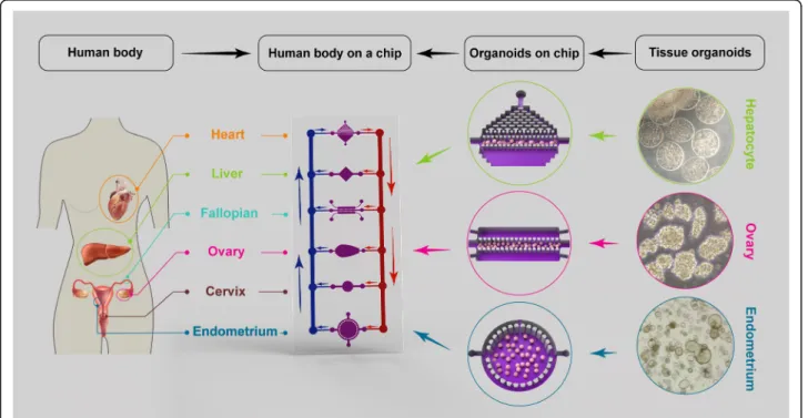
Reproductive system-on-a-chip (repro-on-a-chip): balancing sex differences in preclinical research
pulsatile in males and almost continuous in females [139]. Reproductive steroid hormones can modulate GH action by regulating GH secretion from the pituitary glands and peripherally modulate GH signaling pathways through the GH receptor (GHR) [140]. In addition, a proteomic analysis on total liver protein extracts from male and female rats has demonstrated that liver protein expression is more affected by gender than by nutritional status [141]. There is a paucity of data on sex differences in preclinical and biomedical research and drug development. Traditional in vitro systems such as a monolayer culture of cell lines or primary cells without considering their sex poorly recapitu-late human tissues and lead to inaccuracies in preclinical data and inefficiencies in drug discovery [136]. This is an important biological limitation because tissues and organs that work together in the body communicate and impact each other’s function. Therefore, in order to sufficiently model the human body for biomedical and pharmaceutical researches, a clear need exists for in vitro models that recapitulate the reproductive system and incorporate its endocrine signaling to the other organs of the body. In this regard, OOAC and human-on-a-chip microfluidic technol-ogy devices have recently garnered great attention and offer an alternative platform for animal models to test the effi-cacy and toxicity of new drugs on several cell types, tissues, and organs within a more biologically relevant environment [21,142]. This technology is a promising platform to model human diseases and study tissue development in vitro and may significantly affect the future of medicine. Currently, our view of the OOAC and human-on-a-chip is like a jigsaw puzzle with different connecting pieces. However, the pieces will gradually connect and, in the near future, this complicated puzzle will be completed [21,142].
The reproductive system-on-a-chip (repro-on-a-chip) is promising model to overcome these gaps and provide a new platform that incorporates reproductive sex hormones in preclinical research to address sexual di-morphism in biological processes, disease development, and improve sex-specific drug development [21]. By devel-oping the male and female repro-on-a-chip, an important piece will be added to the human-on-a-chip puzzle, and will raise the hope to provide a suitable in vitro model to study reproductive development, fertility, and reproductive disor-ders such as implantation disordisor-ders or even cancers. In addition, these micro-physiological systems can allow re-searchers to study the effects of reproductive hormones throughout the body. Moreover, an available dynamic flow microenvironment in the OOAC platform mimics chemo-physical signaling between different tissues and provides a controlled microfluidic environment for pharmacokinetic modeling and toxicology studies. This approach is useful for fundamental studies in reproductive biology such as oo-cyte maturation and modeling pathological conditions of infertility, endometriosis, STD, premature ovarian failure,
and polycystic ovary syndrome [143]. Recently, Xiao et al. have integrated female reproductive organs, including the ovaries, fallopian tubes, uterus, and cervix with peripheral organs, including the liver, into a microfluidic system called EVATAR. This EVATAR phenocopies the in vivo female reproductive tract and the hormone profile of the female 28-day menstrual cycle by linking tissues with a sustained circulating flow. Advantages of this technology are the use of human samples (except ovaries that are obtained from rats), long-term culture, and communication between different tissues in a microfluidic platform [21]. However, tissue explants are not expandable and this is a serious challenge for producing scalable chips. The use of female reproductive organoids would allow researchers to fabricate highly scalable and low-cost microfluidic devices for high-throughput assays.
Recent advances in organoid culture systems, including fallopian tubes, endometrium, testicular, and prostate orga-noids hold promise for the combination of male and female reproductive organoids with other technologies, such as microfluidics technology (Fig. 2). This combined technology would create a multi-organoid-on-a-chip plat-form and provide human-on-a-chip for clinical applica-tions, drug discoveries, and toxicology studies. In the multi-organoid-on-a-chip systems, the responses of one organoid to drugs or toxins impact the responses of other organoids, which occurs in actual human physiology. These organoids offer insights into the mechanisms of action of drugs or toxins that cannot be predicted by single-organ models. The combination of primary or stem cell-derived human reproductive organoids with other organoids on-a-chip would provide an emerging in vitro platform to study sex-specific differences and the mecha-nisms of male and female physiology and pathophysiology. This would provide high-value interpretable data to enable the development of more effective and safer drugs, and sex-based therapeutic strategies for precision medicine.
Challenges and future perspectives
blood vessels, innervation, and immune cells could be a hindrance to organoid research. Development of intes-tinal organoids with a functional enteric nervous system [146], human pancreatic cancer organoids with matched stromal and immune cells [147], and human prostate organoid co-cultured with prostate stromal cells [148] are good examples of the development of more complex organoid-based structures. Tackling these challenges will open new avenues for biomedical research. Other inno-vations, such as the use of a bioreactor and optimizing media composition, may minimize variation in culture conditions. Providing a scaffold of biomaterials along with combining microfluidics and organoid technologies may improve tissue architecture. Recent advances in CRISPR/Cas9 and gene editing technologies [149], and single-cell RNA-seq technologies [150] as well as 3D live-cell imaging using light sheet microscopy [151] will greatly accelerate applicability of organoids in biomed-ical research.
Conclusion
Despite current limitations, organoids provide an exciting era as in vitro models of the reproductive system that allow for reproductive biology research, accurate modeling of reproductive organs, clinical decision-making, and indi-vidualized medicine for patients with gynecologic cancers and diseases such as endometriosis. In addition, bio-banked organoids can be used for drug screening or in vitro trials to predict individual patient drug responses. Advances on cutting-edge technologies will doubtless im-prove this novel tool for clinical application.
Abbreviations
STD:Sexually transmitted disease; ECM: Extracellular matrix; PID: Pelvic inflammatory disease; ALI: Air-liquid interphase; OEC: Oviductal epithelial cells; EGF: Epidermal growth factor; FGF: Fibroblast growth factor; FTE: Fallopian tube epithelium; CGH: Comparative genomic hybridization; CTB: Cytotrophoblast; EVT: Extravillous; STB: Syncytiotrophoblast; HLA: Human leukocyte antigen; C19MC: Chromosome 19 miRNA cluster; BMP4: Bone morphogenetic protein 4; HESC: Human embryonic stem cell;VCTB:Villous CTB; PC: Principal component; LC-MS/MS: Liquid chromatography-tandem mass spectrometry; PSG: Pregnancy-specific glycoprotein; EPIL: Early placental insulin-like; HCG: Human chorionic gonadotropin; GDF15: Growth differentiation factor 15; KISS1: Kisspeptin; CSH1: Chorionic somatomammotropin hormone 1; EVTM: EVT
differentiation medium; CF: Cystic fibrosis; CFTR: CF transmembrane conductance regulator; MTOR: Mammalian target of rapamycin; RTI: Reproductive tract infection; HIV: Human immunodeficiency virus; IUD: Intrauterine device; UEC: Uterine epithelial cell; RWV: Rotating wall vessel; OOAC: Organ-on-a-chip; ZIKV: Zika virus; EOC: Epithelial ovarian cancer; HGSC: High-grade serous carcinoma; EMT: Epithelial-to-mesenchymal transition; TCGA: The cancer genome atlas; WGS: Whole genome sequencing; WES: Whole exome sequencing; PDO: Patient-derived organoid; IHC: Immunohistochemistry; VP: Verteporfin;
PDT: Photodynamic therapy; MSI: Microsatellite instability; SCNA: Somatic copy number alteration; cCCC: Cervical clear cell carcinoma;
AS: Asherman’s syndrome; IUA: Intrauterine adhesions; RIF: Repeated implantation failure; EA: Endometrial atrophy; ADR: Adverse drug reactions; CYP: Cytochrome P450; GH: Growth hormone; GHR: GH receptor; PEG: Polyethylene glycol
Acknowledgements
We would like to express our appreciation to our colleagues at Royan Institute for their helpful discussions. This article was extracted from a thesis written by Mr. Heidar Heidari Khoei in the School of Medicine, Shahid Beheshti University of Medical Sciences (registration no. 343).
Authors’contributions
All the authors researched data for this article, substantially contributed to the discussion of the content, wrote, reviewed, and/or edited it before submission. The authors read and approved the final manuscript.
Funding
This work was supported by grants from the National Institute for Medical Research Development (NIMAD, grant no. 987770) and Royan Institute (grant no. 96000189) to H.B., and Shahid Beheshti University of Medical Sciences (grant no. 12466) and the Iran National Science Foundation (INSF, grant no. 96004094) to A.P.
Availability of data and materials
All data supporting the conclusion of this article are included in this published article.
Ethics approval and consent to participate
Not applicable.
Consent for publication
Not applicable.
Competing interests
The authors declare that they have no competing interests.
Author details 1
Department of Stem Cells and Developmental Biology, Cell Science Research Center, Royan Institute for Stem Cell Biology and Technology, ACECR, P.O. Box: 16635-148, Tehran 1665659911, Iran.2Urogenital Stem Cell Research Center, Shahid Beheshti University of Medical Sciences, Tehran, Iran. 3
Department of Biology and Anatomical Sciences, School of Medicine, Shahid Beheshti University of Medical Sciences, P.O. Box: 19395-4719, Tehran, Iran.4Department of Developmental Biology, University of Science and Culture, Tehran, Iran.
Received: 29 February 2020 Accepted: 11 June 2020
References
1. Young AN, Moyle-Heyrman G, Kim JJ, Burdette JE. Microphysiologic systems in female reproductive biology. Exp Biol Med. 2017;242(17):1690–700. 2. Laronda MM, Burdette JE, Kim JJ, Woodruff TK. Recreating the female
reproductive tract in vitro using iPSC technology in a linked microfluidics environment. Stem Cell Res Ther. 2013;4(1):S13.
3. Turco MY, Gardner L, Hughes J, Cindrova-Davies T, Gomez MJ, Farrell L, et al. Long-term, hormone-responsive organoid cultures of human endometrium in a chemically defined medium. Nat Cell Biol. 2017;19(5): 568–77.
4. Boretto M, Cox B, Noben M, Hendriks N, Fassbender A, Roose H, et al. Development of organoids from mouse and human endometrium showing endometrial epithelium physiology and long-term expandability. Development. 2017;144(10):1775–86.
5. Haider S, Meinhardt G, Saleh L, Kunihs V, Gamperl M, Kaindl U, et al. Self-renewing trophoblast organoids recapitulate the developmental program of the early human placenta. Stem cell Rep. 2018;11(2):537–51.
6. Weimar CH, Uiterweer EDP, Teklenburg G, Heijnen CJ, Macklon NS. In-vitro model systems for the study of human embryo–endometrium interactions. Reprod Biomed Online. 2013;27(5):461–76.
7. Truskey GA. Human microphysiological systems and organoids as in vitro models for toxicological studies. Front Public Health. 2018;6:185. 8. Ferraz MA, Henning HH, Stout TA, Vos PL, Gadella BM. Designing
3-dimensional in vitro oviduct culture systems to study mammalian fertilization and embryo production. Ann Biomed Eng. 2017;45(7):1731–44. 9. Łaniewski P, Gomez A, Hire G, So M, Herbst-Kralovetz MM. Human