R E S E A R C H
Open Access
MicroRNA-21 and PDCD4 expression during
in vitro oocyte maturation in pigs
Elane C. Wright, Benjamin J. Hale, Cai-Xia Yang, Josephat G. Njoka and Jason W. Ross
*Abstract
Background:MicroRNA (miRNA) are small non-coding RNA molecules critical for regulating cellular function, and are abundant in the maturing oocyte and developing embryo. MiRNA-21 (MIR21) has been shown to elicit posttranscriptional gene regulation in several tissues associated with rapid cell proliferation in addition to demonstrating anti-apoptotic features through interactions with PDCD4 mRNA and other targets. In many tissues, MIR21 interacts and suppresses PDCD4 due to the strong complementation between MIR21 and the PDCD4 3′UTR.
Methods: The objective of this project was to examine the relationship between MIR21 and PDCD4 expression in porcine oocytes during in vitro maturation and assess the impact of MIR21 inhibition during oocyte maturation on early embryo development. Additionally, we evaluated the effect of gonadotropins in maturation media and the presence of cumulus cells to determine their ability to contribute to MIR21 abundance in the oocyte during maturation.
Results:During in vitro maturation, expression of MIR21 increased approximately 6-fold in the oocyte and 25-fold in the cumulus cell. Temporally associated with this was the reduction of PDCD4 protein abundance in MII arrested oocytes compared with GV stage oocytes, althoughPDCD4mRNA was not significantly different during this transition. Neither the presence of cumulus cells nor gonadotropins during in vitro maturation affected MIR21 abundance in those oocytes achieving MII arrest. However, inhibition of MIR21 activity during in vitro maturation using antisense MIR21 suppressed embryo development to the 4–8 cell stage following parthenogenetic activation.
Conclusions:MIR21 is differentially expressed in the oocyte during meiotic maturation in the pig and inhibition of MIR21 during this process alters PDCD4 protein abundance suggesting posttranscriptional regulatory events involving MIR21 during oocyte maturation may impact subsequent embryonic development in the pig.
Keywords:miRNA, Oocyte, Pig, MIR21
Background
Germinal vesicle breakdown (GVBD) is the first physical sign that an oocyte is committed to maturation and also represents the onset of a period of transcriptional quies-cence which persists until the activation of the embryonic genome. During this period, changes in mRNA and protein abundance within the oocyte can occur through interac-tions with the surrounding cumulus cells and/or through posttranscriptional gene regulation (PTGR) within the oocyte. MicroRNA (miRNA) represent a unique RNA class that function as potent regulators of transcription and
protein abundance through PTGR [12]. MiRNA are small (18–24 nt), non-coding RNA molecules that confer PTGR through several mechanisms, such as impairing translation efficiency and affecting mRNA stability following inter-action with the 3′untranslated region (3′UTR) of target mRNA molecules [5, 6]. Numerous miRNA are expressed in the mouse oocyte and developing embryo and it has been demonstrated that the conditional knockout of DICER, an enzyme involved in miRNA processing, during oocyte development impairs the production of oocytes capable of being fertilized and developing normally [38]. Estimations predict approximately 1,000 miRNA are present in the human genome having the potential ability to impact approximately 30 % of protein coding genes [28]. * Correspondence:[email protected]
Department of Animal Science, Iowa State University, 2356 Kildee hall, Ames, IA 50011, USA
Some miRNA have numerous mRNA targets [27] while others have few predicted targets [8, 34].
Utilizing miRNA microarray analysis and deep sequen-cing we have previously identified microRNA-21 (MIR21) as an up-regulated miRNA during porcine oocyte in vitro maturation [41]. In mice it has been reported that luteinizing hormone may increase the expression of MIR21 in mouse granulosa cells and in vivo MIR21 inhibition has a negative impact on ovulation rate [11]. MIR21 is a well characterized miRNA that has demonstrated the ability to confer PTGR oncogenic cell lines by affect-ing cellular proliferation through controllaffect-ing apoptosis [9, 13, 15, 42]. The MIR21 gene is transcribed via RNA polymerase II and is located in intronic regions of the transmembrane 49 gene (TMEM49; also re-ferred to as VMP1) [10, 17]. The mature MIR21 se-quence was first identified in human HeLa cells [22] and has since been predicted and verified to be present in the transcriptome of several other species including the pig. The anti-apoptotic capabilities of MIR21 in cancer cells are manifested through the ability to suppress critical apoptotic genes including programmed cell death 4 (PDCD4, previously referred to as neoplastic transformation inhibitor) [3, 16, 24, 33]. MIR21 interacts with PDCD4 through binding with complementary sequence in the 3′UTR of PDCD4
mRNA resulting in reduced translation and subse-quently reduced protein abundance in oncogenic cell lines [3, 24]. Importantly the 3′UTR of pig PDCD4
possesses a conserved MIR21 recognition sequence, particularly in the seed sequence. This suggests that if both MIR21 and PDCD4 are present in the oocyte, MIR21 could impact PDCD4 protein abundance as the necessary accessory proteins for miRNA function are present in the oocyte during GVBD and progres-sion to MII arrest.
Our working hypothesis that increased MIR21 abun-dance in the maturing cumulus oocyte complex of the pig is associated with posttranscriptional regulation of
PDCD4 expression in the oocyte and that suppression of MIR21 function during oocyte maturation will com-promise subsequent embryonic development. The objective of this study was to determine expression pat-terns of MIR21 and demonstrate its potential interac-tions with PDCD4 in the cumulus oocyte complex (COC) during oocyte maturation in the pig. Here we demonstrate PDCD4 protein down regulation is tem-porally associated with MIR21 abundance increase dur-ing in vitro oocyte maturation. These data indicate a reduced ability of MIR21 to suppress PDCD4 protein abundance in the presence of a MIR21 inhibitor sug-gesting a biological interaction between MIR21 and PDCD4 mRNA occurs during in vitro oocyte matur-ation in the pig.
Methods
Animal use was in accordance with the Guiding Principles for Care and Animals and procedures were approved by the Iowa State Institutional Animal Care and Use Committee.
In vitro maturation
All chemicals were purchased from Sigma Chemical Co. (St. Louis MO) unless otherwise stated. Sow ovaries were obtained from a local abattoir for isolation of cumulus oocyte complexes (COCs) to be subjected to in vitro mat-uration (IVM) as previously described [41, 44]. Briefly, fol-licles (3–5 mm) were aspirated and COC were collected and washed in TL-Hepes with 0.1 % polyvinyl alcohol (PVA). Cumulus oocyte complexes were cultured in mat-uration media (Tissue Culture Media 199 (TCM-199)) containing 0.57 mM L-cysteine, follicle stimulating hor-mone (0.5 μg/mL), luteinizing hormone (0.5 μg/mL), and epidermal growth factor (10 ng/mL) for 42–44 h at 39.0 °C in 5 % CO2. Prior to in vitro maturation, an ali-quot of GV stage oocytes for each replication were ran-domly selected from the COC pool. GV stage oocytes used for analysis were stripped of cumulus cells via vor-tex (6 to 8 min) in 1 % hyaluronidase in TL-Hepes-PVA. Following in vitro maturation oocytes were stripped of cumulus cells by vortexing 4–6 min in TL-Hepes-PVA supplemented with 1 % hyaluronidase, and Metaphase II arrested (MII) oocytes were identified by the presence of an extruded polar body. Cumulus cells before and after maturation and GV and MII oocytes (25 oocytes per pool) were snap frozen in liquid nitro-gen and stored at −80 °C until used for quantitative reverse transcription PCR (RT-qPCR). Pools of GV and MII arrested oocytes from the same replications (50 oocytes per pool) were utilized for Western blot analysis.
MIR21 expression in oocytes with and without LH and FSH during in vitro maturation
MIR21 expression in oocytes cultured with and without cumulus cells
To determine the effect of cumulus cell presence on MIR21 expression in oocytes during in vitro maturation we subjected COCs to one of three treatments: 1) stand-ard in vitro maturation as described above using intact COCs, 2) in vitro maturation following cumulus cell removal and then utilization of detached cumulus cells for culture with denuded oocytes or, 3) denuded oocytes matured without the presence of cumulus cells. Cumulus cells were removed by gentle vortex in 1 % hyaluronidase in TL-Hepes, washed twice in 200μL of maturation media and then resuspended in 200μL of maturation media and added to the in vitro maturation culture plates which con-tained 300μL maturation media and the denuded oocytes. Final volume of culture media for all plates was 500 μL and each well contained 75–85 oocytes. This experiment consisted of four biological replications. Maturation rates, as defined by the percentage of oocytes achieving MII arrest, were recorded and MII oocytes from each treat-ment and replication were collected in pools and used for MIR21 expression analysis as described above.
MIR21 inhibition during in vitro maturation
Peptide nucleic acids (PNA) are artificially constructed oligonucleotides with strong affinity and specificity to endogenous nucleotides while resistant to nucleases making them ideal for miRNA inhibition [31]. We used an anti-MIR21 PNA (Panagene Inc. Daejeon, Korea) designed to specifically bind to and prevent MIR21 activity. A scrambled PNA with no predicted targets was used as a negative control (Panagene Inc., Daejeon, Korea). PNA oligonucleotides were diluted in matur-ation media at a stock concentrmatur-ation of 100 nM/μL and then added to maturation media on the day of COC col-lection to acquire a final concentration of 2.0 nM and 0.2 nM. A control group without PNA was used to evaluate the potential toxicity of the PNA.
Parthenogenetic activation of MII oocytes, used to remove the confounding effects of miRNA introduced by sperm, was performed with 50 oocytes from each treatment to determine developmental competence of embryos up to 60 h. MII oocytes were washed in a high calcium activation media (Mannitol 0.28 M, CaCl2
1.0 mM, MgCl2 0.1 mM, HEPES 0.5 mM and BSA
1 mg/mL), then placed between two electrodes covered with activation media and activated by two consecutive 30 μsec pulses at 1.2 kV/cm. Following activation, zygotes were washed and cultured in porcine zygote medium-3 (PZM3) at 39 °C in 5 % CO2 [21]. At 60 h embryos were evaluated for development and the num-ber of embryos with four or more uniform blastomeres was recorded.
Quantitative RT-PCR of oocytes and cumulus cells
Oocytes were collected and denuded of cumulus cells as described above. Oocytes from each stage of develop-ment and treatdevelop-ment were collected in pools of exactly 25 oocytes in 5 μL of PBS. As before using a precise number of oocytes per reaction we were able to avoid the introduction of additional variation associated with reference genes [41]. Both PDCD4 and MIR21 ana-lysis were analyzed from the same oocyte sample lysis. TaqMan™ Gene Expression Cells-to-Ct™ Kit (Ap-plied Biosystems, Carlsbad, CA) was used to lyse oocytes and prepare samples for RT-qPCR. Lysis solu-tion and DNase from the Cells-to-Ct kit were added to each oocyte pool at 4.95 and 0.05 μL, respectively, and incubated at RT for 5 min. Stop solution (0.5 μL) was added, incubated for an additional 2 min and placed on ice. Two μl of the sample lysis was added to each RT-qPCR reaction. PDCD4 forward
(5′-ACAGTTGGTGGGCCAGTTTATTGC-3′) and
re-verse (5′-CTTTGCGCCTTCCACCTTTAGACA- 3′) primers were used to determine mRNA abundance of
PDCD4 within each pool. QuantiTect® SYBR® Green RT-PCR Kit (Qiagen) was used for the RT-qPCR reac-tion for PDCD4 according to manufacturer’s recom-mendations. The standard cycling conditions were 50 °C for 30 min, 95 °C for 15 min followed by 45 -cycles of 95 °C for 15 s, 60 °C for 30 s, and 72 °C for 30 s followed by melting curve analysis.
MIR21 was quantified using TaqMan® MicroRNA Reverse Transcription kit (Applied Biosystems Carlsbad, CA) for the reverse transcription (RT) reaction and the primers and probe used were TaqMan® MicroRNA Assay for hsa-MIR21 (Applied Biosystems, Carlsbad, CA) according to manufacturer’s recommendations. The RT reaction was 20μL consisting of 13μL master mix, 3μL primers, and 4 μL sample lysis. Reverse transcription conditions were 16 °C for 30 min, 42 °C for 30 min and 85 ° C for 5 min. The final volume for all RT-qPCR reac-tions was 20 μL which include 1.33 μL of the RT prod-uct, 1 μL TaqMan MicroRNA Assay (20x), 10 μL TaqMan 2X Universal PCR Master Mix and 7.67 nucle-ase free water. The thermal cycling conditions for the TaqMan MicroRNA RT-qPCR were 95 °C for 10 min, followed by 45 cycles of 95 °C for 15 s and 60 °C for 60 s. Fluorescent data acquisition was during the 60 °C extension step.
between stages and statistical analysis. All samples were assayed in duplicate. The comparative CT method was used to calculate relative fold changes between samples as previously described [35].
PDCD4 Western blot analysis
Pools of 50 denuded GV and MII oocytes within a replica-tion were collected, washed in PBS, and stored at−80 °C until used for Western blot analysis. Oocyte pools were lysed in 2.5μL of 5X SDS (total sample volume 12.5μL) at 95 °C for 4 min followed by 1 min on ice and then cen-trifugation at 1000 rpm for 1 min at RT. Samples were then loaded into a 4–20 % Tris glycine gel (Lonza PAGEr® Gold Precast Gels). The BioRad Mini PROTEAN Tetra System was used to run the gel at 60 V for 30 min followed by 120 V for 90 min. The gel was transferred to a nitrocellulose membrane for 1 h at 100 V at 4 °C. In addition to utilizing an exact number of oocytes per lane, Ponceau S staining was used to confirm relative transfer efficiency between lanes and equivalent total protein load-ing per lane. Membrane blockload-ing was conducted usload-ing 5 % milk in PBST (PBS with 0.5 % Tween 20) for 1 h at RT. A rabbit anti-PDCD4 monoclonal antibody (Abcam, ab79405) was added (1:1000 dilution) to the membrane in 0.5 % milk in PBST overnight at 4 °C and a negative control membrane lacking primary antibody was also con-ducted. Following primary antibody incubation, the mem-branes were washed with PBST three times at RT for 10 min each. Donkey anti-Rabbit IgG (Amersham™ECL™ NA934) was incubated (1:2000) with the membrane for 1 h at RT. The membrane was then washed three times for 10 min each at RT. Horseradish peroxidase substrate (Millipore, Billerica, MA) was added to the membrane for 1 min in the dark. The membrane was then exposed to x-ray film and developed for visualization. Average pixel intensity for the protein corresponding to 52 kDa (PDCD4 molecular weight) was conducted using Image J [1].
MIR21 in situ hybridization in the developing follicle
Ovaries were preserved in 4 % paraformaldehyde and utilized for in situ hybridization to determine MIR21 expression. Ovary sections (5 μm) were then mounted on slides for analysis. Each section was subjected to CitriSolv (Fisher Scientific) twice for five minutes rehy-drated in two changes each of 100 % ethanol for 3 min, followed by 95 % ethanol for one minute and finally 80 % ethanol for one minute. Slides were immersed in heated citrate buffer (95 °C) for 30 min and then cooled to RT. Once at RT slides were blocked for 30 min with 5 % BSA. Slides were then placed in hybridization solu-tion for 1 h at 65 °C. The 5′fluorescein labeled miRCURY LNA detection probe (Exiqon) was added and slides were incubated in high humidity overnight at 65 °C. Slides were then washed in saline-sodium citrate (SSC) solution and
PBST at RT. Anti-fade DAPI was added and a cover slip was placed over each section. Primary and secondary folli-cles were imaged at 400X and tertiary follifolli-cles were imaged at 200X with a Leica microscope.
Statistical analysis
PROC MIXED of the Statistical Analysis System was used to determine statistical differences of all data in-cluding percentage maturation and differences in CT value for RT-qPCR data. Significance (P< 0.05) was determined for the model and least-square means was used to determine significant differences between sam-ples. The effect of oocyte stage on MIR21 and PDCD4 expression (CTvalue) was determined. Treatment effect for the PNA inhibitor of MIR21, presence or absence of gonadotropins, and the presence or absence of cumulus cells during in vitro maturation on MIR21 expression was evaluated. Replication was included as a covariate. Graphs depicting percent change were adjusted to reflect the GV stage as 100 % and all other treatments are rela-tive to GV. Data are displayed as mean ± SEM.
Results
MIR21 expression is temporally regulated during porcine cumulus oocyte complex maturation
To determine the relationship between MIR21 and its putative target PDCD4, during in vitro maturation, RT-qPCR and Western blot analysis were utilized to evalu-ate expression of MIR21 and PDCD4 in GV and MII stage oocytes and cumulus cells. During the transition from GV to MII, MIR21 was up-regulated approximately 6-fold in oocytes (P= 0.001, Fig. 1a) and approximately 25-fold in cumulus cells (P= 0.003, Fig. 1b). In the same samples PDCD4 mRNA abundance was not statistically different (P= 0.34, Fig. 1a). Western blot analysis demonstrated PDCD4 protein abundance was reduced (P= 0.02) in oocytes during the transition from GV stage to MII arrested oocytes (Fig. 1c and d).
Gonadotropins in maturation media influence maturation rate but not MIR21 abundance in MII arrested oocytes
Cumulus cells influence oocyte maturation but not MIR21 abundance in MII arrested oocytes
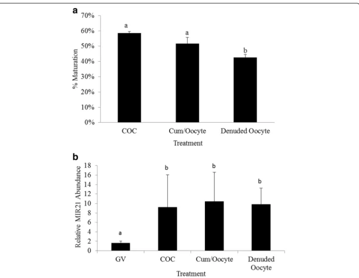
To determine the impact of cumulus cell presence on MIR21 abundance in the oocyte during in vitro matur-ation we compared MIR21 expression in MII arrested oo-cytes following maturation of intact COCs, denuded oocytes cultured in the presence of cumulus cells and de-nuded oocytes cultured without cumulus cells. Maturation
rate for control COCs was 58.4 ± 1.5 % which tended to be greater than denuded oocytes cultured with cumulus cells (51.2 ± 4.0 %, P= 0.07). However, denuded oocytes cultured without cumulus cells had significantly lower (42.5 ± 2.0 %) maturation rates than intact COCs (P <
0.05) and denuded oocytes cultured with cumulus cells (P < 0.05, Fig. 3a). MIR21 expression in MII arrested oocytes was not affected by the presence or absence of cumulus cells during maturation (P= 0.82). GV oocytes had lower MIR21 expression (P< 0.05) compared to MII arrested oocytes for all treatments (Fig. 3b), consistent with data from the experiment presented in Fig. 1.
Inhibition of MIR21 affects oocyte maturation and PDCD4 protein expression
Using a fluorescently labeled MIR21 antagonist we first demonstrated its ability to translocate into both the cumulus cell and the oocytes (data not shown). All oo-cytes exposed to the fluorescently labelled inhibitor demonstrated a measurable level of fluorescence com-pared with oocytes treated with a non-fluorescently tagged control inhibitor, although some variability in the intensity of the fluorescence existed between oocytes. Anti-MIR21 antisense oligonucleotides were added to maturation media during oocyte maturation to deter-mine the effects of MIR21 inhibition on maturation rate and PDCD4 expression in MII arrested oocytes. Inhib-ition of MIR21 using an anti-MIR21 PNA during oocyte maturation decreased (P< 0.01) the percentage of oocytes achieving MII after 42 h of culture (Table 1). Oocytes cultured in the presence of the negative control inhibitor (NC-PNA 2.0 nM) had similar maturation rates compared to control maturation conditions (P> 0.05) and greater (P< 0.05) maturation rates compared to the MIR21 inhibited oocytes (Table 1).
The effect of MIR21 inhibition on PDCD4 protein abundance was analyzed by Western blot with pools of 50 oocytes within each treatment and presented as a percentage of GV stage PDCD4 abundance. Western blot analysis demonstrated significantly lower PDCD4 abundance in control MII arrested oocytes (31.9 % of GV) compared to MII arrested oocytes matured in the presence of 2.0 nM MIR21 antagonist (69.7 % of GV; P < 0.05). PDCD4 abundance in MII oocytes subjected to IVM in the presence of low (0.2 nM) MIR21 inhibitor concentration was similar (26.4 % of GV stage) to control MII oocytes and oocytes cultured with either concentration of NC-PNA possessed a similar quantity or less of PDCD4 compared to GV oocytes (Fig. 4).
MIR21 inhibition during in vitro maturation impacts parthenogenetic embryo development
Metaphase II arrested oocytes were parthenogenetically activated (to eliminate confounding impact of sperm-Fig. 1MIR21 expression is significantly increased in cumulus cells
and oocytes during in vitro maturation.aRT-qPCR analysis for MIR21 andPDCD4mRNA in GV stage and MII arrested oocytes (n= 4).b RT-qPCR analysis for MIR21 in cumulus cells isolated from GV stage and in vitro matured oocytes (n= 4).cPDCD4 Western blot analysis of GV stage and MII arrested oocytes. Pixel intensity was quantified with ImageJ and demonstrates a decrease in PDCD4 protein abundance in MII oocytes compared with GV oocytes (n= 3).dRepresentative Western blot of PDCD4 protein detection in oocytes representing the 52 KDa band.a,bMeans ± SEM with different superscripts are
borne miRNA) following in vitro maturation to test the hypothesis that MIR21 inhibition during in vitro oocyte maturation negatively impacts early embryo develop-ment prior to activation of the embryonic genome at the 4-cell stage of development. The number of embryos achieving the 4-cell stage of development or greater within 60 h post activation was significantly (P <0.05) affected by MIR21 inhibition during in vitro maturation (Table 1). Development to the 4-cell stage or greater within 60 h post activation was greatest for control oocytes (73.0 ± 5.7) compared with oocytes matured in the presence of a MIR21 inhibitor (41.7 ± 12.1) or in the presence of a negative control PNA (60.2 ± 15.8).
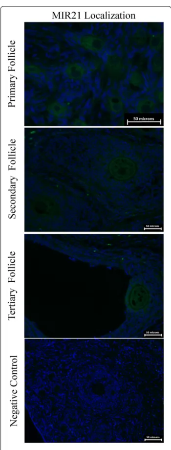
MIR21 abundance in the developing pig follicle
In situ hybridization of MIR21 demonstrated expression throughout the granulosa cells and the oocyte (Fig. 5). In primary follicles expression is present in the oocyte with faint expression in the surrounding cells. However sec-ondary and tertiary follicles express MIR21 more abun-dantly in the oocyte as well as the surrounding cumulus
cells compared to primary follicles. Based on staining intensity, MIR21 abundance appears greatest in oocytes and granulosa cells of tertiary follicles.
Discussion
While abundant in the developing oocyte and mouse ovary [11, 14] miRNA expression and function during pig oocyte maturation and early embryo development is only in the initial stages of characterization [18, 41]. The potential ability of MIR21 to interact withPDCD4 lead-ing to posttranscriptional gene regulation of PDCD4 protein expression in the maturing pig oocyte as demon-strated herein has also been described in several types of cancer cells [3, 15, 16, 29, 33, 42]. The interaction between MIR21 and PDCD4 is likely conserved in pigs as the MIR21 target recognition sequence in the 3′UTR of human and pig PDCD4 is 97 % similar with 100 % similarity in nucleotides responsible for recognition by the MIR21 seed sequence.
miRNA function in maturing oocytes has been debated, primarily the result of the conditional knock-out during oocyte maturation of enzymes needed for both canonical and non-canonical miRNA biogenesis pathways. Loss of DICER function, essential for both siRNA and canonical miRNA, in developing oocytes results in the production of non-viable oocytes [38]. Alternatively, using the same
ZP3-cre conditional knock-out approach with a floxed DGCR8 allele, mice lacking the ability to produce miRNA through the canonical biogenic pathway still produced viable oocytes, despite reduced fecundity [25, 36]. This suggests that while miRNA activity in mature mice oocytes may be suppressed, miRNA may still contribute to the developmental competency of Fig. 3Cumulus cells influence oocyte maturation but not MIR21abundance in MII arrested oocytes.aMaturation rates for intact COC, denuded oocytes cultured with cumulus cells, and denuded oocytes.bExpression of MIR21 in GV oocytes, and MII arrested oocytes from intact COC, denuded oocytes cultured with cumulus cells, and denuded oocytes.a,bMeans ± SEM with different superscripts are different (P< 0.05)
Table 1Oocyte development to MII arrest and 4-cell stage of embryonic development at 60 h following parthenogenetic activation
Treatment
groupc Total oocytesmaturedd Percentage MIIarrested oocytese Percentage 4-cellor greater at 60 hf Average blastomere# at 60 hg
Control 1646 55.4 ± 3.6a 73.0 ± 5.7a 5.8 ± 1.2 (n= 16)
NC-PNA 1136 49.0 ± 2.5a 60.2 ± 15.8ab 6.3 ± 1.4 (n= 12)
Anti-MIR21 PNA 1639 33.7 ± 3.6b 41.7 ± 12.1b 4.8 ± 1.4 (n= 12)
a,b
Values with different superscripts in the same column are significantly different (P< 0.05)
c
Treatments include: control maturation conditions, NC-PNA (2.0 nM), anti-MIR21 PNA (2.0 nM)
d
Total number of GV oocytes matured for each treatment from five replications
e
Percentage of MII arrested oocytes from each treatment. Mean ± SEM
f
Percentage of embryos achieving 4-stage or greater within 60 h following parthenogenetic activation of MII arrested oocytes. Mean ± SEM
g
the subsequently produced embryo. This study tested the hypothesis that increased MIR21 abundance in the maturing cumulus oocyte complex of the pig is associ-ated with posttranscriptional regulation of PDCD4 expression in the oocyte and that suppression of MIR21 function during oocyte maturation would com-promise subsequent embryonic development.
Utilizing miRNA microarray and small RNA sequencing, the expression of numerous miRNA in the maturing porcine cumulus oocyte complex have been previously demonstrated [41]. The current study further characterizes the temporal relationship between increased mature MIR21 abundance and decreased PDCD4 protein abundance dur-ing in vitro maturation of pig oocytes. In cumulus cells, MIR21 abundance increases approximately 25-fold during in vitro maturation. This is consistent with reports in humans [4] and in mice demonstrating increased MIR21 expression in granulosa cells in response to LH [11]. Because MIR21 abundance changes appear to occur rapidly during in vitro maturation, it is of interest if MIR21 expres-sion in the pig cumulus oocyte complex is also responsive to the gonadotropins. To examine this, the ability of both LH and FSH to affect MIR21 expression during in vitro maturation was examined. While exclusion of LH from maturation media negatively impacted oocyte maturation, as demonstrated by others [45], a significant effect of the gonadotropins on MIR21 abundance during in vitro matur-ation was not detected.
achieve MII arrest was not affected by the presence of cumulus cells suggesting the increased MIR21 observed is at least in part, the result of oocyte specific mechanisms.
Taken together neither gonadotropins nor the pres-ence of cumulus cells during IVM is demonstrably responsible for increased MIR21 abundance consistently observed in MII arrested oocytes compared to their GV stage counterparts. It has also been observed that GVBD does not occur in the majority of porcine oocytes until approximately 16–24 h of in vitro maturation [30]. Therefore, it remains possible that the increased abun-dance of MIR21 in the MII arrested oocyte occurs as a transcriptional response in the oocyte during the initial phases of in vitro maturation prior to GVBD in the oocyte.
These findings taken together suggest the potential for MIR21 and miRNA in general, to impact protein expres-sion in the maturing oocyte having implications regarding the developmental ability of subsequently produced embryos. Oocyte growth and development begins prior to antral follicle development and it is unclear when activa-tion of MIR21 expression occurs or what mechanism is primarily responsible for this observation. The cumulus oocyte complexes collected for this study were from 3 to 5 mm antral follicles, prior to GVBD, and may be capable of transcribing primary MIR21 (pri-MIR21) transcript that can be further processed into mature MIR21 during mat-uration. Future studies will include an analysis of pri-MIR21 abundance prior to and during oocyte maturation which may yield insight into the mechanisms contributing to the current observations.
Several promoters have been identified upstream to the pri-MIR21 transcription start site containing pre-dicted consensus sequences for binding of AP-1 and sig-nal transducer and activator of transcription 3 (STAT3) [17]. In certain cancers, the interaction between MIR21 and PDCD4 is necessary for maximal AP-1 activation. AP-1 activation is suppressed by PDCD4, however, AP-1 induction of MIR21 and subsequent posttranscriptional regulation of PDCD4 allows further and more sustained AP-1 activation [17, 37]. In addition to AP-1, STAT3 is another transcription factor that has also been docu-mented to induce pri-MIR21 transcription [23]. It is pos-sible that STAT3 could be related to the expression of MIR21 during oocyte maturation and early embryo development as leptin (an adipokine) has been
demonstrated to increase STAT3 expression during bovine embryo culture and is associated with reduced apoptosis in bovine blastocysts [7]. In addition, leptin has been shown to increase STAT3 expression in both cumu-lus cells and oocytes during in vitro maturation and enhance the oocytes ability to complete meiosis [32].
Conclusion
In summary, these data demonstrate MIR21 miRNA and PDCD4 protein have a reciprocally inverse, temporal expression pattern during oocyte maturation, and that inhibition of MIR21 results in an increased abundance of PDCD4 protein. MIR21 expression in MII arrested oocytes matured in vitro was not affected by gonadotro-pins or the presence of cumulus cells during in vitro maturation. It is possible that increased MIR21 expres-sion in the oocyte occurs earlier during oocyte recruit-ment or during initial in vitro maturation prior to GVBD and warrants further investigation. Obtaining an understanding of MIR21 and its mechanism is necessary to further understand molecular regulation of oocyte maturation and early embryo development in the pig. Understanding molecular mechanisms of early embryo development will provide a more comprehensive under-standing of mammalian reproduction that can be funda-mental in developing strategies to improve reproductive efficiency.
Abbreviations
3′UTR:3′untranslated region; BSA: bovine serum albumin; COC: cumulus oocyte complex; GVBD: germinal vesicle breakdown; IVM: in vitro maturation; MII: metaphase II; MIR21: microRNA-21; miRNA: microRNA; mTBM
medium: modified Tris-buffered medium; PBS: phosphate buffered saline; PBST: phosphate buffered saline containing 0.1 % Tween-20; PDCD4: pro-grammed cell death 4; PNA: peptide nucleic acids; PTGR: posttranscriptional gene regulation; PVA: polyvinyl alcohol; PZM3: porcine zygote medium 3; RT: room temperature; TCM-199: tissue culture media 199.
Competing interests
The authors declare that they have no competing interests.
Authors’contributions
ECW and BJH conducted experiments in the manuscript. CY and JGN assisted with completing experiments. ECW, BJH and JWR were responsible for experimental design and analysis of results. ECW, BJH and JWR prepared the manuscript. All authors read and approved the final manuscript.
Acknowledgements
The authors would like to thank Robyn Scanlon and Ben Selman for their assistance with this project.
Grant support
This project was supported by National Research Initiative Competitive Grant no. 2008-35205-05309 and 2008-35205-18712 from the USDA National Institute of Food and Agriculture.
Received: 28 December 2015 Accepted: 4 April 2016
References
1. Abramoff MD, Magalhaes PJ, Ram SJ. Image processing with ImageJ. Biophotonics Intern. 2004;11(7):36–42.
2. Arellano RO, MartAnezTorres A, Garay E. Ionic currents activated via purinergic receptors in the cumulus cell-enclosed mouse oocyte. Biol Reprod. 2002;67(3):837–46.
3. Asangani IA, Rasheed SAK, Nikolova DA, Leupold JH, Colburn NH, Post S, Allgayer H.. MicroRNA-21 (miR-21) post-transcriptionally downregulates tumor suppressor Pdcd4 and stimulates invasion, intravasation and metastasis in colorectal cancer. Oncogene. 2007;27(15):2128–36. 4. Assou S, Al-edani T, Haouzi D, Philippe N, Lecellier CH, Piquemal D,
Commes T, Ait-Ahmed O, Dechaud H, Hamamah S. MicroRNAs: new candidates for the regulation of the hman cumulus-oocyte complex. Hum Reprod. 2013;28(11):3038–49.
5. Bagga S, Bracht J, Hunter S, Massirer K, Holtz J, Eachus R, Pasquinelli AE. Regulation by let-7 and lin-4 miRNAs results in target mRNA degradation. Cell. 2005;122(4):553–63.
6. Bartel DP. Micro RNAs: genomics biogenesis, mechanism, and function. Cell. 2004;116(281–297):281.
7. Boelhauve M, Sinowatz F, Wolf E, Paula-Lopes FF. Maturation of bovine oocytes in the presence of leptin improves development and reduces apoptosis of in vitro-produced blastocysts. Biol Reprod. 2005; 73(4):737–44.
8. Brennecke J, Stark A, Russell RB, Cohen SM. Principles of microRNA target recognition. PLoS Biol. 2005;3(3):e85.
9. Bueno MJ, Perez De Castro I, Malumbres M. Control of cell proliferation pathways by microRNAs. Cell Cycle. 2008;7(20):3143–8.
10. Cai X, Hagedorn CH, Cullen BR. Human microRNAs are processed from capped, polyadenylated transcripts that can also function as mRNAs. RNA. 2004;10(12):1957–66.
11. Carletti MZ, Fiedler SD, Christenson LK. MicroRNA 21 blocks apoptosis in mouse periovulatory granulosa cells. Biol Reprod. 2010;83:286–95. 12. Carthew RW, Sontheimer EJ. Origins and mechanisms of miRNAs and
siRNAs. Cell. 2009;136(4):642–55.
13. Chan JA, Krichevsky AM, Kosik KS. MicroRNA-21 is an antiapoptotic factor in human glioblastoma cells. Cancer Res. 2005;65(14):6029–33.
14. De Sousa PA, Dobrinsky JR, Zhu J, Archibald AL, Ainslie A, Bosma W, Bowering J, Bracken J, Ferrier PM, Fletcher J, Gasparrini B, Harkness L, Johnston P, Ritchie M, Ritchie WA, Travers A, Albertini D, Dinnyes A, King TJ, Wilmut I. Somatic cell nuclear transfer in the pig: control of pronuclear formation and integration with improved methods for activation and maintenance of pregnancy. Biol Reprod. 2002;66(3):642–50. 15. Dillhoff M, Liu J, Frankel W, Croce C, Bloomston M. MicroRNA-21 is
overexpressed in pancreatic cancer and a potential predictor of survival. J Gastrointest Surg. 2008;12:2171–6.
16. Frankel L, Christoffersen N, Jacobsen A, Lindow M, Krogh A, Lund A. Programmed cell death 4 (PDCD4) is an important functional target of the microRNA miR-21 in breast cancer cells. J Biol Chem. 2008;283(2):1026–33. 17. Fujita S, Ito T, Mizutani T, Minoguchi S, Yamamichi N, Sakurai K, Iba H.
miR-21 gene expression triggered by AP-1 is sustained through a double-negative feedback mechanism. J Mol Biol. 2008;378(3):492–504. 18. Hale BJ, Yang CX, Ross JW. Small RNA regulation of reproductive
function. Mol Reprod Dev. 2014;81(2):148–59.
19. Hawkins SM, Matzuk MM. Oocyte-somatic cell communication and micoRNA function in the ovary. Ann Endocrinol. 2010;71(3):144–8. 20. Hunter MG, Brankin V, Quinn RL, Ferguson EM, Edwards SA, Ashworth CJ.
Oocyte-somatic cell-endocrine interactions in pigs. Domest Anim Endocrinol. 2005;29(2):371–84.
21. Isom SC, Li RF, Whitworth KM, Prather RS. Timing of first embryonic cleavage is a positive indicator of the in vitro developmental potential of porcine embryos derived from in vitro fertilization, somatic cell nuclear transfer and parthenogenesis. Mol Reprod Devel. 2012;79(3):197–207. 22. Lagos-Quintana M, Rauhut R, Yalcin A, Meyer J, Lendeckel W, Tuschl T.
Identification of tissue-specific microRNAs from mouse. Curr Biol. 2002; 12(9):735–9.
23. Loffler D, Brocke-Heidrich K, Pfeifer G, Stocsits C, Hackermuller J, Kretzschmar AK, Burger R, Gramatzki M, Blumert C, Bauer K, Cvijic H, Ullmann AK, Stadler PF, Horn F. Interleukin-6 dependent survival of multiple myeloma cells involves the stat3-mediated induction of microRNA-21 through a highly conserved enhancer. Blood. 2007;110(4): 1330–3.
25. Ma J, Flemr M, Stein P, Berninger P, Malik R, Zavolan M, Svoboda P, Schultz RM. MicroRNA activity is suppressed in mouse oocytes. Curr Biol. 2010;20(3): 265–70.
26. Mattioli M, Gioia L, Barboni B. Calcium elevation in sheep cumulus-oocyte complexes after luteinising hormone stimulation. Mol Reprod Devel. 1998; 50(3):361–9.
27. Mickoleit M, Banisch TU, Raz E. Regulation of hub mRNA stability and translation by miR430 and the dead end protein promotes preferential expression in zebrafish primordial germ cells. Dev Dyn. 2011;240(3):695–703. 28. Miranda KC, Huynh T, Tay Y, Ang Y-S, Tam W-L, Thomson AM, Lim B,
Rigoutsos I. A pattern-based method for the identification of microRNA binding sites and their corresponding heteroduplexes. Cell. 2006;126(6): 1203–17.
29. Mudduluru G, Medved F, Grobholz R, Jost C, Gruber A, Leupold J. Loss of programmed cell death 4 expression marks adenoma-carcinoma transition, correlates inversely with phosphorylated protein kinase B, and is an independent prognostic factor in resected colorectal cancer. Cancer. 2007; 110:1697–707.
30. Ocampo MB, Ocampo LT, Kanagawa H. Timing of sequential changes in chromosome configurations during the 1st meiotic division of pig oocytes cultured in vitro. Jpn J Vet Res. 1990;38(3–4):127–37.
31. Oh SY, Ju Y, Park H. A highly effective and long-lasting inhibition of miRNAs with PNA-based antisense oligonucleotides. Mol Cells. 2009;28(4):341–5. 32. Paula-Lopes FF, Boelhauve M, Habermann FA, Sinowatz F, Wolf E. Leptin
promotes meiotic progression and developmental capacity of bovine oocytes via cumulus cell-independent and -dependent mechanisms. Biol Reprod. 2007;76(3):532–41.
33. Qi L, Bart J, Tan L, Platteel I, Sluis T, Huitema S, Harms G, Fu L, Hollema H, Berg A. Expression of miR-21 and its targets (PTEN, PDCD4, TM1) in flat epithelial atypia of the breast in relation to ductal carcinoma in situ and invasive carcinoma. BMC Cancer. 2009;9(1):163.
34. Rajewsky N, Socci ND. Computational identification of microRNA targets. Dev Biol. 2004;267(2):529–35.
35. Ross JW, Malayer JR, Ritchey JW, Geisert RD. Characterization of the interleukin-1beta system during porcine trophoblastic elongation and early placental attachment. Biol of Reprod. 2003;69(4):1251–9.
36. Suh N, Baehner L, Moltzahn F, Melton C, Shenoy A, Chen J, Blelloch R. MicroRNA function is globally suppressed in mouse oocytes and early embryos. Curr Biol. 2010;20(3):271–7.
37. Talotta F, Cimmino A, Matarazzo MR, Casalino L, De Vita G, D’Esposito M, Di Lauro R, Verde P. An autoregulatory loop mediated by miR-21 and PDCD4 controls the AP-1 activity in RAS transformation. Oncogene. 2009;28(1):73–84. 38. Tang F, Kaneda M, O’Carroll D, Hajkova P, Barton SC, Sun YA, Lee C,
Tarakhovsky A, Lao K, Surani MA. Maternal microRNAs are essential for mouse zygotic development. Genes Dev. 2007;21(6):644–8. 39. Turchinovich A, Weiz L, Langheinz A, Burwinkel B. Characterization of
extracellular circulating microRNA. Nucleic Acids Res. 2011;39(16):7223–33. 40. Wang K, Zhang S, Weber J, Baxter D, Galas DJ. Export of microRNAs and
microRNA-protective protein by mammalian cells. Nucleic Acids Res. 2010; 38(20):7248–59.
41. Yang CX, Du ZQ, Wright EC, Rothschild MF, Prather RS, Ross JW. Small RNA profile of the cumulus oocyte complex and early embryos in the pig. Biol Reprod. 2012;87(5):117.
42. Yang Y, Chaerkady R, Beer MA, Mendell JT, Pandey A. Identification of miR-21 targets in breast cancer cells using a quantitative proteomic approach. Proteomics. 2009;9(5):1374–84.
43. Zhang X, Miao YL, Zhao JG, Spate L, Bennett MW, Murphy CN, Schatten H, Prather RS. Porcine oocytes denuded prior to maturation can develop the to the blastocyst stage if provided a cumulous cell-derived coculture system. J Anim Sci. 2010;88(8):2604–10.
44. Zhao J, Ross JW, Hao Y, Spate LD, Walters EM, Samuel MS, Rieke A, Murphy CN, Prather RS. Significant improvement in cloning efficiency of an inbred miniature pig by histone deacetylase inhibitor treatment after somatic cell nuclear transfer. Biol Reprod. 2009;81(3):525–30.
45. Zuelke KA, Brackett BG. Luteinizing hormone-enhanced in vitro maturation of bovine oocytes with and without protein supplementation. Biol Reprod. 1990;43(5):784–7.
• We accept pre-submission inquiries
• Our selector tool helps you to find the most relevant journal
• We provide round the clock customer support
• Convenient online submission
• Thorough peer review
• Inclusion in PubMed and all major indexing services
• Maximum visibility for your research
Submit your manuscript at www.biomedcentral.com/submit