Address for correspondence
Dr. Muhammad Irfan Anwar, Assistant Professor Dermatology Department,
Bahria University Medical and Dental College, Karachi
Email: [email protected]
Original Article
Frequency of HCV seropositivity in lichen planus
Introduction
Lichen Planus (LP) is an inflammatory disease that affects the skin, mucous membranes, nails, and hair and is classically characterized by pruritic, purple, polygonal, plane-topped papules The association of Lichen planus and HCV is uncertain and controversial in literature because the prevalence of HCV infection in patients with lichen planus varies considerably from one geographical area to another ranging from 22% in brazil to 62% in Japan while studies from
Great Britain failed to reveal any association.1-3 It has been suggested that the geographical origin of patients would be an important factor in HCV prevalence among patient with lichen planus.4,5 This study was conducted to determine the frequency of HCV seropositivity in patients of lichen planus in Pakistani population attending a tertiary care hospital in Karachi.
Methods
This observational cross-sectional study was conducted over a period of 6 months at Dermatology Department PNS Shifa Hospital, Karachi. Hospital ethical committee approval for conducting this study was taken before the commencement of study. A sample size of 82 patients was calculated, taking the prevalence of
Mehreen Javed*, Muhammad Khurram Ahmed*, Moizza Tahir**, Muhammad Irfan Anwar*
* Dermatology Department, PNS Shifa Hospital, Karachi ** Dermatology Department, CMH, Quetta Cantt
Abstract
Objective To determine the frequency of Hepatitis C (HCV) seropositivity in patients of lichen planus (LP).Methods This observational cross-sectional study was conducted over a period of 6 months, at Dermatology Department PNS Shifa hospital Karachi. Eighty-two (n=82) patients with a diagnosis of LP fulfilling the inclusion and exclusion criteria were enrolled using non-probability consecutive sampling technique. Detailed history, physical examination and biochemical measurements were recorded. Outcome variable i.e. HCV seropositivity was determined in laboratory with third generation ELISA technique. All data were entered and analyzed using SPSS Version 20.0.
Results The mean age of study population was 39.32±13.212 years. On analysis of demographics data it was observed that 25 (30.5%) were below 30 years of age and 57 (69.5%) were of age 30 years and above. 39 (47.6%) were males and 43 (52.4%) were females. On analysis of frequency of outcome variable, 15 patients (18.3%) were HCV positive out of 82 patients irrespective of gender, morphological pattern and site of disease.
Conclusion HCV positivity is common in patients with lichen planus in Pakistani population. Age, gender, morphological pattern and site of involvement have no effect of HCV positivity.
Key words
hepatitis C seropositivity in patients with LP as 22% 4, confidence interval at 95% and margin of error at 9%. Non-probability consecutive sampling technique was used to enroll patients. Informed written consent was taken from all patients of LP who were diagnosed by skin biopsy. The histological confirmation was based on the presence of degeneration of the basal layer with band like lymphocytic infiltration of the papillary dermis. The demographic data such as age, gender and information including site of involvement and morphologic types were noted in a designated proforma. All patients were screened for the presence of serological evidence of HCV using the third generation ELISA kit for detection of antibodies to human HCV. Finally, outcome variable i.e. frequency of HCV was analyzed.
Data were analyzed using SPSS Version 20.0. Descriptive statistics were used to calculate mean and standard deviation for quantitative variables like age. Frequencies with percentages were presented for qualitative variables like gender, morphological pattern (hypertrophic,
atrophic or follicular), site of lesion
(generalized, skin, oral, scalp, genital) and outcome variable i.e. HCV seropositivity. Effect modifiers were controlled through stratification
of age, gender, duration of illness,
morphological pattern (hypertrophic, atrophic or follicular) and site of lesion (generalized, skin, oral, scalp, genital) to see effect of these on outcome variable applying chi-squire test taking
p value <0.05 as significant
Results
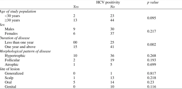
Eighty-two patients fulfilling the inclusion criteria were included in this study. The mean age of study population was 39.32±13.212 years. On analysis of demographics data, it was observed 25 (30.5%) were below 30 years of age and 57 (69.5%) were of age 30 years and above).
39 (47.6%) were males and 43 (52.4%) were females. 26 (30.9%) patients had duration of disease less than one year while 56 (69.1%) had duration of disease for one year and above. On analysis of frequency of morphological pattern 46 (56.1%) had hypertrophic, 21 (25.6%) had follicular and 6 (7.3%) had atrophic pattern and 9 (11%) were of other pattern. On analysis of site of lesion 1 (1.2%) had generalized disease, 72 (87.8%) had skin involvement, 14 (17.1%) had scalp involvement, 19 (23.2%) had oral involvement and 10 (12.1%) had genital involvement.
Among 82 patients of LP, 15 (18.3%) were HCV positive serologically. Stratification of age, gender, duration of disease is mentioned in
Table 1. Morphological pattern and site of disease were not significantly associated with
outcome variable (p value 0.699 and 0.817,
respectively).
Discussion
Table 1 Stratification of age, gender, duration of disease and HCV seropositivity (n=82).
HCV positivity p value
Yes No
Age of study population
<30 years 2 23
0.095
≥30 years 13 44
Sex
Males 9 30
0.217
Females 6 37
Duration of disease
Less than one year 00 25
0.002
One year and above 15 41
Morphological pattern of disease
Hypertrophic 10 36 0.268
Follicular 2 19 0.193
Atrophic 1 5 0.699
Site of lesion
Generalized 0 1 0.817
Scalp 1 13 0.218
Oral 5 14 0.23
Genital 0 10 0.116
differentiated effector cells were identified in close vicinity to HCV (detected by PCR) in lesions of mucosal lichen planus.12
Our study found a significant prevalence of HCV in patients with lichen planus. Findings of our study are consistent with the results found in many studies in the medical literature.13,14,15 Another study showed a statistically significant difference (p=0.040) between the prevalence of HCV infection among patients with LP (7.5%±0.27) and among the control group (0.69%±0.08), but our study showed increased frequency. Nonetheless, a wide variation in data obtained in different countries was observed. The influence of geographic origin of the patients on the study results is remarkable.16 For instance, in Italy, the prevalence of HCV infection reaches high levels when compared to those of the rest of the world population, ranging from 0.7 to 1.3%. Same outcome was observed in a case-control study conducted in Iran, in which patients with LP were compared to blood bank donors.16 On the other hand, studies conducted in Great Britain and in the Netherlands, where the overall prevalence of HCV infection is lower (0.088-0.55% and 0.7%,
respectively), did not show a significant association.17 So a wide variability in the prevalence of HCV infection in the world population may explain such contradictory results.
virus infection. However, this method is known to be subject to errors, and false-positive and false-negative results may occur. Currently, the confirmation of positive serology for HCV has become possible with the detection of viral RNA by PCR. More recent studies have used this technique to confirm HCV infection in the patients studied, and even as an attempt to detect viral RNA in biopsies of LP lesions. Our study also found that there is a significantly increased risk of having positive HCV serology with increased total duration of disease (p=0.002). All 15 patients with positive HCV serology in this study were having disease duration of more than 1 year. It also implies that LP is a chronic inflammatory dermatosis which at times resolves over a period of years.
Another important consideration is the clinical presentation of LP that is the most frequently associated with HCV infection. Literature review reveals that the clinical presentation of LP most frequently associated with HCV infection is the erosive type in oral mucosa. Some studies showed that HCV might be identified in oral mucosa epithelium cells and in oral LP lesions of patients with positive serology. A study with Italian patients found a significant association between HCV and exclusively oral LP and HLA-DR6 allele, suggesting that, more than the virus itself, the host could influence in progression of the disease.20 Our study did not found association of HCV with site of lesion. Another interesting and controversial point is the influence of the treatment of hepatitis C with alpha interferon in the development of LP. This drug has an antiviral and immunomodulating activity. In some patients, it may exacerbate inflammatory skin conditions that were at a low activity level prior to initiation of treatment. This reaction would be triggered by the production of lymphokines and by the expression of adhesion molecules in the skin, induced by the drug.21 In
other patients, however, LP may improve or even resolve after alpha interferon therapy.22
Conclusion
HCV positivity is common in patients with LP. Patients with long duration of disease i.e. more than one year are more likely to be HCV positive. Age, gender, morphological pattern and site have no effect on HCV seropositivity in patients of lichen planus.
References
1. Lodi G, Pellicano R, Carrozzo M. Hepatitis C virus infection and lichen planus: a systematic review with meta-analysis. Oral Dis. 2010;16:601-12.
2. Nagao Y, Sata M. A retrospective case-control study of hepatitis C virus infection and oral lichen planus in Japan: association study with mutations in the core and NS5A region of hepatitis C virus. BMC Gastroenterol.2012;12:31.
3. Ukonu AB, Augustine U. The prevalence of hepatitis C Virus (HCV) among lichen planus patients and its clinical pattern at the University of Abuja Teaching Hospital, Gwagwalada, Abuja, Nigeria. Glob J Health Sci.2012;4(5):113-9.
4. Konidena A, Pavani BV. Hepatitis C virus infection in patients with oral lichen planus. Niger J Clin Pract. 2011;14:228-31. 5. Gupta S, Jawanda MK. Oral lichen planus:
An update on etiology, pathogenesis, clinical presentation, diagnosis and management. Indian J Dermatol. 2015;60:222-9.
6. Yamamoto T, Nakane T, Osaki T. The mechanism of mononuclear cell infiltration in oral lichen planus: the role of cytokines released from keratinocytes. J Clin Immunol. 2000;20:294-305.
8. Musso T, Scutera S, Vermi W, Daniele R, Fornaro M, Castagnoli C et al. Activin A induces Langerhans cell differentiation in vitro and in human skin explants. PLOS One. 2008;3(9):e327137.
9. Zhou XJ, Sugerman PB, Savage NW, Walsh LJ, Seymour GJ. Intra‐epithelial CD8+ T cells and basement membrane disruption in oral lichen planus. J Oral Pathol Med. 2002;31(1):23-7.
10. Zhou XJ, Sugerman PB, Savage NW, Walsh LJ. Matrix metalloproteinases and their inhibitors in oral lichen planus. J Cutan Pathol. 2001;28(2):72-82
11. Ammar M, Mokni M, Boubaker S, El Gaied A, Ben Osman A, Louzir H. Involvement of granzyme B and granulysin in the cytotoxic response in lichen planus. J Cutan Pathol. 2008;35:630-4.
12. Abell E, Presbury DG, Marks R, Ramnarain D. The diagnostic significance of immunoglobulin and fibrin deposition in lichen planus. Br J Dermatol. 1975;93 :17-24.
13. Kulthanan K, Jiamton S, Varothai S, Pinkaew S, Sutthipinittharm P. Direct immunofluorescence study in patients with lichen planus. Int J Dermatol. 2007;46:1237–41.
14. Issa MCA, Gaspar AP, Gaspar NK. Líquen plano e hepatite C. An Bras Dermatol. 1999;75:459-63.
15. Van der Meij EH, Van der Waal I. Hepatitis C virus infection and oral lichen planus: a report from the Netherlands. J Oral Pathol Med. 2000;29:255-8.
16. Tucker SC, Coulson IH. Lichen planus is not associated with hepatitis C virus
infection in patients from North West England. Acta Derm Venereol. 1999;79 :378-9.
17. Ghodsi SZ, Daneshpazhooh M, Shani M, Nikfarjam A. Lichen planus and hepatitis C: a case-control study. BMC Dermatol. 2004;4:6.
18. Ingafou M, Porter SR, Scully C, Teo CG. No evidence of HCV infection or liver disease in British patients with local lichen planus. Int J Oral Maxillofac Surg. 1998;27:65-6.
19. Gruppo Italiano Studi Epidemiologic In Dermatologia (GISED). Lichen planus and liver disease: a multicenter case-control study. Br Med J. 1990;300:227-30.
20. Figueiredo LC, Carrilho FJ, Andrage HF, Milgliari DA. Oral lichen planus and hepatitis C virus infection. Oral Dis. 2002;8:42-6.
21. Carozzo M, Francia Di Celle P, Gandolfo S, Carbone M, Conrotto D, Fasano ME. Increased frequency of HLA-DR6 allele in Italian patients with hepatitis C virus-associated lichen planus. Br J Dermatol. 2001;144:803-8.
22. Lunel F, Cacoub P. Treatment of autoimmune and extra hepatic manifestations of hepatitis C virus infection. J Hepatol.1999;31:210-6.