Ronnachai Viriyataveekul, M.D., Jaruda Kobkitjaroen, M.D.
Department of Clinical Pathology, Faculty of Medicine Siriraj Hospital, Mahidol University, Bangkok 10700, Thailand.
Detection of Specific Autoantibodies in Sera with
Negative Antinuclear Antibody by Indirect
Immunofluorescence Assay but Positive by
Enzyme Immunoassay
Corresponding author: Jaruda Kobkitjaroen E-mail: jaruda.kob@mahidol.ac.th
Received 16 March 2018 Revised 27 November 2018 Accepted 21 February 2019 ORCID ID: http://orcid.org/0000-0002-6553-2185
http://dx.doi.org/10.33192/Smj.2019.36 ABSTRACT
Objective: The aim of this study was to investigate the predictive value of positive enzyme immunoassay (EIA) for detection of specific autoantibodies in sera negative for antinuclear antibody (ANA) by indirect immunofluorescence (IIF) assay, but positive for ANA by EIA.
Methods: Eighty sera that tested negative for ANA by IIF, but positive for ANA by EIA were included. All sera were tested for specific autoantibodies by line immunoassay (LIA). The positive predictive value (PPV) of EIA was calculated using LIA result as a reference standard. Medical records of patients were reviewed. Clinical findings at the time of blood sampling for ANA testing and at 5 years after sampling were obtained.
Results: Twenty-eight sera (35%) were found to be positive by LIA. The PPV of EIA for detection of specific autoantibodies at the manufacturer’s recommended cut-off was 35.0% (95% CI: 24.5-45.5%). The most prevalent antibodies were anti-SSA/Ro60 (64.3%, 95% CI: 46.5-82.0%), anti-Ro52 (25.0%, 95% CI: 9.0-41.0%), and anti-SSB/ La (10.7%, 95% CI: 0-22.2%). A diagnosis of systemic autoimmune rheumatic disease was established in 7 patients (25%) at the time of blood sampling, and 4 patients (14.3%) were diagnosed with non-rheumatic autoimmune disease. Conclusion: EIA testing in IIF-ANA negative sera yielded a chance to detect antinuclear antibodies. However, the poor PPV of EIA may have low benefit in real-life clinical practice. Anti-SSA/Ro60 was the most prevalent antibody detected. A high proportion of LIA-ANA positive patients were not diagnosed as autoimmune disease at the time of antibody detection.
Keywords: Antinuclear antibody; extractable nuclear antigen; enzyme immunoassay; indirect immunofluorescence assay (Siriraj Med J 2019;71: 234-239)
INTRODUCTION
Antinuclear antibodies (ANA) are essential autoantibodies for screening and diagnosis of systemic autoimmune rheumatic diseases (SARD), such as systemic lupus erythematosus (SLE), as well as non-rheumatic
autoimmune diseases.1-5 Certain specific antibodies are
included in the diagnostic criteria for certain specific diseases.6-9 Indirect immunofluorescence (IIF) assay
a substrate, which provides almost all of the antigens that are required for ANA testing. However, enzyme immunoassay (EIA) is more rapid and simple than IIF for detecting ANA. EIA contains disease-relevant native or recombinant antigens that are coated on a microtiter plate. IIF using Hep-2 cells could yield a false-negative result for anti-Ro and anti-Jo-1 antibodies, while EIA could detect them.11,12 However, EIA has been associated
with both false-positive and false-negative results.13
During 2010, the Clinical Pathology Laboratory at Siriraj Hospital implemented a protocol that calls for IIF to be used for initial screening of ANA. In cases of negative result by IIF, secondary screening is performed using EIA. We observed that among sera positive for ANA by EIA, some sera samples were positive and some were negative for specific antibodies subsequently requested by the physician.
Accordingly, the aim of this study was to investigate the predictive value of positive EIA for detection of specific autoantibodies in sera negative for ANA by IIF, but positive for ANA by EIA. The secondary objective was to determine and report the prevalence and clinical findings of certain antibodies.
MATERIALS AND METHODS Patient sera
Sera negative for ANA by IIF, but positive for ANA by EIA were randomly selected from stored sera sent to the Clinical Pathology Laboratory at Siriraj Hospital for ANA testing during 2010. Sera were stored at -20°C before analysis. Siriraj Hospital is Thailand’s largest national tertiary referral center.
Medical records of patients whose sera were included in this study were reviewed. Clinical findings at the time of blood sampling for ANA testing and at 5 years after sampling were obtained and recorded.
The protocol for this study was approved by the Siriraj Institutional Review Board (SIRB), Faculty of Medicine Siriraj Hospital, Mahidol University, Bangkok, Thailand (Si 211/2557).
IIF assay
IIF assay was performed using a Mosaic HEp-20-10/Liver (Monkey) Kit (Euroimmun AG, Lübeck, Germany). Briefly, sera were diluted to 1:100 and then incubated with Hep-20-10 cells. The Hep-20-10 cells contained an increased spectrum of Hep-2 cells in the mitotic phase. After staining with fluorescent conjugate, slides were visualized under a fluorescent microscope at 400x. Fluorescence intensity was compared with that of the end-point positive control. Fluorescence intensity
equal to or greater than the fluorescence intensity of the end-point positive control was considered a positive result.
EIA testing
EIA testing was performed using an IMTEC-ANA Screen Kit (Human Diagnostics, Wiesbaden, Germany). The microtiter strips came coated with HeLa cell nuclei. Sera were diluted to 1:101 before being added to the plates. The manufacturer defined any value above 55 U/ml as being a positive result.
Antigen-specific assays
Line immunoassay (LIA) was performed using an IMTEC-ANA-LIA Kit (Human Diagnostics). The strips contain the following antigens: nucleosomes, histones, SmD1, nRNP, SSA/Ro60, Ro52, SSB/La, Scl-70, CENP-B, Jo-1, and ribosomal P proteins (Rib-P). Sera showing positive reactivity on the Human Diagnostics LIA were tested again using a EUROLINE ANA Profile 3 Kit (Euroimmun AG) to confirm the result. The strips in that kit contain the following antigens: nRNP, Sm, SSA/Ro60, Ro-52, SSB/La, Scl-70, PM/Scl, Jo-1, CENP-B, PCNA, dsDNA, nucleosomes, histones, Rib-P, and AMA M2. Sera were diluted to 1:101 before incubation with the strips. Positive reactivity on both LIA kits for at least 1 identical antibody (Ab) type was considered a positive result.
Statistical analysis
All data were collected and analyzed using Microsoft Excel 2016 spreadsheet software (Microsoft Corporation, Redmond, WA, USA). Rate of positive or negative LIA, predictive value of positive EIA, prevalence of each specific antibody, and 95% confidence interval (CI) were calculated.
RESULTS
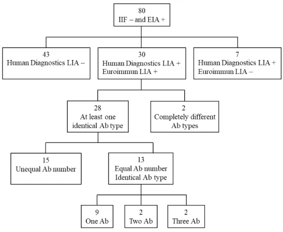
Eighty sera that tested negative for ANA by IIF, but that tested positive for ANA by EIA were included. An overview of LIA results from both LIA test kits is shown in Fig 1. Seven sera with positive LIA by only the Human Diagnostics LIA (samples 31-37), and 2 sera positive for different antibody types using either the Human Diagnostics LIA or the Euroimmun LIA (samples 29-30) were considered negative LIA (Table 1). Therefore, 28 sera (35%) were considered positive LIA (samples 1-28).
The manufacturer’s recommended cut-off value of 55 U/mL could not discriminate positive from negative LIA
Fig 1. Overview of LIA results of 80 sera negative for ANA by IIF, but positive for ANA by EIA.
Abbreviations: Ab = antibody; EIA = enzyme immunoassay; IIF = indirect immunofluorescence assay; LIA = line immunoassay, - = negative; + = positive
Fig 2. Comparison of EIA values between positive and negative LIA.
Abbreviations: EIA= enzyme immunoassay; LIA= line immunoassay; Neg= negative; Pos= positive
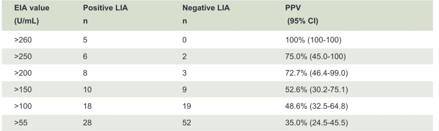
positive predictive value (PPV) of EIA for the detection of specific autoantibodies at this value was 35.0% (95% CI: 24.5-45.5%). The PPV of different EIA values are shown in Table 2.
The most prevalent antibodies were anti-SSA/Ro60 (64.3%, 95% CI: 46.5-82.0%), anti-Ro52 (25%, 95% CI:
9.0-41.0%), and anti-SSB/La (10.7%, 95% CI: 0-22.2%)
TABLE 1. EIA values, specific antibodies, and clinical findings of 37 sera showing reactivity on LIA.
Sample EIA Human Diagnostics Euroimmun
no. (U/ml) LIA LIA Clinical findings
1 95 Ro60 Ro60 Urticarial vasculitis
2 98 Ro60 Ro60 Chronic urticaria, allergic rhinitis
3 94 Ro60 Ro60 Tubulointerstitial disease of unknown cause
4 305 Ro60, Ro52 Ro60, Ro52 Dead fetus in utero at 13 weeks 5 252.57 Ro60, Ro52, La Ro60, Ro52, La SLE
6 113.63 Ro60, SmD1, Rib-P Ro60, Sm, Rib-P Discoid LE, lupus profundus
7 >500 Ro60 Ro60, Ro52 SLE
8 139 Ro60 Ro60, Ro52 Amaurosis fugax
9 114 Ro60 Ro60, Ro52 RA, secondary Sjogren syndrome
10 193.41 Ro60 Ro60, AMA-M2 Primary biliary cirrhosis 11 >500 Ro60 Ro60, histone Nonalcoholic steatohepatitis
12 486 Ro60 Ro60, Rib-P AIH
13 206 Ro60 Ro60, Ro52, AMA-M2 Nephrotic syndrome (FSGS) 14 198.6 Ro60 Ro60, Ro52, La, CENP-B RA, leukopenia
15 120.3 Ro60, nRNP Ro60 DM, AKI on top of CKD
16 211 Ro60, Ro52 Ro60, Ro52, PM/Scl Chronic ITP 17 90 Ro60, SmD1, nucleosome Ro60, Ro52 Alopecia areata 18 288.3 Ro60, histone, nucleosome Ro60, dsDNA Lupus nephritis type IV
19 58.41 Ro52 Ro52 Myofascial pain syndrome
20 106 Ro52 Ro52, Ro60 Common variable immune deficiency
21 63 Ro52 Ro52, AMA-M2 Chronic hepatitis C
22 117 Ro52, Jo-1 Ro52, Jo-1 RA
23 148.54 CENP-B CENP-B Suspected AIH
24 72 Jo-1 Jo-1 Progressive obstructive jaundice of
unknown cause
25 88.9 La La Beta-thalassemia/Hb E disease, AIHA
26 83 La La, Jo-1 Erythema nodosum
27 124.29 Scl-70 Scl-70 Chronic urticaria
28 96.87 SmD1 Sm Unilateral posterior uveitis
29 78.9 SmD1 Ro60 N/A
30 146 nRNP Sm N/A
31 68 Ro60 - N/A
32 72 SmD1 - N/A
33 77.8 SmD1 - N/A
34 135 Histone - N/A
35 158 nRNP - N/A
36 107.4 SmD1, Ro60 - N/A
37 81 Nucleosome, histone, nRNP - N/A
Five years later, 13 of those (76.5%) had not developed autoimmune disease, and 4 patients (samples 19, 21, 23, and 28) had missing data.
DISCUSSION
The present study set forth to investigate for specific antibodies in sera negative for ANA by IIF, but positive for ANA by EIA. We found a higher rate of positive specific antibodies than previous study (35% vs. 16.9%).14
Compared to previous studies that performed antigen-specific assay in all IIF-ANA negative sera and who found a rate of positivity of 0.7-14%15-17, we demonstrated that
screening with EIA-ANA before performing antigen-specific assay in IIF-ANA negative sera may increase the probability of identifying specific antibodies.
The manufacturer’s recommended cut-off value yielded a low PPV. Even though the EIA value was as high as 250 U/mL, the PPV of 75% was not sufficiently high. Therefore, a positive result from this EIA kit may not be applicable to real-life clinical practice. However, reporting the EIA result along with its specific PPV may provide benefit relative to a decision to request or not request antigen-specific assay.
Similar to previous studies14,16, the most prevalent
antibodies identified in this study were anti-SSA/Ro60, anti-Ro52, and anti-SSB/La, and these antibodies have been recognized as being associated with Sjogren syndrome and SLE.18 Anti-SSA/Ro60 was shown to have association
with both subacute cutaneous lupus erythematosus19
and neonatal lupus erythematosus.20,21 In the present
study, a large proportion of patients with positive LIA were not diagnosed as autoimmune disease by the 5-year follow-up. However, the presence of autoantibodies often portends the development of disease that does not fully clinically manifest until years into the future. Anti-SSA/
Ro60 could be detected before the onset of symptoms and diagnosis of SLE at intervals as long as 9.1 and 10.4 years, respectively.22,23
This study has some mentionable limitations. First and consistent with the retrospective nature of this study, some data was missing or incomplete. Second, the number of included samples was relatively small. Third, random samples were selected from routine ANA testing; therefore, our findings may not be generalizable to patients tested in specialized clinic. Fourth and last, we investigated only one EIA kit that uses HeLa cell nuclear extract, which may be different from other EIA kits that use different combinations of defined native or recombinant antigens.
CONCLUSION
EIA testing in IIF-ANA negative sera yielded a chance to detect antinuclear antibodies. However, the poor PPV of EIA may have low benefit in real-life clinical practice. Anti-SSA/Ro60 was the most prevalent antibody detected. A high proportion of LIA-ANA positive patients were not diagnosed as autoimmune disease at the time of antibody detection.
ACKNOWLEDGMENTS
The authors gratefully acknowledge Ms. Jintana Jaiyen for assistance with specimen collection, and the laboratory technicians in the Clinical Pathology Serology Laboratory of Siriraj Hospital for performing the LIA test.
Conflict of interest: The LIA kits used in this study
were provided free of charge by their distributors. The authors declare no other conflicts of interest.
TABLE 2. Positive predictive values of different EIA values.
EIA value Positive LIA Negative LIA PPV
(U/mL) n n (95% CI)
>260 5 0 100% (100-100)
>250 6 2 75.0% (45.0-100)
>200 8 3 72.7% (46.4-99.0)
>150 10 9 52.6% (30.2-75.1)
>100 18 19 48.6% (32.5-64.8)
>55 28 52 35.0% (24.5-45.5)
REFERENCES
1. Tan EM, Cohen AS, Fries JF, Masi AT, McShane DJ, Rothfield NF, et al. The 1982 revised criteria for the classification of systemic lupus erythematosus. Arthritis Rheum. 1982;25: 1271-7.
2. Hochberg MC. Updating the American College of Rheumatology revised criteria for the classification of systemic lupus erythematosus. Arthritis Rheum 1997;40:1725.
3. Solomon DH, Kavanaugh AJ, Schur PH, American College of Rheumatology Ad Hoc Committee on Immunologic Testing G. Evidence-based guidelines for the use of immunologic tests: antinuclear antibody testing. Arthritis Rheum 2002;47:434-44.
4. Alvarez F, Berg PA, Bianchi FB, Bianchi L, Burroughs AK, Cancado EL, et al. International Autoimmune Hepatitis Group Report: review of criteria for diagnosis of autoimmune hepatitis. J Hepatol 1999;31:929-38.
5. Hennes EM, Zeniya M, Czaja AJ, Pares A, Dalekos GN, Krawitt EL, et al. Simplified criteria for the diagnosis of autoimmune hepatitis. Hepatology 2008;48:169-76.
6. Alarcon-Segovia D, Cardiel MH. Comparison between 3 diagnostic criteria for mixed connective tissue disease. Study of 593 patients. J Rheumatol 1989;16:328-34.
7. Tanimoto K, Nakano K, Kano S, Mori S, Ueki H, Nishitani H, et al. Classification criteria for polymyositis and dermatomyositis. J Rheumatol 1995;22:668-74.
8. Shiboski SC, Shiboski CH, Criswell L, Baer A, Challacombe S, Lanfranchi H, et al. American College of Rheumatology classification criteria for Sjogren’s syndrome: a data-driven, expert consensus approach in the Sjogren’s International Collaborative Clinical Alliance cohort. Arthritis Care Res (Hoboken) 2012;64:475-87.
9. van den Hoogen F, Khanna D, Fransen J, Johnson SR, Baron M, Tyndall A, et al. 2013 classification criteria for systemic sclerosis: an American college of rheumatology/European league against rheumatism collaborative initiative. Ann Rheum Dis 2013;72:1747-55.
10. Agmon-Levin N, Damoiseaux J, Kallenberg C, Sack U, Witte T, Herold M, et al. International recommendations for the assessment of autoantibodies to cellular antigens referred to as anti-nuclear antibodies. Ann Rheum Dis 2014;73:17-23.
11. Blomberg S, Ronnblom L, Wallgren AC, Nilsson B, Karlsson- Parra A. Anti-SSA/Ro antibody determination by enzyme-linked immunosorbent assay as a supplement to standard immunofluorescence in antinuclear antibody screening. Scand J Immunol 2000;51:612-7.
12. Bahrt KM, McCarty GA. A negative fluorescent antinuclear
antibody test in a patient with Jo-1 antibody. J Rheumatol 1985;12:624-5.
13. Bayer PM, Fabian B, Hubl W. Immunofluorescence assays (IFA) and enzyme-linked immunosorbent assays (ELISA) in autoimmune disease diagnostics--technique, benefits, limitations and applications. Scand J Clin Lab Invest Suppl 2001;235:68-76.
14. Dahle C, Skogh T, Aberg AK, Jalal A, Olcen P. Methods of choice for diagnostic antinuclear antibody (ANA) screening: benefit of adding antigen-specific assays to immunofluorescence microscopy. J Autoimmun 2004;22:241-8.
15. Hoffman IE, Peene I, Veys EM, De Keyser F. Detection of specific antinuclear reactivities in patients with negative anti-nuclear antibody immunofluorescence screening tests. Clin Chem 2002;48:2171-6.
16. Lee SA, Kahng J, Kim Y, Park YJ, Han K, Kwok SK, et al. Comparative study of immunofluorescent antinuclear antibody test and line immunoassay detecting 15 specific autoantibodies in patients with systemic rheumatic disease. J Clin Lab Anal 2012;26:307-14.
17. Sener AG, Afsar I, Demirci M. Evaluation of antinuclear antibodies by indirect immunofluorescence and line immunoassay methods’: four years’ data from Turkey. APMIS 2014;122:1167- 70.
18. Provost TT. Anti-Ro(SSA) and anti-La(SSB) antibodies in lupus erythematosus and Sjogren’s syndrome. Keio J Med 1991;40: 72-7.
19. McCauliffe DP. Cutaneous diseases in adults associated with anti-Ro/SS-A autoantibody production. Lupus 1997;6:158-66.
20. Cimaz R, Spence DL, Hornberger L, Silverman ED. Incidence and spectrum of neonatal lupus erythematosus: a prospective study of infants born to mothers with anti-Ro autoantibodies. J Pediatr 2003;142:678-83.
21. Jaeggi E, Laskin C, Hamilton R, Kingdom J, Silverman E. The importance of the level of maternal anti-Ro/SSA antibodies as a prognostic marker of the development of cardiac neonatal lupus erythematosus a prospective study of 186 antibody- exposed fetuses and infants. J Am Coll Cardiol 2010;55:2778- 84.
22. Arbuckle MR, McClain MT, Rubertone MV, Scofield RH, Dennis GJ, James JA, et al. Development of autoantibodies before the clinical onset of systemic lupus erythematosus. N Engl J Med 2003;349:1526-33.