How to cite:
Setiawati A (2017) Cytotoxic Selectivity of Ganoderma lucidum in Colon Cancer through Cyclooxygenase 2 (COX-2) as Its Molecular Target. J. Trop. Life. Science 7 (2): 177 – 183. *Corresponding author:
Agustina Setiawati
Department of Pharmaceutical Biology, Faculty of Pharmacy, Sanata Dharma University
Campus III, Maguwoharjo, Depok, Yogyakarta, Indonesia 55284 E-mail: nina@usd.ac.id
VOL. 7, NO. 2, pp. 177 – 183, April 2017 Submitted February 2017; Revised March 2017; Accepted April 2017
Cytotoxic Selectivity of
Ganoderma lucidum
in Colon Cancer through
Cyclooxygenase 2 (COX-2) as Its Molecular Target
Agustina Setiawati *
Department Pharmaceutical Biology, Faculty of Pharmacy, Sanata Dharma University, Yogyakarta, Indonesia
ABSTRACT
Many studies were designed explore chemopreventive activity of natural products on colon cancer especially ad-dressing COX-2 as molecular target. Another promising source of natural product that potentially exhibit anticancer activity on colon cancer is Ganoderma lucidum. This study assessed selectivity of cytotoxic effect of G. lucidum
extract on WiDr to Vero cells and investigated molecular mechanism on COX-2. G. lucidum extract was prepared
by reflux extraction method; in vitro anticancer was assayed by MTT method on WiDr and Vero cell line. This study applied apoptosis induction assay to observe cell death mechanism using double staining method; further
COX-2 expression was stained by immunocytochemistry method. G. lucidum extract has cytotoxic effect on WiDr
cells with IC50 135 µg/mL. However, the cytotoxic effect had low selectivity towards Vero cells with Selectivity Index (SI) 3.66. The extract induced apoptosis and suppressed COX-2 expression in WiDr cells. G. lucidum extract was potential to be developed as anticancer agent towards colon cancer.
Keywords: Ganoderma lucidum, selectivity, colon cancer, COX-2
INTRODUCTION
Colon cancer has high mortality case in developing countries [1] and it was ranked as the third most fre-quently diagnosed cancer in both of male and female in all of the countries [1, 2, 3], as well as in the South East Asian Nations (ASEAN) countries [2, 3]. Since its inci-dence rapidly increases [4, 5], many studies are em-ployed to prevent and cure colon cancer. There are many possible clinical treatments of colon cancer; such as sur-gery, chemotherapy, radiotherapy, and adjuvant chemo-therapy [5, 6]. However, painful, nausea, vomiting, skin changes, diarrhea, hair loss, bleeding and fatigue are se-vere side effects of the treatment [7]. The side effects arise because of non-selective and specific target of the treatments. Therefore, more studies set to discover spe-cific and selective colon cancer treatment [8, 9, 10] to kill colon cancer cells without endangering normal cells.
Colon cancer cells excessively produce cyclooxygen-ase 2 (COX-2) in both of carcinoma and adenoma [11,12, 13]. COX-2, an enzyme that is responsible for
converting arachidonic acid to prostaglandins (PGs) [14], plays an important role in inflammation, cell pro-liferation and apoptosis regulation of colon cancer [15]. One of COX-2 product, prostaglandin E2 (PGE2), pro-motes angiogenesis and stimulates colon cancer growth by preventing apoptosis [16, 17]. Thus, the inhibition activity of COX-2 is an alternative to suppress colon cancer. Previous studies concluded that COX-2 inhibi-tors successfully prevented colon cancer growth and polyp formation [17, 18, 19, 20, 21, 22]. Therefore, COX-2 is an important molecular target for anticancer chemopreventive screenings on colon cancer.
[12, 27]. Among them, there was curcumin, a yellow pigment isolated from turmeric that successfully inhib-ited COX-2 on WiDr, a model of COX-2 overexpressing cancer cells [24].
Another promising source of natural product that potentially exhibit anticancer activity on colon cancer is
Ganoderma lucidum. G. lucidum is well known as tra-ditional medicine use against cancer, viral and bacterial infection, diabetes, and liver injury. Among its activi-ties, its anticancer properties have been the most inter-esting studies [28]. It revealed cytotoxic activity of sup-pressed inflammatory breast cancer [28, 29, 30], ovarian cancer [31], gastric cancer [32] and liver cancer [33]. A large number of chemical compounds can be extracted from the body or spores. There are two major com-pounds that had anticancer activity in G. lucidum; pol-ysaccharides and triterpenoids. Polpol-ysaccharides of G. lu-cidum boosted cytotoxic immune cells so that it could induce apoptosis in cancer cells [34]. Other compounds in G. lucidum, terpenoid compounds revealed cytotoxic, antioxidant as well as antiinflammation activity on can-cer cells through immune modulation [35-37]. Terpe-noids isolated from G. lucidum activated NF-κB, a tran-scription factor that regulated COX-2 expression in can-cer cells [38, 39]. They excessively induced cell cycle ar-rest on G1 phase through immune boosting in colon cancer xenograft model [40]. Another study underlined that ethanolic extract of G. lucidum decreased COX-2 expression and increased nitric oxide synthesis in colon HT-29 cells [41]. Therefore, G. lucidum is a potential to be developed as chemopreventive agent on colon cancer with COX-2 as the target. Moreover, the molecular pathway involving COX-2 of G. lucidum will give a pos-itive contribution to assess its specificity on colon cancer cells.
However, the selectivity of G. lucidum as chemo-preventive agent is still remaining questioned. Besides the specificity, the selectivity of a chemopreventive agent is also important to investigate. Valadan et al [42] re-ported G. lucidum had cytotoxicity on Vero cells, a model of normal cells. Selectivity Index (SI) (IC50 Vero cells/IC50 cancer cells) indicated cytotoxicity selectivity of a compound or extract to kill normal cells versus in cancer cells [42, 43]. Furthermore, investigating selectiv-ity of cytotoxic effect of G. lucidum is an interesting to be further studied.
This study assessed cytotoxic effect of ethanolic ex-tract of G. lucidum on WiDr as well as Vero cells and determined SI of the extract. Furthermore, the molecu-lar mechanism of death cells was investigated with apop-
tosis assay using double staining method and immuno-cytochemistry of COX-2.
MATERIALS AND METHODS
Plant material
G. lucidum was harvested in June, 2015 from Pan-dowoharjo, Sleman, Yogyakarta, Indonesia.
Chemicals
Celecoxib was purchased from Sigma-Aldrich with
≥ 98% purity. Vero and WiDr cells were cultivated in
Fetal Bovine Serum (FBS) 10% (v/v) (Gibco) and 1% (v/v) streptomycin and penicillin (Gibco) in Roswell Park Memorial Institute Medium (RPMI) (Gibco). To dissolve the extract and celecoxib, this study used dime-thyl sulfoxide (DMSO) (Merck).
Immunocytochemistry used COX-2 primary anti-body from Thermo Scientific Lab Vision and secondary antibody was diluted from Starr Trek HRP universal de-tection system No.901-STUHRP700-090314. All plates and micro plate in this study were Iwaki®, moreover blue, yellow and white tips were supplied by Axygen®.
Extraction
G. lucidum was dried using oven and homoge-nously powdered. 10 g of its powder was extracted in 90% ethanol using reflux method for 2 hours. Liquid extract was evaporated under 80°C until reached con-stant weight.
Cytotoxic assay
Cytotoxic assay towards Vero cells as well as WiDr cells were designed based on our previous studies [45-46]. Cells were seeded in a culture tissue flask until 80% confluent, then 5×10³ cells were planted into a 96-well
The plate was wrapped in dark paper to avoid light ex-posure and incubated 12-24 hours, then formazan com-plex was measured in 595 nm visible wavelength using ELISA reader.
Apoptosis induction assay
Apoptosis induction assay was conducted only in WiDr cells. The cells were cultured 5×10⁴ on coverslips
in 24-well plate. WiDr cells were adapted in 37°C and 5% CO2 for 24 hours. Then, the medium was discarded and twice soaked in PBS. The extract and celecoxib at IC50 concentration (135 µg/mL and 68 µM) were poured into the plate then it was incubated in the same as previous condition. IC50 concentration of celecoxib was calculated from our previous study [47]. The me-dium was removed from WiDr cells and they were twice soaked in PBS. The coverslips were taken from the plate and moved to glass objects, and then acridine orange-ethidium bromide (AE) was added into the coverslips. The glass objects were immediately investigated under fluorescence microscope.
Immucytochemistry assay
WiDr cells were spread in 6-well plate and placed in incubator with 5% CO2 and 37°C for 24 hours. The extract at 135 µg/mL and celecoxib at 68 µM were treated on cells and incubated for 24 hours. At the end of the incubation time, cells were collected and washed in PBS three times. The cells were suspended in me-dium, placed and fixed in object glass for 5 minutes. Hy-drogen peroxidase was added into the object glass and placed in room temperature for 10-15 minutes. Then, it was gently washed by PBS twice and monoclonal anti-body of COX-2 was dropped into the object glass at least for an hour. The object glass was washed in PBS and secondary antibody was dropped into the glass. It was incubated in room temperature for ten minutes, later it was washed four times in PBS. The solution of 3, 3'-diaminobenzidine (DAB) as chromogen was poured into the cells and incubated for 3- 8 minutes. Finally, the object glass was washed by aquadest and hematoxylin solution was added. It was incubated for 3-4 minutes and dried. The expression of COX-2 was observed under inverted microscope.
Data analysis
Cell viability was calculated from MTT data using equation:
Sample treatment absorbance − Medium absorbance Untreated cells absorbance − Medium absorbance × 100%
Cell viability data was analyzed using linier regres-sions to calculate IC50 of extract and celecoxib. This SI was calculated by dividing IC50 towards Vero cells/IC50 towards Vero cells [19]. The number of apoptosis, ne-crosis and living cells was semi-quantitative analyzed by blind observed by blind counting cell distribution in three areas of object glass.
RESULTS AND DISCUSSION
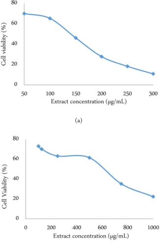
G. lucidum extract demonstrated dose-depend cy-totoxic activity both in WiDr cells (Figure 1a) and Vero cells (Figure 1b). It had IC50 135 µg/mL towards WiDr cells and 494 µg/mL towards Vero cells. Cell morpholo- gy observation revealed G. lucidum extract IC50 con-centration on slightly effected cell population otherwise celecoxib dramatically effected cell population than the extract (Figure 2). By comparing IC50 in Vero cells to IC50 in WiDr cells, the SI was determined 3.66 respec-tively.
(a)
(b)
Figure 1. The effect of G. lucidum extract on WiDr cells (a) and Vero cells (b)
0 20 40 60 80
50 100 150 200 250 300
Ce
ll v
iab
ilit
y
(%
)
Extract concentration (µg/mL)
0 20 40 60 80
0 200 400 600 800 1000
Ce
ll Vi
ab
ilit
y
(%
)
Tabel 1. Cell distribution in apoptosis assay
Treatment Apoptosis (%) ± SD Necrosis (%) ±SD Living (%)± SD
G. lucidum extract 46.4 ± 0.56 5.21 ± 1.12 48.40 ± 5.95
Celecoxib 81.80 ± 0.88 3.48 ± 1.43 14.68 ± 0.65
Untreated cells 1.42 ± 0.65 0.72 ± 0.04 97.86 ± 0.61
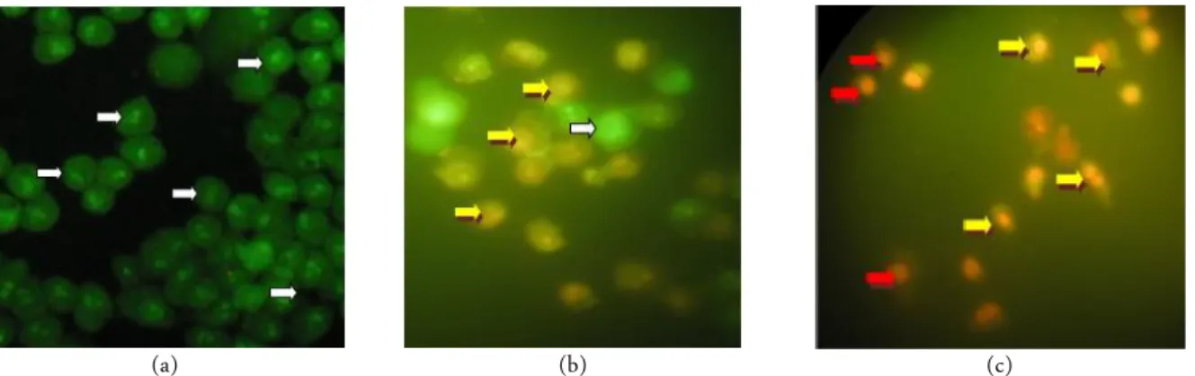
(a) (b) (c)
Figure 2. Morphology observation of WiDr cells under light microscope using 400 × magnification in untreated cells (a), cells treated by 135 µg/mL extract (b), and cells treated by 68 µM celecoxib (c). Living and dead cells are indicated by white arrow and red arrow.
(a) (b) (c)
Figure 3. Apoptotic observation in WiDr cells using double staining method under flourenscence microscopy using 400 × magnification in untreated cells (a), cells treated by 135 µg/mL extract (b), and cells treated by 68 µM celecoxib (c). Living, dead, and apoptotic cells are indicated by white arrow, red arrow, and yellow arrow.
(a) (b) (c)
Furthermore, this study investigated apoptosis in-duction in WiDr cells using double staining method. The nuclei showed condensed yellow-green fluorescence by acridine orange in early stage of apoptosis. On the other hand, nuclei of cells showed condensed orange flu-orescence by ethidium bromide in late apoptosis stage [47]. Apoptosis cells were showed in orange colour with condensed nuclei while necrosis cells was stained in or-ange colour with uncondensed nuclei (Figure 3). G. lu-cidum extract as well as celecoxib induced apoptosis in WiDr cell (Table 1). It confirmed from our previous re-search that celecoxib strong induced apoptosis in WiDr cells [47]. Molecular mechanism of extract in WiDr cells was further investigated through COX-2 expression analysis. COX-2 suppressing cells were stained in brown color while non-suppressing cells were stained in purple color. Immunocytochemistry staining revealed extract suppressed COX-2 expression. This suppression of the extract was lower than celecoxib (Figure 4).
Overall, this study evaluated cytotoxic activity of
G. lucidum extract on WiDr colon cancer cells and its selectivity on normal cells. This study was designed to discover new therapeutic agent of colon cancer that has less side effects to cancer patient. This study used Vero cells, as model of normal mammalian cell line that de-rived from the kidney of the African green monkey (Cercopithecus aethiops). This cell line was recom-mended for screening chemical toxicity in vitro [48, 49].
Selectivity Index (SI) is parameter that indicates se-lectivity of a compound or an extract. Based on Moustafa et al. (2014) [43], the SI value of G. lucidum
extract (SI value < 10) was consider as unselective extract towards normal cells. However, other studies mentioned that SI value < 2 indicating high selectivity [50, 51]. G. lucidum extract induced apoptosis in WiDr cells.
Instead of its selectivity and apotosis induction,
molecular mechanism of G. lucidum extract on WiDr
cells must be further investigated. This study focused on COX-2 protein that produced PGE2 that increased cell proliferation and escaped from apoptosis [16, 17]. Therefore, COX-2 is important target to screen chemo-preventive agent on colon cancer. The result of this study showed that G. lucidum extract suppressed COX-2 expression in WiDr. The suppression effect was lower than celecoxib, a selective COX-2 inhibitor, as positive control. Celecoxib is one of prospective drug against co-lon cancer [52] and our previous research showed it pressed COX-2 expression in WiDr cells [47]. The sup-pression effect of the extract may be activated by terpe-noids in G. lucidum to NF-κB, a transcription factor
that regulated COX-2 expression in cancer cells [38, 39].
G. lucidum extract offers a new promising for se-lective chemopreventive agent on colon cancer. Our data revealed it inhibited COX-2 expression, even though de-tail molecular mechanism of these benefit remain to be established in the future study
CONCLUSION
G. lucidum extract has selective cytotoxic effect to-wards WiDr cells than Vero cells. It molecular mecha-nism in WiDr cells involved COX-2 suppression.
ACKNOWLEDGMENT
The author is grateful to Skolastika who help in technical work during the G. lucidum extraction.
REFERENCES
1. Torre LA, Bray F, Siegel RL et al. (2015) Global Cancer Statistics. CA: Cancer Journal for Clinicians 65: 87- 107. doi: 10.3322/caac.20107.
2. World Health Organization (2013) Latest world cancer sta-tistics Global cancer burden rises to 14.1 million new cases in 2012: Marked increase in breast cancers must be ad-dressed. https://www.iarc.fr/. Accessed: March 2015. 3. Kimman M, Norman R, Jan S et al. (2012) The Burden of
cancer in member countries of the association of Southeast Asian Nations (ASEAN). Asian Pacific Journal of Cancer Prevention 13 (2): 411-420.
4. Jemal A, Center MM, DeSantis C, Ward EM (2010) Global pattern of cancer incidence and mortality rates and trends. Cancer Epidemiology, Biomarkers and Prevention 9 (8): 1893- 1907. doi: 10.1158/1055-9965.EPI-10-0437. 5. Labianca R, Nordinger B, Beretta GD et al. (2010) Primary
colon cancer: ESMO clinical practical guidelines for diag-nosis, adjuvant treatment and follow-up. Annals of Oncol-ogy 21 (Supplement_5): v70-v77. doi: 10.1093/an-nonc/mdq168.
6. Association of Coloproctology of Great Britain and Ireland (2007) Guideline for the Management of Colorectal Can-cer. London. 3rd Edition. http://www.uhb.nhs.uk/. Ac-cessed: April 2015.
7. American Cancer Society. Colon Cancer: Facts and Figure 2014-2016. https://www.cancer.org/ Accessed: April 2015. 8. Jiang Y, Zhang C, Chen K et al. (2014) The clinical signif-icance of DC-SIGN and DC-SIGNR, which are Novel markers expressed in human colon cancer. PLoS ONE. 9 (12): e114748. doi: 10.1371/journal.pone.0114748. 9. Wang XW, Zhang YJ (2014) Targeting mTOR network in
10. Mishra J, Dromund J, Quazi SH et al. (2013) Prospective of colon cancer treatments and scope for combinatorial ap-proach to enhanced cancer cell apoptosis. Critical Reviews in Oncology/Hematology 86 (3): 232-250. doi: 10.1016/j.critrevonc.2012.09.014.
11. Dixon DA, Blanco FF, Bruno A, Patrignani P (2013) Mech-anistic Aspects of COX-2 Expression in Colorectal Neo-plasia. In: Chan AT, Detering E (eds) Prospect. Chemopre-vention Color. Neoplasia. Berlin, Springer, pp 7–37. doi: 10.1007/978-3-642-30331-9_2.
12. Qi J, Dong Z, Liu J, Zhang JT (2014) EIF3i promotes colon oncogenesis by regulating COX-2 protein synthesis and β -catenin activation. Oncogene 33 (32): 4156–4163. doi: 10.1038/onc.2013.397.
13. Roelofs HMJ, Te Morsche RHM, van Heumen BWH et al. (2014) Over-expression of COX-2 mRNA in colorectal can-cer. BMC Gastroenterology 14 (1): 1- 6. doi: 10.1186/1471-230X-14-1.
14. Park J, Conteas CN (2010) Anti-carcinogenic properties of curcumin on colorectal cancer. World Journal of Gastroin-testinal Oncology 2 (4): 169-176. doi: 10.4251/wjgo.v2.i4. 169.
15. Sobolewski C, Cerella C, Dicato M et al. (2010) The Role of Cyclooxygenase-2 in cell proliferation and cell death in human malignancies. International Journal of Cell Biology 2010 (2010): 1-21. doi: 10.1155/2010/215158.
16. Nakanishi M, Rosenberg DW (2013) Multifaceted roles of PGE2 in inflammation and cancer. Seminars in Immuno-pathology 35 (2): 123–137. doi: 10.1007/s00281-012-0342-8.
17. Li HJ, Reinhardt F, Herschman HR, Weinberg RA (2012) Cancer-stimulated mesenchymal stem cells create a carci-noma stem-cell niche via Prostaglandin E2 signaling. Can-cer Discovery 2 (9): 840- 855. doi: 10.1158/2159-8290.CD-12-0101.
18. Ghosh N, Chaki R, Mandal V et al. (2011) COX-2 as a target for cancer chemotherapy. Pharmacological Reports. 62: 233-244.
19. Rahman M, Selvarajan K, Hasan MR et al. (2012) Inhibi-tion of COX-2 in colon cancer modulate tumor growth and MDR-1 expression to enhance tumor regression in therapy-refractory cancers in vivo. Neoplasia 14 (7): 624-633. 20. Kodela R, Chattopadhyay M, Goswami S et al. (2013)
Po-sitional isomers of Aspirin are equally potent in inhibiting colon cancer cell growth: Differences in mode of cyclooxy-genase inhibition. The Journal of Pharmacology and Exper-imental Therapeutics 345 (1): 85–94. doi: 10.1124/jpet.112. 201970.
21. Makar KW, Poole EM, Resler AJ et al. (2013) COX-1
(PTGS1) and COX-2 (PTGS2) polymorphisms, NSAID in-teractions, and risk of colon and rectal cancer in two inde-pendent populations. Cancer Causes and Control 24 (12): 2059–2075. doi: 0.1007/s10552-013-0282-1.
22. Kuno T, Tsukamoto T, Hara A, Tanaka T (2012) Cancer chemoprevention through the induction of apoptosis by natural compounds. Journal of Biophysical Chemistry 3 (2): 156- 173. doi: 10.4236/jbpc.2012.32018.
23. Rosas C, Sinning M, Ferreira A et al. (2014) Celecoxib de-creases growth and angiogenesis and promotes apoptosis in a tumor cell line resistant to chemotherapy. Biological Re-search 4 (1): 27. doi: 10.1186/0717-6287-47-2.
24. Jaganathan SK, Vellayappan MV, Narasimhan G, Su-priyanto E (2014) Role of pomegranate and citrus fruit juices in colon cancer prevention. World Journal of Gastro-enterology 20 (16): 4618-4625. doi: 10.3748/wjg.v20.i16.46 18.
25. Amado NG, Predes D, Moreno MM et al. (2014) Flavo-noids and Wnt/β-Catenin signaling: Potential role in colo-rectal cancer therapies. International Journal of Molecular Science 15: 12094- 12106. doi: 10.3390/ijms150712094. 26. Nassar ZD, Aisha AF, Idris N et al. (2012) Koetjapic acid,
a natural triterpenenoids, induces apoptosis in colon cancer cells. Oncology Reports 27: 727- 733. doi: 10.3892/or.2011. 1569.
27. Patel VB, Misra S, Patel BB, Majumdar APN (2010) Colo-rectal cancer: Chemopreventive role of curcumin and resveratrol. Nutrition and Cancer 62 (7): 958-967. doi: 10.1080/01635581.2010.510259.
28. Galor SW, Yuen J, Buswell JA, Benzie IFF (2011) Ganoderma lucidum (Lingzhi or Reishi), a medicinal mushroom in herbal medicine: Biomolecular and clinical aspects 2nd edition. Florida, CRC Press/Taylor and Fran-cis.
29. Arroyo IJ, Acevedo RR, Perez AA et al. (2013) Anti-tumor effects of Ganoderma ludicum (Reishi) in inflammatory breast cancer in in vivo and in vitro models. PLoS One 8 (2): e57431. doi: 10.1371/journal.pone.0057431.
30. Zhao H, Zhang Q, Zhao L et al. (2012) Spore powder of Ganoderma lucidum improves cancer-related fatigue in breast cancer patients undergoing endocrine therapy: a pi-lot clinical trial. Evidence-Based Complementary and Alter-native Medicine. doi: 10.1155/2012/809614
31. Dai L, Sun W (2014) Ganoderma lucidum inhibits prolif-eration of human ovarian cancer cells by suppressing VEGF expression and up-regulating the expression of connexin 43. BMC Complementary and Alternative Medicine 14: 434. 14:434. doi: 10.1186/1472-6882-14-434.
tosis by ethanol extracts of Ganoderma lucidum in human gastric carcinoma cells. Journal of Acupuncture and Merid- ian Studies 3 (1): 24-31. doi: 10.1016/S2005-2901(10)60004-0.
33. Cao LZ, Lin ZB (2003) Regulatory effect of Ganoderma lucidum polysaccharides on cytotoxic T-lymphocytes in-duced by dendritic cells in vitro. Acta Pharmacologica Sinica 24 (4): 312-326.
34. Weng CJ, Chau CF, Yen GC et al. (2009) Inhibitory effects of Ganoderma lucidum on tumorigenesis and metastasis of human hepatoma cells in cells and animal models. Journal of Agricultural and Food Chemistry 57 (11): 5049–5057. doi: 10.1021/jf900828k.
35. Kao CHJ, Jesuthasan AC, Bishop KS et al. (2013) Anti-cancer activities of Ganoderma lucidum: active ingredients and pathways. Functional Food in Health and Disease 3 (2): 48-65.
36. Calvino E, Pajuelo L, Casas OE et al. (2011) Cytotoxic Ac-tion of Ganoderma lucidum on Interleukin-3 Dependent Lymphoma DA-1 cells: Involvement of apoptosis protein. Phytotherapy Research 25 (1): 25-32. doi: 10.1002/ptr.3202. 37. Wu GS, Guo JJ, Bao JL et al. (2013) Anti-cancer properties of triterpenoids isolated from Ganoderma lucidum - a re-view. Expert Opinion on Investigational Drugs 22 (8): 981-92. doi: 10.1517/13543784.2013.805202.
38. Montemayor MM, Acevedo RR, Franqui EO et al. (2011) Ganoderma lucidum (Reishi) inhibits cancer cell growth and expression of key molecules in inflammatory breast cancer. Nutrition and Cancer 63 (7): 1085-1094. doi: 10.1080/01635581.2011.601845.
39. Jiang J, Slivova V, Harvey K et al. (2004) Ganoderma lu-cidum suppresses growth of breast cancer cells through the inhibition of Akt/NF-κB signaling. Nutrition and Cancer 49 (2): 209- 216. doi: 10.1207/s15327914nc4902_13. 40. Thyagarajan A, Jedinak A, Nguyen H et al. (2010)
Triter-penes from Ganoderma lucidum induce autophagy in colon cancer through the inhibition of p38 mitogen-activated ki-nase (p38 MAPK). Nutrition and Cancer 62 (5): 630-640. doi: 10.1080/01635580903532390.
41. Hong KJ, Dunn DM, Shen CL, Pence BC (2004) Effects of Ganoderma lucidum on apoptotic and anti-inflammatory function in HT-29 human colonic carcinoma cells. Phyto-therapy Research 18 (9): 768–770. doi:10.1002/ptr.1495. 42. Valadan S, Ahmadi R, Sadri M, Mahdavi E (2014) Anti-proliferative effects of Ganoderma lucidum in cell culture on Vero cells. In proceedings of International Conference
on Chemical, Environment and Biological Sciences (CEBS – 2014): 17- 18 September 2014; Kuala Lumpur. Edited by Rahman A, Ahmadi R.
43. Moustafa SMA, Menshawi BM, Wassel GM et al. (2014) Screening of some plants in egypt for their cytotoxicity against four human cancer cell lines. International Journal of PharmTech Research 6 (3): 1074-1084.
44. Irungu BN, Adipo N, Orwa JA et al. (2015) Antiplasmodial and cytotoxic activities of the constituents of Turraea ro-busta and Turraea nilotica. Journal of Ethnopharmacology 174: 419-425. doi: 10.1016/j.jep.2015.08.039.
45. Setiawati A (2016) Celecoxib, A COX-2 selective inhibitor, induces cell cycle arrest at the G2 / M phase in hela cervical cancer cells. Asian Pacific Journal of Cancer Prevention 17 (4): 1655–1659.
46. Calderón-arancibia J, Espinosa-bustos C, Cañete-molina Á et al. (2015) Synthesis and pharmacophore modelling of 2,6,9-Trisubstituted Purine derivatives and their potential role as apoptosis-inducing agents in cancer cell lines. Mol-ecules 20 (4): 6808-6826. doi: 10.3390/molMol-ecules20046808. 47. Setiawati A, Immanuel H, Utami MT (2016) The Inhibition of Typhonium flagelliforme Lodd. Blume leaf extract on COX-2 expression of WiDr colon cancer cells. Asian Pacific Journal of Tropical Biomedicine 6 (3): 251–255. doi: 10.1016/j.apjtb.2015.12.012.
48. U.S. Department of Health and Human Services - Food and Drug Administration (2016) Biological evaluation of medi-cal devices - Part 5: Tests for cytotoxicity: in vitro meth-ods.3: 42. https://www.fda.gov/. Accessed: April 2017. 49. Menezes C, Valerio E, Dias E (2013) The Kidney Vero-E6
Cell Line: A Suitable Model to Study the Toxicity of Mi-crocystins. New Insights into Toxic Drug Test. doi: 10.5772/54463
50. Awang N, Aziz ZA, Kamaludin NF, Chan KM (2014) Cy-totoxicity and mode of cell death induced by Triphenyltin (IV) compounds in vitro. OnLine Journal of Biological Sci-ences 14 (2): 84-93. doi: 10.3844/ojbsci.2014.84.93. 51. Machana S, Weerapreeyakul N, Barusrux S et al. (2011)
Cytotoxic and apoptotic effects of six herbal plants against the human hepatocarcinoma (HepG2) cell line. Chinese Medicine 6 (1): 39. doi: 10.1186/1749-8546-6-39.