Original Research Article
Clinico etiological profile of seizures in adults attending
a tertiary care hospital
Ashwin T.
1*, Tumbanatham A.
2, Siva Ranganathan Green
3, Jaya Singh K.
4INTRODUCTION
Man has witnessed and known seizures for thousands of years. Hans Berger a German psychiatrist in 1929 discovered the human electroencephalogram. Research has been done in the field of epilepsy and seizures since the beginning of twentieth century. As the research progressed epilepsy was known to be a network disease and not only a symptom of local brain abnormalities. Seizures can arise in neocortical, thalamo cortical, limbic, brainstem network from networks perspective. Dreifuss and Penry classified seizures into generalized and partial.1 The aim of 2001 and 2006 reports on
reclassification was to identify unique diagnostic entities with etiologic, therapeutic and prognostic implications. This was done because when a syndromic diagnosis was made, then the therapy and diagnosis will be made on seizure type.
There are very limited studies on the etiology of seizures. The etiology of seizures broadly includes genetic, structural or metabolic causes and seizures of unknown aetiology. ILAE has also modified the definition of epilepsy in 2014 as at least two unprovoked seizures occurring more than 24 hours apart; One unprovoked seizure and a probability of further seizures similar to the
1Resident, 2Assistant Professor, 3Associate Professor, 4Professor and HOD, Department of Medicine, Mahatma Gandhi
Medical College and Research Institute, Pondicherry, India
Received: 26 January 2017
Accepted: 23 February 2017
*Correspondence:
Dr. Ashwin T.,
E-mail: ashwinthangavel615@gmail.com
Copyright: © the author(s), publisher and licensee Medip Academy. This is an open-access article distributed under the terms of the Creative Commons Attribution Non-Commercial License, which permits unrestricted non-commercial use, distribution, and reproduction in any medium, provided the original work is properly cited.
ABSTRACT
Background: Seizures are one of the important causes of morbidity and mortality in adults. There are many studies based on the old seizure and epilepsy classification system but there are only few studies on the clinical profile and cause of seizures. The present study attempted to explore the clinical and etiological profile of seizures in adults above 18 years of age in our tertiary care hospital.
Methods: This was a cross sectional study on 100 seizure participants having EEG abnormality. Patient seizure type was classified according to clinical features. History taking and physical examination of all selected participants were done and investigations were done and recorded to find the etiology according to proforma.
Results: GTCS was the most common seizure type accounting for 57%. Ischemic infarct was most common cause of post stroke seizures. Hypoglycemia was most common metabolic cause of seizures. Bacterial meningitis was most common cause of seizures due to infections. Stroke was the most common aetiology accounting for 21%. Focal seizures were seen predominantly in participants with calcified granuloma. In participants with alcohol withdrawal GTCS was the predominant seizure type.
Conclusions: GTCS was the most common type of seizure in present study. Stroke was the most common cause of seizures and rheumatic heart disease was most common cause of seizures due to stroke in young participants. Seizures due to alcohol withdrawal were on the rise.
Keywords: Alcohol withdrawal, Focal, Hypoglycemia, Seizures
general recurrence risk after two unprovoked seizures, occurring in the next 10 years; diagnosis of an epilepsy syndrome.2 Therefore there is need for epidemiologic and
incidental studies. Many participants termed epileptic previously might not be termed as suffering from epilepsy according to newer definition and classification. The aetiology of seizures in adults also varies according to geographic distribution and the culture prevalent in the area. For example, in some states due to easy availability of alcohol seizures due to alcohol withdrawal are more prevalent.
Therefore this study was done with the intention of studying the clinical and etiological profile of seizures in adults pertaining to area of Puducherry and Cuddalore district of Tamil Nadu, India.
Objectives
To study the clinical profile of seizures in adults attending our hospital and naming seizures according to the 2010 ILAE classification of seizures and to determine the aetiology of seizures in adults attending our hospital.
METHODS
Study design
This study was carried out as an observational cross sectional study.
Study area
The study area was Mahatma Gandhi Medical College and Research Institute Hospital, a rural tertiary care hospital situated in Puducherry, India.
Study population
All adults who were admitted to the neurology department of Mahatma Gandhi Medical College and Research Institute Hospital with history of new onset seizures were the study population.
Study period
The period of study was 21 months from December 2014 to August 2016.
Sample size
Based on intensive literature review, the prevalence of seizures in a study done in Bangalore was reported to be 1.1%.3 This was taken for sample size calculation. At
95% level of significance, and at 2.25% absolute precision, the sample size was estimated to be 83.55. Accounting 10% for non-response, the sample size was calculated as 91.9. The final sample size was rounded off to 100.
Inclusion criteria
All the participants with age more than 18 years of age admitted with new onset of seizures by history and having EEG abnormality. Type of seizure was established according to 2010 ILAE classification system of seizures.4
Exclusion criteria
• Hyperventilation syndrome
• Movement disorders
• Syncope
Sampling method
The sampling technique used in this study was purposive sampling.
Data collection tools
Data was collected with the help of a structured interview schedule which consisted of the background information, history of seizures. EEG was recorded and the findings were documented.
Operational definition2
Epilepsy is a disease of brain. It should be defined by any one of the following.
• Minimum of 2 unprovoked seizures occurring more than 24 hours apart.
• At least one unprovoked seizure and a probability of further seizures similar to general recurrence risk after 2 unprovoked seizures, occurring over the next 10 years.
• Diagnosis of an epilepsy syndrome.
Ethical committee approval
Ethical approval was obtained from the Institutional Ethics Committee prior to the data collection.
Informed consent
The participants were clearly explained about the objectives of the study and informed consent was obtained in local language (Tamil) prior to the administration of the interview schedule.
Data collection
• Hemoglobin, Total count, Differential count, Platelet count
• Random blood sugar.
• Renal function test.
• Liver function test
• Serum electrolytes
• Electroencephalogram
• CT brain, contrast &MRI
• ECG.
Statistical analysis
Statistical analysis was carried out using SPSS version 19.0 (IBM SPSS, US) software with Regression Modules installed.
Descriptive analyses were reported as mean and standard deviation of continuous variables. Chi square test and Fischer exact test were used to establish association.
RESULTS
Type of seizure among study participants
This study was carried out among 100 participants who were admitted for seizures. The distribution of the participants based on seizure type is depicted in Figure 1. It was observed that GTCS was the most predominant type of seizure (57%).
Figure 1: Type of seizure among the study participants.
Past medical illness among study participants
Around 41 participants had no previous illness. Figure 2 shows the previous illness pattern among the patents admitted with seizures. Six participants suffered from systemic hypertension. Eight participants suffered from diabetes and eight participants had history of recent or old CVA. Moreover, five patents suffered from alcohol dependence. Two participants were pregnant; five
participants suffered from rheumatic heart disease and two patents suffered from coronary artery disease. Also, two participants suffered from pulmonary tuberculosis and two suffered from mental retardation. One patient suffered from polycystic ovarian disease. Rheumatic heart disease was found in young participants and seizure occurred secondary to CVA which was in turn due to cardio embolism.
Figure 2: Past medical illness among the study participants.
Table 1: Clinical profile and associated symptoms of seizures.
Clinical features Frequency
(n=100) Percentage
Post ictal confusion 27 27
Aura 20 20
Focal neurological deficit 20 20
Prodrome 18 18
Urinary incontinence 14 14
Tongue bite 8 8
Neck stiffness 8 8
Focal neurological deficit
Hemiparesis 8 8
Monoparesis 3 3
Cranial nerve palsy 7 7 Slurring of speech 5 5
Clinical profile
The clinical profile and associated symptoms of seizures is given in Table 1. Among 18 participants who had prodromal symptoms 10 suffered from headache. Two participants had fearfulness, two participants had mood changes and two participants had generalized body pain. About 20 participants with partial seizures had aura. Five participants among them had disturbance in smell. Around 10 participants suffered from motor aura like lip smacking and chewing. Five patents suffered from
0 10 20 30 40 50 60
GTCS focal seizure without cognitive imparment
focal seizure with secondary
generalisation
focal seizure with cognitive
impairment Type of seizure
cognitive disturbances. Moreover, 27 participants suffered from post ictal confusion and it was predominantly seen in GTCS. Tongue bite was seen in 8 participants. Urinary incontinence was seen in 14 participants. About 20 participants suffered from focal neurological deficit in the form of hemiparesis, hemiplegia, monoparesis, monoplegia, cranial nerve palsies.
Table 2: Haematological and biochemical parameters.
Parameters MeanSD
Haemoglobin 10.71.9 g/dl
Total count 103003848cells/cumm Platelets 2.711.18 cells/cumm Serum calcium 8.90.6 mg/dl Serum sodium 136.65.6 Meq/L Serum potassium 40.6 Meq/L Serum magnesium 1.70.3 Meq/L Serum creatinine 1.10.7 mg/dl Blood urea 3626 mg/dl
Table 3: Fundus changes among the study participants.
Fundus changes Frequency
(n=100) Percentage
Normal 83 83
Diabetic retinopathy 12 12 Hypertensive retinopathy 2 2 Mixed retinopathy 1 1
Papilloedema 2 2
Biochemical parameters
The mean values of the biochemical parameters are given in Table 2. The mean Hemoglobin was 10.7g/dl and mean calcium was 8.9mg/dl. The mean serum creatinine was found to be 1.1mg/dl.
Fundus examination
Fundus examination was done for all the participants and is given in Table 3. About two participants suffered from papillo edema while 12 participants had diabetic retinopathy. Only two participants suffered from hypertensive retinopathy and one patient had both hypertensive and diabetic retinopathy.
Etiology
The various etiologies of seizures in present study are shown in Table 4. Stroke was seen as a major contributor to seizures in present study. Various types of strokes causing seizures in our study show that ischemic stroke was the most common among all the causes of stroke (66%). Second most predominant cause was infections,
amongst which bacterial meningitis was the most common (47%). There was a total of 15 participants affected by seizures due to metabolic cause in present study. Various metabolic disorders causing seizures in present study are shown below. For 16 participants etiologies could not be ascertained. All these participants were included in Table 1 under the category of other causes of seizures. Six participants suffered from tumours and space occupying lesions. Two participants suffered from medial temporal lobe sclerosis and five participants had gliotic lesions. One patient had seizures secondary to ciprofloxacin drug intake for bronchial asthma. In one patient seizures was secondary to organochlorine pesticide poisoning. Idiopathic seizures were seen most commonly in 18-29 years’ age group. Post stroke epilepsy and seizures due to metabolic cause were seen in large numbers n 50-59 years’ age group.
Imaging findings
CT brain and MRI was done in all participants, and is depicted in Table 5. In participants in whom lesions could not be picked up by CT brain inspite of clinical evidence MRI brain with or without contrast was done. Abnormal results were found among 50% of the participants. In 21% stroke was detected, of which 18% were infarcts and 3% were hemorrhagic stroke. In 8% of the participants, calcified granuloma was present.
Table 4: Aetiology of seizures among the study participants.
Aetiology Frequency
(n=100) Percentage
Stroke 21 21
Ischemic 14 66.6
Cortical vein
thrombosis 4 19
Haemorrhagic 2 9.5 Sub dural haemorrhage 1 4.7
Unknown 18 18
Infection 17 17
Bacterial meningitis 8 47 Neurocysticercosis 4 23.5 Tubercular meningitis 3 17.6 Tuberculoma 1 5.8 Meningo encephalitis 1 5.8
Metabolic 15 15
Hypoglycaemia 30 30 Hyponatremia 30 30 Hypocalcemia 20 20 Mixed metabolic
disorders 20 20
Calcified granuloma 8 8 Alcohol withdrawal 5 5
Table 5: Imaging findings among the study participants.
Imaging findings Frequency
(N=100) Percentage
Infarct 18 18
Calcified granuloma 8 8 Atrophic and gliotic lesions 6 6 Single CT enhancing lesion 5 5 Space occupying lesions 4 4
CVT 4 4
Haemorrhage 3 3
Leptomeningeal
enhancement 2 2
Aetiology and type of seizures
The association between etiology of seizures and the types of seizures is given in Table 6. Out of 100 seizure
participants 8 participants had seizures due to calcified granuloma. All of them had focal seizures. Chi square test was applied for association between calcified granuloma and focal seizures and chi square value was 11.526 and p-value<0.05 and it was found to be statistically significant.
Out of 100 seizure participants 17 participants had seizures due to infection. Out of those 17 participants 14 of them suffered from GTCS and 4 of them suffered from focal seizures.
Chi square test was applied to establish association between infection and type of seizure and chi square value was found to be 5.37 and p-value was <0.05 and was found to be statistically significant. Out of 100 seizure participants 15 participants had seizures due to metabolic cause.
Table 6: Type of seizure and etiology.
Etiology Type Total Chi-square
P-value
GTCS Focal
Stroke 11 10 21 0.2314 0.6305
19.3% 23.3% 21.0%
Infection 14 3 17 5.3714 0.0205*
24.6% 7.0% 17.0%
Alcohol withdrawal 4 1 5 1.1359 0.2865
7.0% 2.3% 5.0%
Metabolic 12 3 15 3.8088 0.0510
21.1% 7.0% 15.0%
Calcified granuloma 0 8 8 11.5268 0.0007*
0.0% 18.6% 8.0%
Idiopathic 11 8 19 0.0077 0.9303
19.3% 18.6% 19.0%
8.8% 23.3% 15.0%
12 out 15 participants suffered from GTCS and 3 participants had focal seizures. Participants with seizures due to metabolic cause were predominantly affected by GTCS. Chi square test was applied to establish the association and chi square value was 3.808 and p-value<0.05 and was found to be statistically significant. Out of 100 seizure participants 5 participants had seizures due to alcohol withdrawal. Out of those 5 participants 4 of them had GTCS and 1 patient had focal seizure. Participants with alcohol withdrawal were predominantly affected by GTCS but it was not statistically significant because of sample size.
DISCUSSION
Our medical college is a tertiary care hospital located in Pondicherry near Cuddalore border with a large number
of referral cases from government sector hospitals like PHC, Taluk hospitals, District head quarter hospitals and many private hospitals in and around Pondicherry and neighboring districts of Tamil Nadu, India like Cuddalore. Seizures are important cause of morbidity and mortality in these areas.
Mode of presentation
In present study for 57% of participants the type seizure was GTCS. About 31% of participants presented with focal seizures without cognitive impairment nine percent of participants with focal seizures with secondary generalization and 3% of participants had focal seizures with cognitive impairment. In similar studies Narayanan JT et al, Sendil et al, Hirani MM et al GTCS was found to be the pre-dominant seizure type.5-7 However Chalasani S
their study accounting for 46% and GTCS accounting for 44%.8 Out of 100 seizure cases in present study 27
participants had postictal confusion, 14 participants suffered from bladder incontinence. Around 20 participants had focal neurological deficit. Eight participants suffered from tongue bite. Among 57participants who had GTCS 18 of them had prodromal symptoms. Among 43 participants who had focal seizures 20 of them had aura and eight participants had tongue bite. Participants with tongue bite were predominantly affected by GTCS. A similar study by Benbadis et al and in the he showed that tongue bite was highly specific for GTCS.9 A meta-analysis by Francesco Brigo also favored
that tongue bite is more specific of epileptic seizures particularly GTCS.10 However this finding was not
statistically significant in our study. A larger sample would make this finding statistically significant. Table 7 gives a summary of clinical profile given in various studies.
Participants with headache were predominantly affected by GTCS. Participants with motor auras like lip smacking and automatisms were predominantly affected by focal seizures. Participants having post-ictal confusion were predominantly affected by GTCS. These findings were found to be statistically significant.
Table 7: Associated signs and symptoms.
Study Aura Tongue bite Prodromal symptoms Bladder incontinence Focal deficit Post ictalconfusion
Present 20 8 18 14 20 27
Sendil6 20 10 11 15 9 24
Hirani7 4 - 8 - 12 32
Table 8: Etiology of seizures.
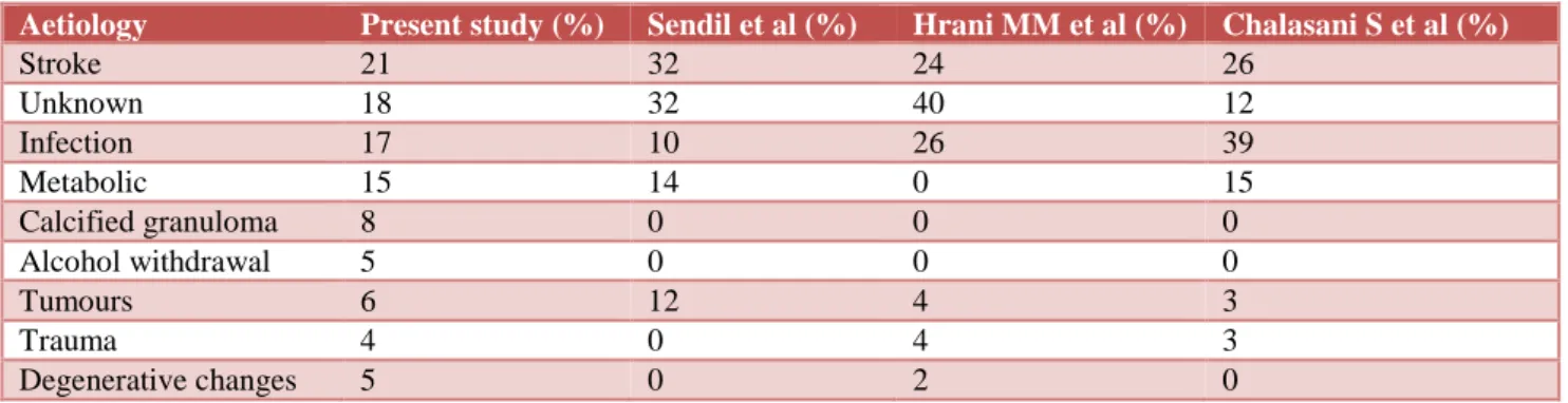
Aetiology Present study(%) Sendil et al (%) Hrani MM et al(%) Chalasani S et al(%)
Stroke 21 32 24 26
Unknown 18 32 40 12
Infection 17 10 26 39
Metabolic 15 14 0 15
Calcified granuloma 8 0 0 0
Alcohol withdrawal 5 0 0 0
Tumours 6 12 4 3
Trauma 4 0 4 3
Degenerative changes 5 0 2 0
Etiology
Most common cause of seizure in present study was stroke followed by infection followed by metabolic cause. Stroke was leading cause of seizures in present study accounting for 21%. Infection was next leading cause of seizures accounting for 17%. Metabolic cause accounts for 15%. Calcified granuloma accounts for 8% and alcohol withdrawal accounts for 5%. Tumours accounts for 6%. Gliotic changes accounts for 5%. This was contrast to study by Hirani et al where seizure due to unknown cause was most common 40%.7 This may be
because the study involved only 50 participants. However, Sendil et al did a similar study with 50 cases and found that post stroke seizures and seizures due to unknown cause were most common and shared equal proportion.6 However Chalasani S et al in his study has
shown infections were common cause of seizures in their study.8 Among infections Neurocysticercosis was most
common. This was a bit contrast to present study where bacterial meningitis was most common cause of seizure
due to infections. Table 8 gives a summary of etiology of seizures among various studies.
Idiopathic seizures were seen commonly in 19-29 years’ age group. Post stroke epilepsy and seizures due to metabolic cause were seen commonly in 50-59 years’ age group. Seizures due to unknown cause were seen commonly in females. Seizures due to alcohol withdrawal and calcified granuloma were seen commonly in males. Majority of the Indian studies have no mention of alcohol withdrawal seizures. This may be due to their low incidence in the pertaining areas. In present study 5% of seizures were due to alcohol withdrawal. Sander et al with a larger sample size than present study in UK have reported around 9% of seizures due to alcohol withdrawal.11 A larger sample size might have shown a
Karnataka, India showed that road traffic accidents constituted for 24% of seizures.12 This difference may be
partly due to the reason that our centre is located in rural area and in the study done by Joseph et al urban centre was the base.12
CONCLUSION
Based on the results and the methodology employed, we have concluded that the predominant seizure type in present study was GTCS. Participants with cardioembolic stroke had rheumatic heart disease and they developed seizures. Ischemic infarct was major cause of post-stroke seizures in present study. Hypoglycemia was major cause seizures due to metabolic cause in present study. Patents with alcohol withdrawal were predominantly affected by GTCS. Participants with calcified granuloma were predominantly affected by focal seizures. Bacterial meningitis was major cause of seizures due to infections in present study. Eight participants suffered from age related epileptic seizures during infancy. They were off seizure medications for more than 10 years. They were considered having new onset seizures and were not started on treatment as they had only single seizures.
ACKNOWLEDGEMENTS
Authors would like to thank Almighty and professors and colleagues for helping them in this work.
Funding: No funding sources Conflict of interest: None declared
Ethical approval: The study was approved by the institutional ethics committee
REFERENCES
1. Fisher RS, Cross JH, French JA, Higurashi N, Hirsch E, Jansen FE, et al. Operational classification of seizure types by international league against epilepsy (Internet). Available from http://www.ilae.org/visitors/centre/documents/Classi ficationSeizureILAE-2016.pdf.
2. International league against epilepsy. Definition of epilepsy 2014. (Internet). Available from http:// www.ilae.org/ visitors/ centre/ Definition-2014.cfm. 3. Santhosh NS, Sinha S, Satishchandra P. Epilepsy: Indian Perspective. Ann Indian Acad Neurol. 2014;17(1):S3-11.
4. Shinnar S. The new ILAE classification. Epilepsia. 2010;51(4):715-7.
5. Murthy J, Narayanan J. New-onset acute symptomatic seizure in a neurological intensive care unit. Neurol India. 2007;55(2):136.
6. Sendil G, Kumar AN, Kumar MV. Late onset shake-etiology at stake- a prospective study. Int J Sci Stud. 2014;2(1):20-4.
7. Hirani DMM, Shrivastava DS. Clinical profile of new onset seizures in adults. Indian J Appl Res (Internet). 2016;5(5)19-21.
8. Chalasani S, Kumar MR. Clinical profile and etiologic evaluation of new onset seizures after age 20 years. IOSR-JDMS. 2015;14(2):97-101.
9. Benbadis SR, Wolgamuth BR, Goren H, Brener S, Fouad-Tarazi F. Value of tongue biting in the diagnosis of seizures. Arch Intern Med. 1995;155(21):2346-9.
10. Brigo F, Nardone R, Bongiovanni LG. Value of tongue biting in the differential diagnosis between epileptic seizures and syncope. Seizure- Eur J Epilepsy. 2012;21(8):568-72.
11. Sander JW, Hart YM, Johnson AL, Shorvon SD. National general practice study of epilepsy: newly diagnosed epileptic seizures in a general population. Lancet Lond Engl. 1990;336(8726):1267-71. 12. Joseph N, Kumar GS, Nelliyanil M. Pattern of
seizure cases in tertiary care hospitals in Karnataka state of India. Ann Indian Acad Neurol. 2013;16(3):347-51.