Original Research Article
Histopathological spectrum of ophthalmic lesions in Chhattisgarh:
study from a tertiary care centre
Kujur P., Mulkalwar M.*, Kosam S.
INTRODUCTION
Ophthalmic pathology is a subspecialty of cellular pathology which deals with the diagnosis and characterization of eye diseases.1 Malignant orbital tumors are generally treated by orbital exenteration. The term enucleation applies to the removal of the eye alone, sparing the conjunctiva and eyelids. The term evisceration refers to the evacuation of intraocular contents and replacement with space occupying material to preserve motility of a cosmetically acceptable
prosthesis.2 Ophthalmic pathologists study tissues excised by ophthalmologists through evisceration and enucleation and other ophthalmic biopsies.3 The orbit is composed of the globe, extra ocular muscles, fat, vascular, nerve, glandular and connective tissues of neuroectodermal origin.4 The neoplastic orbital lesions show wider range of pathological findings and further compounded by the patients’ fear of loss of vision pose as a major challenge to ophthalmologists.5 Ophthalmic histology techniques differ from those of normal tissue in fixation, processing and sectioning. Ophthalmic pathologist should be trained
ABSTRACT
Background: Ophthalmic pathology is a subspecialty of cellular pathology which deals with the diagnosis and characterization of eye diseases. Ophthalmic pathologists study tissues excised by ophthalmologists through evisceration and enucleation and other small biopsies from various ophthalmic surgeries. Ophthalmic lesions include a wide spectrum of lesions ranging from benign, premalignant to malignant lesions. The diagnosis of these lesions is based on the clinical as well as histopathological features.
Methods: The present study was an observational prospective study conducted on a 62 ophthalmic biopsies received in Histopathology laboratory, Department of Pathology, Pt. J.N.M. Medical College and Dr. B.R.A.M. Hospital, Raipur, Chhattisgarh, India, between March 2016 and October 2016. Institutional ethics committee of Pt. J.N.M. Medical College, Raipur, India has approved this study. Informed written consent was collected from the patients or their relatives.
Results: Out of 62 cases of ophthalmic biopsies, 38 (61.29%) were non-neoplastic and 24 (38.71%) were neoplastic lesions. Among non-neoplastic lesions, inflammatory lesions of lacrimal sac were more common 16 (42%) followed by conjuctival lesions 10 (26.3%). Among the neoplastic lesions, retinoblastoma was the most common malignancy 11 (45.83%).
Conclusions: The most common ocular malignancy was retinoblastoma; among non-neoplastic disorder, chronic dacryoadenitis followed by benign lesions of conjunctiva. Some ocular masses composed of rhino sporangium involving conjunctiva, lid or lacrimal sac.
Keywords: Adnexal, Leukocoria, Ocular lesions, Retinoblastoma
Department of Pathology, Pt. J. N. M. Medical College, Raipur, Chhattisgarh, India
Received: 05 November 2016
Accepted: 09 November 2016
*Correspondence:
Dr. Mulkalwar Madhubala,
E-mail: mulkalwarmadhu@gmail.com
Copyright: © the author(s), publisher and licensee Medip Academy. This is an open-access article distributed under the terms of the Creative Commons Attribution Non-Commercial License, which permits unrestricted non-commercial use, distribution, and reproduction in any medium, provided the original work is properly cited.
to identify disease processes unique to eye and to demonstrate them on a microscopic slide along with the pupil and optic nerve.6 The present study is aimed to correlation morphological and clinico-pathological features of ophthalmic lesions, to know the pattern of prevalence of various ophthalmic lesions in Chhattisgarh, India.
METHODS
A total of 62 ophthalmic biopsy specimens referred Histopathology and Cytology laboratory, Department of Pathology, Pt. J. N. M. Medical College and Dr. B. R. A. M. Hospital, Raipur, Chhattisgarh, India, between March 2016 and October 2016 included in this study. Institutional ethics committee of Pt. J. N. M. Medical College, Raipur, India has approved this study. Informed written consent was collected from the patients or their relatives. All specimens were subjected to the fixation, grossing, processing and staining with Harris haematoxylin and Eosin stain. Microscopic evaluation was done on each specimen along with the clinical data on hand. The study was done to report the spectrum and frequency of different ocular and adnexal lesions in a tertiary care hospital in Chhattisgarh, India.
RESULTS
The site specific distributions of the ophthalmic biopsies were given in Figure 1. Of the 62 ophthalmic biopsies, 17
(27.42%) specimens were from globe lesions including retina and choroid, 16 (25.8%) from lacrimal gland, 14 (22.58%) from conjunctiva lesions, 13 (20.97%) from eye lid and 2 (3.23%) cases from cornea. The age range was from 5 months to 86 years with the mean age of 32.53 years.
Out of 62 biopsies evaluated, 33 (53.22%) were of male and 29 (46.78%) were of female [Figure 2(A)]. About 38 (61.29%) ophthalmic biopsies were found to be non-neoplastic and 24 (38.71%) were non-neoplastic in nature [(Figure 2(B)]. Frequencies of distribution of various non- neoplastic and neoplastic ocular lesions were documented in Table 1.
Figure 1: Site specific distributions of the ophthalmic biopsies.
Table 1: Frequency of distribution of non neopalstic and neoplastic ocular lesions.
Diagnosis Site of biopsy
Globe Conjunctiva Cornea Eye- lid Lacrimal apparatus Non-neoplastic
Inflammatory 2 4 - - -
Cystic - 2 - 4 -
Infectious (rhinosporidium) - 2 - 1 1
Granulomatous (tubercular) - 1 - - -
Chalazion - - - 2 -
Retinal detatchment 2 - - - -
Pterigium - 1 - - -
Ulcer - - 1 - -
Chronic dacryoadenitis - - - - 14
Recurrent dacryoadenitis - - - - 1
Neoplastic
RB 11 - - - -
Sebaceous cell carcinoma - - - 3 -
SCC - 2 - - -
BCC - - - 1 -
NHL 1 - - - -
MM 1 - - - -
Squamous papilloma - 1 - - -
Neurofibroma - - - 1 -
Dysplastic changes - 1 1 1 -
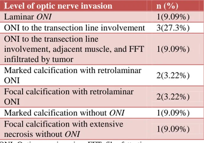
Among non-neoplastic lesions, chronic daryoadenitis was found highest 14 (36.8%). Among all malignancies 11 (45.83%) specimens were diagnosed as retinoblastoma. Complete histopathological spectrum of retinoblastoma based on optic nerve invasion was shown in Table 2.
Figure 2: Distributions of the ophthalmic biopsies (A) based on sex; (B) neoplastic status.
Table 2: Histopathological spectrum retinoblastoma based on optic nerve invasion.
Level of optic nerve invasion n (%)
Laminar ONI 1(9.09%)
ONIto the transection line involvement 3(27.3%) ONIto the transection line
involvement, adjacent muscle, and FFT infiltrated by tumor
1(9.09%)
Marked calcification with retrolaminar
ONI 2(3.22%)
Focal calcification with retrolaminar
ONI 2(3.22%)
Marked calcification without ONI 1(9.09%)
Focal calcification with extensive
necrosis without ONI 1(9.09%)
ONI: Optic nerve invasion; FFT: fibrofatty tissue.
Photomocrographs of some the lesions showed focal to marked calcification and necrosis with or without optic nerve infiltration by tumor as depicted in Figure 3, 4 and 5. Second most common malignancy was squamous in origin; of which two cases of frank WDSCC (Figure 6), one squamous papilloma; remaining showed the spectrum of range from mild, moderate to markedly dysplastic epithelium. Sebaceous cell carcinoma was the commonest malignancy in patients with eye lid mass which is simulating the lipogranulomatous lesions like chalazion mainly when presented recurrently (Figure 7), the other malignancy was the basal cell carcinoma of eye lid. One case of NHL (Figure 8) and another case of malignant melanoma of choroid were diagnosed in excised eye ball specimens. A single case was presented as firm to hard mass over upper eye lid and patient
complaining of foreign body sensation; on
histopathlogical examination it proved out to be a neurofibroma. One specimen of lacrimal sac lesion showed mass composed of rhinosporangim at various
maturation stages and background elicited intense granulomatous-giant cell reaction (Figure 9).
Figure 3: Photomicrograph of retinoblastoma shows tumor invasion.(A) Up to lamina cribrosa (marked
with star); (B) another case showing retrolaminar optic nerve invasion (H and E-100X).
Figure 4: Photomicrograph of retinoblastoma shows malignant cell arrangement.(A) Basophilic cells are
arranged around the lumen of tumor vessels, perivascular pseudorosettes with necrosis; (B) Another case showing fleurettes (H and E-400X).
Figure 5: Photomicrograph of retinoblastoma shows advance changes.(A) calcifications involving choroid and retina; (B) extensive necrosis and hemorrhages
Figure 6: Photomicrograph shows well differentiated squamous cell carcinoma. Squamous cell carcinoma
demonstrates enough differentiation to tell that the cells are of squamous origin. The cells are pink and
polygonal in shape show pleomorphism, with hyperchromatic nuclei and malignant squamous
pearls (H and E-400X).
Figure 7: Photomicrograph of sebaceous cell carcinoma. Intracytoplasmic vacuolation within the
neoplastic cells were seen (H and E-400X).
Figure 8: Photomicrograph of non-hodgkin’s lymphoma. Monomorphic population of malignant
round cells were seen (H and E-400X).
The conjuctival lesions attributed mainly by inflammatory infiltrates, some were cystic in nature showing sebaceous cyst, inclusion cyst etc. Among infectious, two cases were showing conjuctival mass composed of rhinosporidium and one case composed of chronic inflammatory cells along with Langhan’s type of giant cells with granuloma (tubercular in nature). Eye lid mass mainly cystic in nature namely two cases of epidermal inclusion cyst; sebaceous cyst and dermoid cyst had reported one case each. Chalazion was reported in two cases of lid and its most strong differential was sebaceous cell carcinoma; one lid mass was also showing rhinosporidiosis. Two cases involving whole globe, composed of mixed chronic inflammatory infiltrates, one
diagnosed as panohthalmitis and other as
endophthalmitis. One case of glaucoma showed retinal detachment and another traumatic case showed retinal detachment along with detachment of sclera. One case of corneal ulcer was also diagnosed showed complete destruction of corneal membrane.
Figure 9: Photomicrograph biopsy from lacrimal sac mass. Rhinosporidiosis at various stages with
granulomatous (giant cell) reaction was seen (H and E-100X).
DISCUSSION
Neoplastic lesions of ocular and adnexa have relatively high morbidity and mortality. Diagnosing the accurate lesions and for commenting on extent and type of lesion along with surgical margin which can guide for further therapy of patients. In the present study non-neoplastic lesions were outnumbered of neoplastic lesions. Non-neoplastic lesions constituted by chronic dacryoadenits followed by benign and inflammatory lesions of conjunctiva. Out of all non -neoplastic lesions, four cases (6.45%) were showed infection from Rhinospordium Seeberi. Ocular rhinosporidiosis due to this organism are not so common in India, occur particularly in endemic areas. It affects predominantly one of the mucous membranes of the nose and nasopharynx but occasionally other structures, such as conjunctiva, lacrimal sac, eyelids etc. Our study document different ocular and adnexal structures involved by rhinosporidium i.e. conjunctiva in two cases, lid mass in one case and lacrimal sac in one case. A great deal of research related ocular biopsies revealed that the ocular rhinosporidiosis is one of the major ocular infections in different areas. The most common site of ocular rhinosporidiosis is the lower palpebral conjunctiva in Tamil Nadu nasal and nasopharyngeal sac in Orissa, lacrimal sac in Chhattisgarh.7-10
loss of vision and proptosis similar to other studies.13-16 Our study report extensive necrosis in 9.09% cases of retinoblastoma, but much higher frequency of necrosis (25.58%) was reported in another study.17 On histopathology optic nerve invasion was found in 81.8% cases of retinoblastoma, but much lower frequency of optic nerve invasion (32%) was reported.18 Other histopathlogical findings like rosette, perivascular pseudorosette, fleurette formation, calcification, necrosis and haemmorrhage etc. were similarly found as other studies. Squamous origin of neoplasic lesions were found in our study is showing coherence with the other studies.19-21
CONCLUSION
In summary our study revealed that the retinoblastoma is the commonest ocular malignancy and most of them are presented with either lekocoria or blindness. Furthermore, diagnosing sebaceous cell carcinoma is difficult because it simulates chalazion which is a benign lesion. Among non-neoplastic disorder, chronic dacryoadenitis (adnexal) followed by benign lesions of conjuctiva (ocular) and few of them get infected due a fungal organism called
Rhinosporidium Seeberi, which is endemic in some
regions of our Chhattisgarh, India. So in our region, all ophthalmic lesions removed surgically should be sent for histopathological examination to establish the correct diagnosis which helps in further guidance of therapy to patients.
Funding: No funding sources Conflict of interest: None declared
Ethical approval: The study was approved by the institutional ethics committee
REFERENCES
1. Li YP, Li B. Development of ophthalmic pathology and precision medicine. Chinese J Ophthalmol. 2016;52(10):724-7.
2. Fletcher CDM. Diagnostic histopathology of tumors. 4th Edition. Elsevier Saunders, Philadelphia. 2013. 3. Lu X, Ng DS, Zheng K, Peng K, Jin C, Xia H. Risk
factors for endophthalmitis requiring evisceration or enucleation. Scientific Reports. 2016;6:281-300. 4. Ali SM, Shin GA. Abnormal extraocular muscle
anatomy in a case of Williams-Beuren syndrome. Am Asso Pedia Ophthalmol Strabismus. 2009;13(2):196-7. 5. Dymshits LA. Orbital lesions in acute neoplastic
leukemias in children (chloroma, sarcoleukemia of the orbit). Vestnik Oftalmologii. 1961;74:35-45.
6. Oria AP, Oliveira AV, Pinna MH, Martins Filho EF, Lima EA, Peixoto TC. Ophthalmic diagnostic tests, orbital anatomy, and adnexal histology of the broad-snouted caiman (Caiman latirostris). Veterinary Ophthalmology. 2015;18(1):30-9.
7. David SS, Sivaramasubrahmanyan P. Ocular rhinosporidiosis - (a study of twenty one cases). Indian J Ophthalmol. 1973;21(4):204-7.
8. Moses JS, Balachandran C, Sandhanam S, Ratnasamy N, Thanappan S, Rajaswar J. Ocular rhinosporidiosis in Tamil Nadu, India. Mycopathologia. 1990;111(1):5-8.
9. Nanda BK, Naik UP, Panda GK, Behera G. Rhinosporidiosis in western Orissa. J Indian Med Asso. 1969;53(10):489-92.
10. Mukherjee P, Shukla I, Deshpande M, Kher P. Rhinosporidiosis of lacrimal sac. Indian J Ophthalmol. 1982;30(5):513-6.
11. Moll AC, Imhof SM, Meeteren AYNS, Boers M. At what age could screening for familial retinoblastoma be stopped? A register based study 1945-98. British J Ophthalmol. 2000;84(10):1170-2.
12. Park SJ, Woo SJ, Park KH. Incidence of retinoblastoma and survival rate of retinoblastoma patients in Korea using the Korean National Cancer Registry database (1993-2010). Investigative Ophthalmol Visual Sci. 2014;55(5):2816-21.
13. Waddell KM, Kagame K, Ndamira A, Twinamasiko A, Picton SV, Simmons IG. Clinical features and survival among children with retinoblastoma in Uganda. British J Ophthalmol. 2015;99(3):387-90.
14. Chebbi A, Bouguila H, Boussaid S, Aleya N, Zgholi H, Malek I, et al. Clinical features of retinoblastoma in Tunisia. Journal Francais Ophtalmol. 2014;37(6):442-8.
15. Saakian SV, Miakoshina EB, Krichevskaia GI,
Slepova OS. Retinoblastoma and
"pseudoretinoblastoma" in children: clinical, tomographic and serological features. Vestnik Oftalmologii. 2014;130(1):18-24.
16. Sethi S, Pushker N, Kashyap S, Sharma S, Mehta M, Bakhshi S, et al. Extraocular retinoblastoma in Indian children: clinical, imaging and histopathological features. Int J Ophthalmol. 2013;6(4):481-6.
17. Chong EM, Coffee RE, Chintagumpala M, Hurwitz RL, Hurwitz MY, Barrios P. Extensively necrotic retinoblastoma is associated with high-risk prognostic factors. Archives Pathol Lab Med. 2006;130(11):1669-72.
18. Reddy SC, Anusya S. Clinical presentation of retinoblastoma in Malaysia: a review of 64 patients. Int J Ophthalmol. 2010;3(1):64-8.
19. Sjo LD. Ophthalmic lymphoma: epidemiology and pathogenesis. Acta Ophthalmol. 2009;87(1):1-20. 20. Shields CL, Shields JA. Ocular surface squamous
neoplasia: from blue skies to blue dyes we still need our ophthalmic pathologists. JAMA Ophthalmol. 2015;133(11):1321-2.
21. Matsuo T, Ohara N, Namba Y, Koshima I, Ida K, Kanazawa S. Ophthalmic artery embolization as pretreatment of orbital exenteration for conjunctival
squamous cell carcinoma. Cardiovascular
Interventional Radiol. 2009;32(3):554-7.