PCR and RFLP Analysis for Identification and Typing of
Helicobacter pylori Strains from Gastric Biopsy Specimens
F. Navabakbar
1,*and R. Salehi
21
Department of Bacteriology, School of Medicine, Isfahan University of Medical Sciences, Isfahan 81744-176, Islamic Republic of Iran
2
Department of Genetics and Molecular Biology, School of Medicine, Isfahan University of Medical Sciences, Isfahan 81744-176, Islamic Republic of Iran
Abstract
Helicobacter pylori
is the causative agent of chronic gastritis and peptic ulcer
diseases and is also a risk factor for gastric cancer. Since culture of
Helicobacter
pylori
is relatively insensitive and cumbersome, PCR-based molecular detection
and typing of this organism is gaining importance for strain differentiation. The
aim of present study was to apply molecular methods for detection and
genotyping of
H. pylori
directly on gastrointestinal biopsy. For this purpose 79
samples were tested by bacteriological methods including Gram stain, urease test
and culture in a selective medium. Genomic DNA was directly extracted from
gastric biopsy specimens followed by PCR amplification using
ureC
primers.
PCR products from positive samples were purified and digested using three
different restriction endonucleases, AluI, MboI
and CfoI. The numbers of positive
samples detected by bacteriological methods were 71, 40 and 73 using Gram
stain, culture and urease test respectively. Through PCR assay 64 samples were
positive with sensitivity of about 92 and specificity of 100%. The specificity and
sensitivity of the PCR assay was higher than that of all bacteriological methods
applied. RFLP performed on the positive samples and five patterns for AluI, five
patterns for MboI and four patterns for CfoI detected. The patterns were classified
based on the number of produced bands in gel electrophoresis. From our study it
is concluded that the PCR-based RFLP technique would be an useful approach for
identification and genotyping of specific
H. pylori
strains directly in gastric
biopsy specimens without culture.
Keywords:Helicobacter pylori; UreC; PCR; RFLP
* E-mail: [email protected]
Introduction
Helicobacter pylori is a gram-negative
microaero-philic organism that colonizes human gastric mucosa [13]. It has been proved that H. pylori is an etiologic
is also a risk factor for gastric cancer [15]. The World Health Organization has classified H. pylori as a class I
carcinogen [6]. Epidemiological studies have shown that H. pylori infection occurs world wide at a high
prevalence rate [7].
Recurrence of infection after apparent eradication has also been reported and is associated with recurrence of ulcers. However, it is unclear whether the recurrence of ulcers following H. pylori eradication therapy is due to
recrudescence of the previous infection or to exogenous re-infection by another strain [17]. An accurate method for the detection and differentiation of H. pylori strains
in patients both before and after therapy is therefore of great importance for diagnosis, monitoring of treatment, and reduction of the long-term consequences of continued but undetected disease [2].
Attempts to differentiate H. pylori strains have been
made using a variety of conventional typing schemes, like haemagglutination, biotyping, cytotoxin activity, plasmid profiles and immunobloting. However, each of these methods due to some inherent difficulties did not gain widespread use for precise differentiation of H. pylori strains [2,8,11].
Several nucleic acid techniques, such as restriction endonuclease analysis of genomic DNA and southern blot hybridization with rRNA gene probes have been applied to type H. pylori clinical isolates from different
patients. The patterns obtained by these methods were complex and difficult to interpret, particularly for large scale analysis of clinical isolates [5,14,16]. Recently, PCR-based restriction fragment length polymorphism (RFLP) analysis and randomly amplified polymorphic DNA (RAPD) methods have been developed for typing of H. pylori clinical isolates. PCR based RFLP schemes have been used to analyze H. pylori genes, which
encode urease and its accessory proteins, including UreA, UreB, UreC and UreD [1,5,9].
The published works have shown that PCR-based RFLP typing is a rapid, sensitive method and capable of discriminating among clinical isolates, but the studies were limited to H. pylori isolates cultured from gastric
biopsy specimens [13].
In this study, we report a PCR-based RFLP analysis to differentiate H. pylori strains directly from gastric
biopsy specimens without any attempt to culture the bacteria, which is a time consuming and labor intensive task.
Materials and Methods
Bacterial Strains
The type strain ATCC 26694 (kindly donated by Dr.
Leo. Smmeet) was used as positive control in this study.
Proteus mirabilis and Klebsiella pneumoniae (clinical
isolate) as an urease positive and E. coli (clinical
isolate) as an urease negative micro organism, were used as negative control to test the specificity of PCR assay.
Gastric Biopsy Speciemens
Gastric biopsy specimens were obtained from 79 patients, 36 women and 43 men with age range 16 to 80 years (mean age 42.2 years), who underwent endoscopy for upper gastrointestinal complains at the Alzahra hospital center. Two gastric biopsy specimens were collected from similar location in the antrum and fundus of the stomach from each patient. One piece of each specimen was examined by bacteriologic tests (Gram stain, urease test and culture). Another biopsy specimen was preserved immediately at −70°C for molecular analysis.
For bacteriological tests, the gastric biopsy sample was homogenized and one part cultured on BHI agar containing antibiotic supplement (Merck) with 5% pack red blood cell (obtained from Blood Transfusion Organization) plus 7% fetal calf serum (Biogen). Plates were incubated at 37°C in microaerophilic atmosphere with 10% CO2, 5% O2 and 85% N2. Plates were opened
after 5 days. Organisms were identified as H. pylori based on colony morphology, Gram stain, and positive catalase and rapid urease tests.
Processing of the Samples for PCR Assay
The frozen biopsy specimen was thawed, crushed and then DNA was extracted using high pure DNA extraction kit (Roche, Germany) according to the manufacturer instruction. Same kit was used for extraction of genomic DNA from standard strains and negative controls. The extracted DNA then quantified through measurement of its OD260 by Biophotometer
(Eppendorf, Germany).
Primers
UreC (glmM) gene in H. pylori, which encodes a
U Amplitaq Gold (Perkin Elmar, USA) and 0.2 µg of template DNA sample. Thirty cycles of amplification were performed in a thermal cycler (Omnigen, UK). Each cycle consisted of 45 seconds denaturation step at 94°C, 30 sec annealing step at 59°C and 90 sec extension step at 72°C followed by final extension step for 10 min at 72°C to ensure full extension of the partially polymerized products.
Subsequently PCR products were loaded on 1.5% agarose gel, containing 0.5 µg ethidium bromide/ml, electrophoresed for 60 min at 100 V, and then visua-lized under UV light using gel documentation system.
Restriction Digestion of Amplified DNA
The amplified products were purified using PCR products purification kit (Roche, Germany) according to the user manual. Fifteen µl of purified PCR products were digested with 10 U of CfoI, MboI and AluI restriction enzymes separately in an appropriate buffer solution recommended by the manufacturer for at least 3 h at 37°C.
Ten µl of digested products analyzed by electrophoresis on 2.5% agarose gels (Gibco), and the corresponding bands were examined by phototrans-illumintor and compared with a standard 100 bp ladder (XIII Roche) for size estimation. The number of bands produced by each restriction enzymes used grouped together by using the first letter of enzyme name followed by a number which was indicative of the corresponding fragments produced after digestion.
Results
Sixty four out of 79 cases were positive by PCR assay at sensitivity of 91.4% and specificity of 100%. 73 cases were positive using rapid urease test and sensitivity and specificity of 100% and 66.7% were achieved respectively. 71 cases detected with Gram stain with sensitivity of 88.9% and specificity of 100% and 40 cases were positive on culture at sensitivity of 57.1% and specificity of 100%.
PCR amplification of H. pylori type strain 26694
genomic DNA with ureC primers produced a 417 bp
fragment. The gastric biopsy specimens DNA also yielded a comparable products same as that of H. pylori
type strains 26694 (Fig. 1). DNA from urease positive species not related to H. pylori (Klebsiella pneumoniae
and Proteus mirabilis) and urease negative species (E. coli) subjected to PCR amplification using the same
primer sets and PCR conditions. No amplified products were detected in any of above mentioned cases.
Figure 1. PCR amplification products of H-pylori ureC gene. Lanes: 1-11 biopsy samples, 12 positive control, 13 negative control and M, molecular weight marker (XIII Roche).
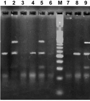
Figure 2. PCR-based RFLP patterns from the gastric biopsy specimens digested with two different restriction enzymes. Lanes: 1-6 RFLP patterns obtained through application of AluI, M, molecular weight marker (100 bp ladder) and 7-9 RFLP patterns for MboIs.
RFLP Analysis
The products of 60 PCR Positive biopsy specimens were digested with three restriction enzymes, AluI, MboI and CfoI separately. Five patterns for MboI, five patterns for AluI and 4 restriction patterns for CfoI were observed. The patterns were classified based on the number of bands detectable by gel electrophoresis (Fig. 2).
A1M1C1 patterns, six samples A2M2C2, for two patterns
of A3M4C2 and A2M3C2 each one included five samples,
four samples A3M2C2 and there samples A3M3C2.
Those RFLP products that were not matched with any of the above categories classified as distinct RFLP patterns. 29 samples were included in this group.
Discussion
In recent years, many investigators using molecular techniques have revealed that H. pylori possess a
remarkable degree of genetic diversity, which is closely related with its epidemiological and pathological characteristics and dynamic of transmission [13]. The PCR-based RFLP analysis has been widely accepted for typing and differentiation of H. pylori strains from
clinical specimens [13,19]. This method has been used to analyze conserved H. pylori genes, especially those
encoding urease structural and accessories proteins [9,10,16,18].
In our study, a PCR assay using primer pairs derived from the ureC gene employed to amplify a 417 bp
fragment from 64 gastric biopsy specimens. Other urease positive bacterial species such as Klebsiella pnumonia and Proteus mirabilis genomes were not
amplified by this PCR assay. We developed a successful PCR based RFLP assay for differentiation of H. pylori
strains directly from gastric biopsy specimens without culture. By using this method we have detected five, five, and four distinct patterns by application of three different restriction endonuclease enzymes, CfoI, MboI and AluI, respectively on 58 biopsy specimens.
In one of patient with chronic gastritis, in both obtained specimens, one from antrum and the other from fundus. In both the bacteriological tests comprising of Gram stain, rapid urease and culture were positive. However in the bacterial culture two different colonies were observed and the RFLP patterns demonstrated two distinct patterns indicating the presence of two different strain or subtype.
It has been previously reported that two strains of H. pylori have been found in the stomach of some patients,
or even three distinct strains reported to be found in some patient’s clinical isolates [4,10,16]. Examination of large numbers of isolates from individual patients will permit a better quantitation of the extent of multiple infections.
Although several PCR-based RFLP protocols have been developed for the differentiation of H. pylori
strains, most of the studies have been carried out on H. pylori isolates from gastric biopsy specimens. Culture of H. pylori is time consuming and not very sensitive. Our
PCR-based RFLP protocol provides a means for direct
detection and differentiation of H. pylori strains in
gastric biopsy specimens without culture. Clayton, et al.
[5] also applied the PCR-RFLP analysis method directly on biopsy samples and stated that it is a reliable method for epidemiological investigation on the transmission of this pathogen. This shows that PCR-based RFLP assay maybe useful as a primary approach for identification of specific H. pylori strains in gastric biopsy specimens
without culturing. Several studies have confirmed that PCR-based RFLP analysis of the ureC gene can
differentiate H. pylori clinical isolates. Fujimoto et al.
[10] have shown that digestion of 820-bP PCR amplified portion of H. pylori ureC gene by the
restriction enzymes HhaI, MboI and MseI resulted in 10, 10, and 11 different patterns, respectively. The 25 H. pylori clinical isolates, however, could be grouped in to
25 distinct RFLP patterns when the three restriction enzymes were combined.
Chuanfu et al. [4] have also shown that digestion of a
1,179 bp PCR amplified fragment of ureC gene of H. pylori digested by the same restriction endonuclease
generated 11, 10, and 6 digestion patterns respectively. From our study it is concluded that, the PCR-based RFLP technique is a useful approach for identification of specific H. pylori strains in gastric biopsy specimens
without any need of time taking and labor-intensive culture method.
It is also helpful for epidemiological studies. In addition, by using this method we can determine reinfection, infection with new strain, recrudescence or drug resistance in H. pylori infected patients.
References
1. Akopynaz N., Bukanov N.O., Westblom T.U., and Berg
D.E. PCR-based RFLP analysis of DNA sequence diversity in the gastric pathogen Helicobacter pylori. Nucleic Acid Res., 20: 6221-6225 (1992).
2. Brenner H., Bode G., and Boeing H. Helicobacter pylori infection among offspring of patients with stomach cancer. Gastroenterology, 118: 31-35 (2000).
3. Burnie J.P., Lee W., Dent J.C., and McNulty C.A.M.
Immunoblot fingerprinting of Campylobacter pylori. J. Med. Microbiol., 27: 153-159 (1988).
4. Chuanfu L.I., Tuanzhu H.A., Chi D.S., Ferguson D.A., et al. Differentiation of Helicobacter pylori strains directly from gastric biopsy specimens by PCR-based restriction fragment length polymorphism analysis without culture. Ibid., 31: 1420-1425 (1997).
5. Clayton C.L., Kleanthous H., et al. Evaluation of
fingerprinting methods for identification of Helicobacter pylori strains. Eur. J. Clin. Microbiol. Infect. Dis., 10: 1040-1047 (1991).
6. Emad M, Omar El., et al. Increased prevalence of
22-30 (2000).
7. Fawcett P.T. and Gibney K.M. Helicobacter pylori can be induced to assume the morphology of Helicobacter heilmannii. Journal of Clinical Microbiology, 37(4): 1045-10480 (1999).
8. Figura N., Guglielmett P., et al. Cytotoxin production by Campylobacter pylori strains isolated from patients with peptic ulcers and from patients with chronic gastritis only. Ibid., 27: 225-226 (1989).
9. Foxall P.A., Hu L.T., and Mobley H.L.T. Use of
polymerase chain reaction-amplified Helicobacter pylori urease structural genes for differentiation of isolates. Ibid., 30: 739-741 (1992).
10. Fujimoto S., Marshall B., and Blaser M.J. PCR-based
restriction fragment length polymorphism typing of Helicobacter pylori. Ibid., 32: 331-334 (1994).
11. Huang J., Symth C.J., et al. Hemagglutinating activity of Campylobacter pylori. FEMS Microbiol, 56: 109-112 (1998).
12. Kansau I., Raymond J., Bingen E., et al. Genotyping of Helicobacter pylori isolates by sequencing of PCR products and comparison with the RAPD technique. Res. Microbiol., 147: 661-669 (1996).
13. Mishra K.K., Srivastava S., Dwiredi P.P., et al. PCR-Based RFLP analysis of ureC gene for typing of Indian Helicobacter pylori strains from gastric biopsy specimens and culture. The Joumal of Microbiology, 40(4): 282-288
(2002).
14. Majewski S.I.H. and Good win C.S. Restriction
endonuclease analysis of the genome of Campylobacter pylori with a rapid extraction method: evidence for considerable genomic variation. J. Infect. Dis., 157: 465-471 (1988).
15. Nomura A., Stemmemann C., et al. Helicobacter pylori infection and gastric carcinoma among Japanese Americans in Hawaii. N. Egy J. Med., 325: 1132-1136 (1991).
16. Owen R.J., Bickley J. et al. Comparison of PCR-based restriction length polymorphism analysis of urease gene with rRNA gene profiling for monitoring Helicobacter pylori infection in patients on triple therapy. J. Clin. Microbiol., 32: 1203-1210 (1994).
17. Queiroz D.M., Dani R., Silval D., et al. Factors associated with treatment failure of Helicobacter pylori infection in a developing country. J. Clin. Gastroenterology, 35(4): 315-320 (2002).
18. Shortridge V.D., Stone G.G., et al. Molecular typing of Helicobacter pylori isolates from a multicenter U. S. Clinical trial by UreC restriction fragment length polymorphism. J. Clin. Microbiol., 35: 471-473 (1997). 19. Simsek I.S., Menevse S., and Sahin F.I. PCR and RFLP