C A S E R E P O R T
Open Access
Selective laser melted titanium implants: a new
technique for the reconstruction of extensive
zygomatic complex defects
Horatiu Rotaru
1*, Ralf Schumacher
2, Seong-Gon Kim
3and Cristian Dinu
1Abstract
The restoration of extensive zygomatic complex defects is a surgical challenge owing to the difficulty of accurately restoring the normal anatomy, symmetry, proper facial projection and facial width. In the present study, an extensive post-traumatic zygomatic bone defect was reconstructed using a custom-made implant that was made with a selective laser melting (SLM) technique. The computer-designed implant had the proper geometry and fit perfectly into the defect without requiring any intraoperative adjustments. A one-year follow-up revealed a stable outcome with no complications.
Keywords:Selective laser melting; Custom-made; Titanium implant; Zygoma reconstruction
Background
Craniofacial trauma, tumor resection and congenital de-formities can result in zygomatic bone deficiencies. The re-construction of the zygomatic bone is essential for the restoration of function and esthetics. The reduction of psy-chosocial morbidity is also an important issue [1]. Accurate restoration of the normal anatomy, symmetry, proper facial projection and facial width are the key points in orbito-zygomatic reconstruction [1].
Different surgical approaches had been described for the reconstruction of the zygomatic complex. These ap-proaches include osteotomy, autologous bone graft, free tissue transfer and the use of different alloplastic im-plants [2]. Autologous bone grafts are still considered the gold standard for the reconstruction of these defects [3]. However, donor site morbidity, limited bone avail-ability, unpredictable resorption rates, and residual de-formities remain important challenges [4]. Different types of alloplastic implants, such as metals [5], silicone [6], polymers [7], and hydroxyapatite-based products [8], have been used to replace autologous bone grafts. How-ever, the ideal alloplastic material has not yet been iden-tified [7].
Although stock-made implants are commercially avail-able in different sizes, these implants are of limited value for repairing acquired and unusual bony defects. Such im-plants fail to accurately fit the defects and hence result in outcomes that are associated with high revision rates [2,7]. In contrast, custom-made patient-specific implants that are produced using computer-aided design and manufac-turing (CAD/CAM) overcome these drawbacks [7]. Patient-specific implants shorten the operative time, re-duce the need for intraoperative implant adjustments and improve the clinical outcomes [9].
In this article, we present a case of post-traumatic zygomatic deficiency that has been successfully treated using a custom-made implant that was made with a se-lective laser melting (SLM) technique. After one year of follow-up, the implant exhibited good integration with no signs of infection or exposure. To the best of our knowledge, this case report is the first to describe a
zygomatic reconstruction utilizing a custom-made
implant that was created with the SLM technique.
Case presentation
A 43-year-old male patient presented to our department with a severe left midfacial post-traumatic deformity due to road traffic accident that occurred 6 years prior (Figure 1). Clinical examination of the left midface revealed the loss of the antero-posterior and medio-lateral (transverse) * Correspondence:[email protected]
1Department of Oral and Cranio-Maxillofacial Surgery,“Iuliu Hatieganu” University of Medicine and Pharmacy, Str. Motilor Nr. 33, 400001 Cluj-Napoca, Romania
Full list of author information is available at the end of the article
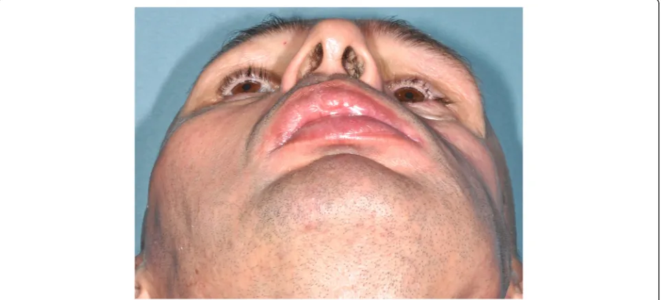
projections of the left zygomatic bone. A slight enophthal-mos was also present. The soft tissues of the area were hypotrophic in response to the initial injury (Figure 2). The clinical findings were confirmed on computerized tomo-gram (CT) images in axial and coronal plane (Figure 3).
The zygomatic-orbito-maxillary defect was recon-structed using a custom-made titanium implant for esthetic reasons. Fine-cut CT scanning of the region with 3-dimensional (3D) reconstruction was performed (Siemens Somatom Sensation, Erlangen, Germany). The CT data were imported into the MIMICS® software (Materialise,
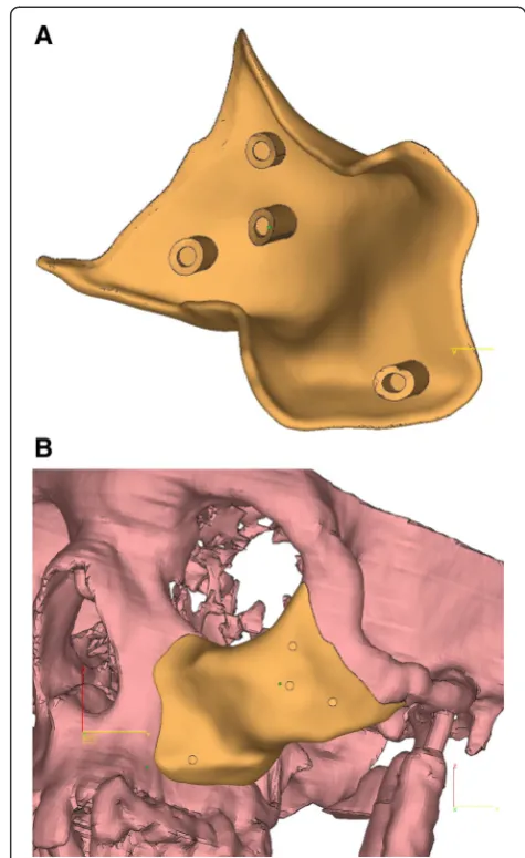
Leuven, Belgium), and a 3D virtual model of the im-plant was produced by “mirroring” the healthy side using Freeform Modeling Plus® (3D Systems, Sensable, Valencia, CA, USA) platform software. Because a full-density titanium SLM implant would have been too heavy for implantation, we decided to produce an im-plant in the form of a shell that was supported by the residual bone and fixation rods (Figure 4).
The virtual model was then printed into the 3D im-plant by SLM using commercially pure titanium Grade 2 (SLM-Solutions, Luebeck, Germany) and an SLM
Figure 1Initial trauma event during which the bone segments were lost.
250HL machine (SLM-Solutions). The physical model of the skull was printed in white acrylic resin using Multi-Jet-Printing (Objet Eden 250, Stratasys, Eden Prairie, MN,USA). The SLM implant was placed on the plastic model of the skull to verify proper matching and seating. No further mechanical processing was needed. Finally, the produced implant was post-processed by sand-blasting and drilling the screw holes and then cleaned and sterilized by autoclaving.
The implant was inserted into the planned position using a combination of mid-tarsal lower eyelid, hemicoro-nal and intraoral incisions. Proper seatings at the infraor-bital rim, zygomatic body and zygomatico-alveolar buttress were confirmed. The fixation was performed with three 2.0-mm titanium screws (Stryker®, Michigan, MI, USA) using the lag-screw principle (Figure 5). The space between the shell-shaped implant and the residual zygo-matic bone was filled with a cortico-cancellous iliac crest bone graft. The facial soft tissues were resuspended, and
the left temporal hollowing was corrected with a titanium mesh. The wounds were sutured in layers and dressed appropriately.
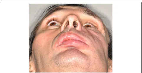
The patient received 1 g of ceftriaxone, 80 mg of genta-mycin, 0.5 g of metronidazole, and 100 mg of ketoprofen b.i.d. for 7 days. The postoperative course was uneventful, and the patient was discharged 8 days after the operation. The follow-ups at 1 month, 6 months and 1 year revealed no complications. At one year, the clinical examination re-vealed the persistence of a slight asymmetry in the zygo-matic regions (Figure 6), and a CT scan supported the good projection of the reconstructed site and the sym-metry between the two zygomas (Figure 7). We believe the residual asymmetry resulted from soft tissue atrophy. The CT scan also revealed good implant integration with ossification of the cortico-cancellous chips that were placed between the implant and the residual bone and no resorption of the residual zygoma.
Figure 3CT scan confirming the left zygomatic deficiency: (A)
Axial, (B) Coronal. Figure 4The virtual zygoma implant. (A)Internal side with
Discussion
Three-dimensional reconstruction of the zygomatic-orbito-maxillary complex is one of the most challenging procedures in craniofacial surgery. The normal anatom-ical contour and position of the zygomatic bone are crit-ical for the appearance of the face [10]. Here, we described a successful use of a custom-made SLM titan-ium implant for the reconstruction of a post-traumatic zygomatic bone defect. Although the bony symmetry was maintained at one-year follow-up (Figure 7), the pa-tient continued to exhibited soft tissue asymmetry. This asymmetry was most likely the result of buccal and zygomatic fat pad atrophy in response to the initial trauma and atrophy of the facial expression muscles sec-ondary to facial nerve palsy.
Because the residual zygomatic bone was deficient in volume and shape, an osteotomy alone would have not properly corrected the projections. The patient rejected the option of free tissue transfer. Thus the only available option was alloplastic implantation. Different materials can be used in cases of post-traumatic zygomatic defi-ciency, the advantages and drawbacks of each of these materials have been thoroughly documented in the lit-erature [6,8].
The development of CAD/CAM technology has opened new perspectives in the field of alloplastic implant produc-tion [11]. After three-dimensional reconstrucproduc-tion of a skull containing the defect, the future implant can be produced by“mirroring”the healthy side. Thus, the projection and symmetry of the zygomatico-maxillary complex can be re-established [7,11]. SLM is one of the CAD/CAM
Figure 5Intraoperative placement and fixation of the implant using 2.0-mm titanium lag-screws(arrows).
techniques that allows for the production of porous titan-ium parts that mimic bone structure [12]. Titantitan-ium is the most commonly used material in medical implants because it is highly biocompatible and integrates very well into tis-sues [13]. The mechanical properties of SLM titanium products are also within the ranges of the properties of bone [12]. These similarities are particularly important be-cause implant materials that are much stiffer than the bone can generate stress shielding, which can potentially lead to bone resorption or hinder bone regeneration [14]. Bone re-sorption caused by stress shielding is believed to contribute to the aseptic loosening of implants [15]. In contrast, the porous surfaces of SLM titanium parts have been demon-strated to be favorable for cell adhesion, migration and in-growth, and these properties result in strong bone-implant contact. When an implant is populated with osteogenic cells, these cells not only migrate on the surface of the im-plant but also inside the pores of the imim-plant [13]. Due to advantages of the titanium structures produced by SLM,
we designed and produced a patient-specific implant for zygoma recontouring using this technology (Figure 4). The implant fit perfectly into the defect, no corrections being needed at the time of surgery (Figure 5). Similar findings have been reported in the literature [7,9]. The implant was designed in the form of a shell and filled with cortico-cancellous chips from the anterior iliac crest to stimulate its integration. This implant behaved as reported in the lit-erature [13], no complications or side-effects occurred.
The left eye enophthalmos was not corrected because an-terior repositioning of the globe would have exposed a larger part of the cornea due to the presence of lagophthal-mos. The limitation of the presented reconstructive proced-ure is that it addressed only the bony deficiency, leaving the soft tissue atrophy to be dealt with later. This atrophy could be corrected with structural fat grafting.
The SLM technique was an expensive procedure. However, the preoperative investment in time and technology was worthwhile due to the proper geometry of the implant, reduced operative time and the lack of donor site morbidity. These characteristics are consist-ent with those reported in the literature [7,9]. The drawback of the current study is the nature of single-patient case report, lacking sufficient follow-up. To reach definitive conclusions, extensive clinical studies should be conducted.
Conclusions
In conclusion, custom-made alloplastic implants are par-ticularly useful for zygoma recontouring making consider-able contributions to the improvement of the final cosmetic and functional results. SLM titanium implants might be a promising alternative approach to alloplastic craniomaxillofacial bone reconstruction due to their geo-metrical, biological, and mechanical properties.
Consent
Written informed consent was obtained from the patient for the publication of this report and any accompanying images.
Competing interests
The authors declare that they have no competing interests.
Authors’contributions
HR surgically treated the case and wrote the manuscript, RS made the design and production of the SLM implant, SGK gave important input and carefully reviewed the manuscript, CD contributed significantly to the treatment of the patient. All authors read and approved the final manuscript.
Author details 1
Department of Oral and Cranio-Maxillofacial Surgery,“Iuliu Hatieganu” University of Medicine and Pharmacy, Str. Motilor Nr. 33, 400001 Cluj-Napoca, Romania.2School of Life Sciences, Institute for Medical and Analytical Technologies, University of Applied Sciences and Arts Northwestern Switzerland, Muttenz, Switzerland.3Department of Oral and Maxillofacial Surgery, Gangneung-Wonju National University, Gangneung, South Korea.
Received: 9 January 2015 Accepted: 9 January 2015
References
1. Ranganath K, Hemanth Kumar HR (2011) The correction of post-traumatic pan facial residual deformity. J Maxillofac Oral Surg 10:20–24
2. Quatela VC, Chow J (2008) Synthetic facial implants. Facial Plast Surg Clin North Am 16:1–10
3. Tessier P, Kawamoto H, Matthews D, Posnick J, Raulo Y, Tulasne JF, et al (2005) Autogenous bone grafts and bone substitutes-tools and techniques: I. A 20,000-case experience in maxillofacial and craniofacial surgery. Plast Reconstr Surg 116:6S–24S
4. Fan KL, Federico C, Kawamoto HK, Bradley JP (2012) Optimizing the timing and technique of Treacher Collins orbital malar reconstruction. J Craniofac Surg 23(Suppl 1):2033–2037
5. El-Khayat B, Eley KA, Shah KA, Watt-Smith SR (2010) Ewings sarcoma of the zygoma reconstructed with a gold prosthesis: a rare tumor and unique reconstruction. Oral Surg Oral Med Oral Pathol Oral Radiol Endod 109:e5–e10 6. Ivy EJ, Lorenc ZP, Aston SJ (1995) Malar augmentation with silicone
implants. Plast Reconstr Surg 96:63–68
7. Scolozzi P (2012) Maxillofacial reconstruction using polyetherketone patient-specific implants by“mirroring”computational planning. Aesthetic Plast Surg 36:660–665
8. Hoffmann J, Cornelius CP, Groten M, Proebster L, Phannenberg C, Schwenzer N (1998) Orbital reconstruction with individually copy-milled cer-amic implants. Plast Reconstr Surg 101:604–612
9. Rotaru H, Stan H, Florian IS, Scumacher R, Park YT, Kim SG, et al (2012) Cranioplasty with custom-made implants: analyzing the cases of 10 patients. J Oral Maxillofac Surg 70:e169–e176
10. Herlin C, Doucet JC, Bigorre M, Khelifa HC, Captier G (2013) Computer-assisted midface reconstruction in Treacher Collins syndrome part 1: skeletal reconstruction. J Craniomaxillofac Surg 41:670–675
11. Benazzi S, Senck S (2011) Comparing 3-dimensional virtual methods for reconstruction in cranio-maxillofacial surgery. J Oral Maxillofac Surg 69:1184–1194 12. Yavari SA, Wauthle R, van der Stok J, Riemslag AC, Janssen M, Mulier M, et al
(2013) Fatigue behavior of porous biomaterials manufactured using selective laser melting. Mater Sci Eng C Mater Biol Appl 33:4849–4858 13. Matena J, Gieseke M, Kampmann A, Petersen S, Murua Escobar H, Sternberg
K, et al (2013) Characterisation of Cell Growth on Titanium Scaffolds Made by Selective Laser Melting for Tissue Engineering. Biomed Tech (Berl) 2013 Sep 7. pii:/j/bmte.2013.58.issue-s1-C/bmt-2013-4047/bmt-2013-4047.xml. doi:10.1515/bmt-2013-4047.
14. Burg KJ, Porter S, Kellam JF (2000) Biomaterial developments for bone tissue engineering. Biomaterials 21:2347–2359
15. Bauer TW, Schils J (1999) The pathology of total joint arthroplasty. II. Mechanisms of implant failure. Skeletal Radiol 28:483–497
Submit your manuscript to a
journal and benefi t from:
7Convenient online submission
7Rigorous peer review
7Immediate publication on acceptance
7Open access: articles freely available online
7High visibility within the fi eld
7Retaining the copyright to your article