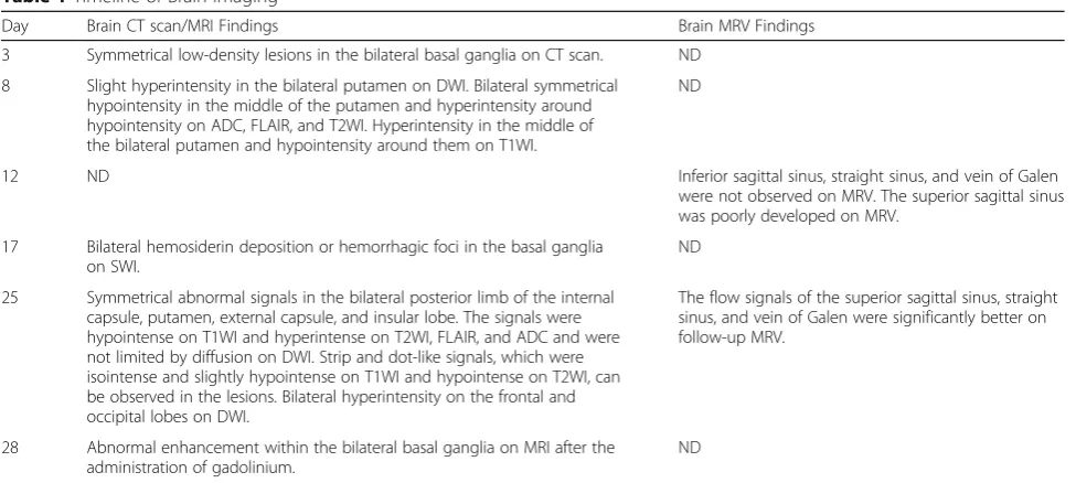
Magnetic resonance imaging and magnetic resonance venography features in heat stroke: a case report
Full text
Figure


Related documents
Variabel nilai fungsional pada penelitian ini tidak berpengaruh terhadap niat beli, dikarenakan fungsi yang diharapkan dari virtual item dari game online pada umumnya
The International Monetary Fund (IMF) loaned 250 million dollars. The political pressure on RPP and antidemocratic altitude towards press, university and military led to
Keywords — Wireless Sensor Network, Humidity, Discrepancies of humidity..
The results obtained showed that GLV extracts inhibited the corrosion process by a physical adsorption mechanism that followed the Langmuir, Freundlich, and Temkin adsorption
We cannot simply apply national security risk assessment to Scotland to discern how its paticular risks and threats might differ from the UK as a whole.. Risks and threats are, to an
The client is communicating to the server through load balancer [3],[5]. So every communication is allocated to the server by the load balancer. If any of the servers
In this paper, multiplication of Sinning e -Gaussian balance beam of light in strongly nonlocal nonlinear media has been stimulated by using paraxial group
