subarachnoid hemorrhage
in vivo
and
in vitro
Sun
et al.
R E S E A R C H
Open Access
Early release of high-mobility group box 1
(HMGB1) from neurons in experimental
subarachnoid hemorrhage
in vivo
and
in vitro
Qing Sun
1†, Wei Wu
1†, Yang-Chun Hu
1†, Hua Li
1, Dingding Zhang
1, Song Li
2, Wei Li
1, Wei-De Li
2, Biao Ma
1,
Jian-Hong Zhu
1, Meng-Liang Zhou
1and Chun-Hua Hang
1,3*Abstract
Background:Translocation of high-mobility group box 1 (HMGB1) from nucleus could trigger inflammation. Extracellular HMGB1 up-regulates inflammatory response in sepsis as a late mediator. However, little was known about its role in subarachnoid hemorrhage-inducible inflammation, especially in the early stage. This study aims to identify whether HMGB1 translocation occurred early after SAH and also to clarify the potential role of HMGB1 in brain injury following SAH.
Methods:Sprague-Dawley (SD) rats were randomly divided into sham group and SAH groups at 2 h, 12 h and on day 1, day 2. SAH groups suffered experimental subarachnoid hemorrhage by injection of 0.3 ml autoblood into the pre-chiasmatic cistern. Rats injected by recombinant HMGB1(rHMGB1) solution were divided into four groups according to different time points. Cultured neurons were assigned into control group and four hemoglobin (Hb) incubated groups. Mixed glial cells were cultured and stimulated in medium from neurons incubated by Hb. HMGB1 expression is measured by western blot analysis, real-time polymerase chain reaction (PCR), immunohistochemistry and immunofluorescence. Downstream nuclear factor kappa B (NF-κB) subunit P65 and inflammatory factor Interleukin 1β (IL-1β) were measured by western blot and real-time PCR, respectively. Brain injury was evaluated by cleaved caspase-3 staining.
Results:Our results demonstrated HMGB1 translocation occurred as early as 2 h after experimental SAH with mRNA and protein level increased. Immunohistochemistry and immunofluorescence results indicated cytosolic HMGB1 was mainly located in neurons while translocated HMGB1 could also be found in some microglia. After subarachnoid injection of rHMGB1, NF-κB, downstream inflammatory response and cleaved caspase-3 were up-regulated in the cortex compared to the saline control group.In-vitro, after Hb incubation, HMGB1 was also rapidly released from neurons to medium. Incubation with medium from neurons up-regulated IL-1βin mixed glial cells. This effect could be inhibited by HMGB1 specific inhibitor glycyrrhizic acid (GA) treatment.
Conclusion:HMGB1 was released from neurons early after SAH onset and might trigger inflammation as an upstream inflammatory mediator. Extracellular HMGB1 contributed to the brain injury after SAH. These results might have important implications during the administration of specific HMGB1 antagonists early in order to prevent or reduce inflammatory response following SAH.
Keywords:HMGB1, SAH, Early brain injury
* Correspondence:[email protected] †Equal contributors
1
Department of Neurosurgery, Jinling Hospital, School of Medicine, Nanjing University, Nanjing, Jiangsu Province, China
3
Department of Neurosurgery, Jinling Hospital, 305 East Zhongshan Road, 210002 Nanjing, People's Republic of China
Full list of author information is available at the end of the article
Introduction
Subarachnoid hemorrhage (SAH), especially aneurysm subarachnoid hemorrhage, is a life-threatening disease of the central nervous system (CNS). The incidence of SAH is about 22.5 cases per 100,000 in the population according to a World Health Organization study [1]. Al-though it has relatively low incidence, the early age of onset and poor outcome results in a many life-years lost. Toll like receptors (TLRs), myeloid differentiation primary response protein 88 (MyD88), NF-κB, IL-1β and TNF-αhave been proved to participate in the dam-aging inflammation process after SAH [2-5]. Moreover, clinical studies have shown that increased levels of pro-inflammatory factors in cerebrospinal fluid (CSF) and serum of SAH patients are associated with poor out-come [6,7]. Elevated inflammatory factors contribute to the breakdown of the blood brain barrier (BBB), brain edema, neuroglia cells apoptosis and death [8]. Adminis-tration of the antagonists of the pro-inflammatory factors confers a neuroprotective effect in library experimental studies [2,5]. However, how the inflammatory response is initiated and upstream of the inflammation response is still poorly investigated.
High-mobility group box 1(HMGB1), as a nonhis-tone protein binding with DNA, is widely expressed in the nucleus of nearly all eukaryotic cells, including brain cells, and stabilizes nucleosome formation, facili-tates gene transcription [9]. Growing evidence shows that HMGB1 is passively released from necrotic cells or actively secreted from immune cells or non-immune parenchymal cells under various pathological condi-tions [9]. Extracellular HMGB1 serves as alarmin or damage-associate molecular pattern (DAMP) that mediates cross-talk between damaged cells and rela-tive healthy cells and triggers inflammatory response after interaction with TLR2, TLR4 and receptors for advanced glycation end-products (RAGE) [10,11]. Extracellular HMGB1 has been regarded as a late inflammatory mediator in sepsis and as an early medi-ator in ischemia-inducible models [10,12]. Individual research into HMGB1 in the late stage of SAH has shown that HMGB1 is highly expressed in a day-5 group in brain stem tissue in the rabbit SAH model [13]. However, previous study suggested that brain parenchymal cells were damaged in the early period after SAH onset [14]. Further, HMGB1 passive trans-location usually occurred in the damaged cells [9]. Thus, we supposed that the HMGB1 might translo-cate early from nucleus to cytoplasm after SAH. Thus, this study aims to identify whether HMGB1 transloca-tion occurred early after SAH and also to detect the expression level of HMGB1 in the early stage and clarify the potential role of HMGB1 in brain injury following SAH.
Method and material
Animal preparation
Male Sprague-Dawley rats (280 to 320 g) were purchased from the Animal Center of Jinling Hospital. The rats were raised in a 12-h dark-light cycle with free access to food and water. All procedures were approved by the Animal Care and Use Committee of Nanjing University and con-formed to Guide for the Care and Use of Laboratory Ani-mals by National Institutes of Health. Forty-five aniAni-mals were divided randomly into a sham group and SAH groups at 2 h and 12 h, and on day 1 and day 2 respect-ively (nine rats per group). Six rats from each group were randomly selected for the analysis of western blot and real-time PCR. In the following step, another 27 rats were prepared for immunohistochemical and immunofluores-cence study in the sham group, the 2-h, and the day-1 group (nine rats per group). Six rats each of the selected groups were randomly chosen and sacrificed for immuno-histochemical and immunofluorescence study. As for sub-arachnoid injection of recombinant HMGB1 (rHMGB1), 45 rats were randomly divided into a control group and rHMGB1 injection groups including 2-h, 12-h, day-1 and day-2 groups (9 rats per group). Meanwhile, another 18 rats were prepared for immunofluorescent analysis (9 rats in the control and day-1 group, respectively).
SAH model
The prechiasmatic injection model was used [15]. Briefly, after intraperitoneal anesthesia with pentobarbital sodium (50 mg/kg) (Sigma, St Louis, MO, USA),then they were positioned prone in a stereotactic frame. After careful dis-infection, a midline scalp incision was made and a 1-mm hole was drilled 7.5 mm anterior to the bregma in the midline, at an angle of 30°E caudally. Then they were posi-tioned supine. After careful disinfection again we used an insulin injection needle (BD Science, Franklin Lakes, New Jersey, USA) to obtain 300 μl blood of themselves from femoral artery. The needle was advanced 11 mm into the prechiasmatic cistern through this burr hole, and the 300 μl blood was injected into the prechiasmatic cistern over 20s. Sham rats experienced the same procedure except for injection of 300μl blood. Cerebral blood flow was moni-tored for 45 minutes and 60 minutes after SAH. After com-pleting these procedures, 1 ml of 0.9% NaCl was injected subcutaneously to prevent dehydration and the rats were arranged in the recovery cage. It took about 30 minutes to one hour to reach recovery. After the rats started to move around and eat some semi-fluid food, they were returned to their clean and new cages and housed at 23 ± 1°C.
Subarachnoid prechiasmatic injection of recombinant HMGB1
HMGB1 (rHMGB1) saline solution. RHMGB1 extracted from Human Embryonic Kidney 293 cells (HEK-293) was prepared from Novoprotein(catalog number: C357, purity >95%, PH 7.4, Summit, NJ, USA). The content of endotoxion was tested by the Novoprotein Company and found to be less than 0.1 ng/μg. This result was also confirmed by our endotoxin Limulus amebocyte lysate test (catalog number: KC48, Chinese Horseshoe Crab Reagent Maunfactory, Xiamen, China). Western blot analysis was designed to exclude Histone 3 protein con-tamination. 50 μg rHMGB1 was diluted to 1,500 μl (33μg/ml) with saline and sterilized by filtration through a 0.22-μm sterile filter in case of bacterial contamin-ation. The dose of rHMGB1 was determined according to Qiu’s research (3.3μg rHMGB1/kg) [16] and adjusted the total volume of injection to be 150 μl. Rats in the control group were injected with 150 ul saline. Tissue was prepared for western blot and immunofluorescent analysis.
Perfusion-fixation and tissue preparation
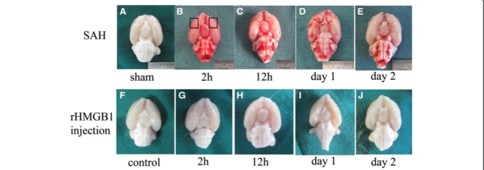
Animals were sacrificed according to the time points of different groups. In our pilot study, we found that there was no statistical difference in any detected variables among sham groups at any time point (data not shown). Therefore, animals in the sham group were sacrificed at 24 h after the sham operation. Animals were anesthetized as above, and perfused through the left cardiac ventricle with 0.9% a NaCl solution until effluent from the right atrium was clear. Animals that had obvious clots in the prechiasmatic cistern were selected to further analyze. The temporal lobe tissue (black box in Figure 1B), which was near the hematoma, was harvested on ice after blood clots on the tissue were carefully cleared. The tissue was
stored at -80°C till further use for western blot, real-time PCR. For immunohistochemistry and immunofluores-cence study, the rats were perfused with 0.9% NaCl so-lution followed by 4% buffered paraformaldehyde. A coronal block cut from 4 mm to 6 mm and 6 mm to 8 mm anterior to the groove between forebrain and cerebellum was prepared and immersed in 4% buffered paraformaldehyde overnight and then embedded in paraffin for immunohistochemistry study and frozen in optimal cutting temperature (OCT) medium for im-munofluorescence study, respectively.
Primary cortical neuron culture, hemoglobin (Hb)-incubated neuron injury model and experimental design
The primary cortical neuron culture was prepared and cultured as per the established technique in our labora-tory. Specifically, timed-pregnant female rats (16 to 18 days gestation) were sacrificed with deep anesthesia, and put in 75% alcohol disinfectant for sterilizing. Then 10 to 14 em-bryos were removed by Caesarean section using sterile techniques. The cortex was separated with the aid of a dis-section microscope and rinsed with pre-cooling PBS and treated by 0.1% trypsin for 5 minutes at 37°C, and then the supernatant containing trysin was discarded and washed by pre-cooling PBS. Subsequently, cells were tritu-rated with fire-polished glass pipettes. Then the neuron suspension was filtered through a 22 μm-filter into a 15 ml conical tube and sedimented at 1500 r/minute for 5 minutes at 4°C. After centrifugation, cells were resus-pended in neurobasal media with B27 (GIBCO, Rockville, MD, USA) plus antibiotics and were dissociated by re-peated pipetting through a 1-ml blue pipette tip. Then the cells were planted at approximately 100 × 104 cells per well in 6-well ploy-D-lysine-coated plates. Cultures were
Figure 1Schematic observation of the brain after surgery.Top: rat brains harvested at different times after subarachnoid hemorrhage (SAH) onset. Rat brains in the sham group were harvested one day after the surgical procedure(A). We can conclude that blood clot in the
maintained at 37°C in a humidified atmosphere of 5% CO2and 95% air. Subsequently, half of the medium was replaced every 2 days during the first 8 daysin vitro. The cultures were used on day 8when the cultures were essen-tially free of astrocytes. Hemoglobin (Hb) provided react-ive oxygen species (ROS) and heme, which caused neuron cell injury. Thus, Hb incubation in cultured neurons is often used as a neuron injury model in the researches of hemorrhage disease, including SAH. In this study, this model was also employed as the experimental SAH in the
in vitromodel. Hb (sigma, St. Louis, MO, USA) were pre-pared and resolved into 10μM with culture medium and sterilized by filtration through a 0.22-μm sterile filter. Then the neurons were treated with Hb at a concentration of 10μM, which was determined from prior studies [17]. After 4, 8, 16 and 24 h, the media of neurons were con-centrated for protein analysis and cultured neurons were arranged for immunofluorescence staining.
Primary mixed glial cells culture and cell medium stimulation experimental design
Primary mixed glial cells cultures were prepared as pre-vious study [10]. Briefly, cerebral hemispheres of 1- to 3-day-old postnatal rat brains (Sprague-Dawley rats) were separated with the aid of a dissection microscope and rinsed with pre-cooling PBS and treated by 0.125% trypsin for 5 minutes at 37°C, and then DMEM contain-ing 10% FBS(Hyclone, Logan, Utah, USA) were added to stop the digestion process. Subsequently, cells were trit-urated by repeated pipetting through a 1-ml blue pipette tip. Then the suspension was filtered through a 22μm-filter into a 15-ml conical tube and sedimentedat 1,500 r/minute for 5 minutes at 4°C. After centrifugation, cells were resus-pended and planted at approximately 100 × 104cells per well in 6-well plates in DMEM (Hyclone, Logan, Utah, USA) containing 10% FBS(Hyclone, Logan, Utah, USA). Culture media were renewed after 24 h and then twice per week. After 1 week, cells were subjected to different treatments.
Cell medium preparation: neuron cells were cultured as was described above. After incubation with neuroba-sal medium containing 20μmol Hb for 2 h, the medium was removed and replaced with fresh DMEM. After neu-rons with DMEM were cultured for 22 h, the DMEM medium was collected as the neuron medium. The con-trol medium was prepared from neurons treated with neurobasal containing 0 μmol Hb and incubated with DMEM medium for 22 h.
Groups and experiment design: cultured mixed glial cells were arranged into three groups. The control group: mixed glial cells treated with control medium; the medium group: mixed glial cells treated with neuron medium; the glycyrrhizic acid (GA) group: after mixed glial cells were treated with neuron medium, GA (Sigma,
catalog number:50531, purity >95%, St. Louis, MO, USA) diluted in PBS and adjusted PH to 7.4, then added to medium, the final concentration of GA in medium was 2 mM), a special inhibitor of HMGB1 was added in the medium to silence the activity of HMGB1 [18,19]. Mixed glial cells in all the groups were cultured for another 24 h. Then, glial cells were collected for real-time PCR analysis.
Preparation of tissue protein for western blot analysis Total protein extraction
Proper size of tissues (50 ~ 100 mg) were completely ho-mogenized using buffer and centrifuged at 14,000 × g for 15 minutes at 4°C. The supernatant was collected as the total protein extraction of tissue.
Cytosolic/nuclear fraction extraction
Rat brain-tissue cytosolic/nuclear fraction extraction was performed following the methods used in our la-boratory [20]. The brain tissue (about 100 mg) was ho-mogenized in 1 ml ice-cold buffer A composed of 10 mM HEPES (pH 7.9), 2 mM MgCl2, 10 mM KCl,0.1 mM EDTA, 1 mMdithiothreitol (DTT) and 0.5 mM phenyl-methylsulfonyl fluoride (PMSF) (all from Sigma Chemical Co).The homogenate was incubated on ice for 20 minutes, and then 30 μl of 10% NonidetP-40 solution was added (Sigma, St. Louis, MO, USA); the mixture was vortexed for 30 s and spun by centrifugation for 10 minutes at 5,000 g, 4°C. The cytosolic fraction extracts were collected and stored at -80°C for western blot analysis. The crude nuclear pellets were suspended in 200μl ice-cold buffer B containing 20 mM HEPES (pH 7.9), 25% (v/v) glycerol, 1.5 mM MgCl2, 20 mMKCl, 0.1 mM EDTA, 0.5 mM PMSF, and 1 mM DTT, and incubated on ice for 30 mi-nutes with intermittent mixing and centrifuged at 12,000 g at 4°C for 15 minutes. The supernatant contain-ing nuclear proteins was collected and stored at -80°C for western blot analysis. Protein concentration was deter-mined using a bicinchoninic acid assay kit with bovine serum albumin as the standard (Pierce Biochemicals, Rockford, IL, USA).
Western blot analysis
subunit) (Santa cruz, USA, 1:200 dilution), anti-Histone 3 (Cell signaling technology, Beverly, MA, USA 1:1000 dilu-tion)and β-actin (Catalog no.AP0060, Bioworld, USA, 1: 3000 dilution) After that the membrane was washed for 10 minutes each for four times in TBS + Tween 20 (TBST), followed by incubated in the appropriate HRP-conjugated secondary antibody (Catalog no. BS13278, Bio-world, USA, diluted 1:5000 in TBST) for 2 h at room temperature. The blotted protein bands were visualized by enhanced chemiluminescence (ECL) western blot detec-tion reagents (Catalog number NCI5079, Thermo Scien-tific, Rockford, IL, USA) and were exposed to x-ray film. Relative changes in protein expression were estimated from the mean pixel density using UN-SCAN-IT, normal-ized toβ-actin, and calculated as target protein expression/
β-actin expression ratios.
RNA isolation and quantitative real-time PCR
Rat brain tissues were isolated using TRIzol Reagent (TAKARA Biotechnology, Japan) as per the manufac-turer’s instructions. The concentration of the RNA was determined by spectrophotometric analysis (OD260/280). The quantity of RNA was measured using OD260. The isolated RNA was stored at-80°C until analyzed. RNA was reverse-transcribed to cDNA using Reverse Transcriptase Reagent (TAKARA Biotechnology, Japan) and oligodT primers. Quantitative real-time PCR analysis was performed using the Agilent Technologies Stratagene Mx3000P real-time PCR system (Genereal-times Technology, Inc), applying real-time SYBR Green PCR technology. The reaction mix-tures contained 1μl cDNA, 12.5μl SYBR Green (TAKARA Biotechnology, Japan), 1 μl of each forward and reverse primer (10μM) and nuclease-free water to a final volume of 25μl. The primers were synthesized by Life Technolo-gies (Invitrogen, Shanghai, China) and the sequences used were from a database at NCBI for rat HMGB1, TNF-Θ, IL-1β and β-actin. HMGB1 forward and reverse primers were 5’-ATGGGCAAAGGAGATCCTA-3’ and 5’-ATTC ATCATCATCATCTTCT-3’; TNF-Θ forward and reverse primers were 5’-TGCCTATGTCTCAGCCTCTTC-3’and 5’-GAGGCCATTTGGGAACTTCT-3’; IL-1βforward and reverse primers were 5’-TGAGCACCTTCTTTTCCTTC A-3’and 5’-TTGTCTAATGGGAACGTCACAC-3’;β -actin forward and reverse primers were 5’-AGGG AAATCGTGCGTGAC-3’ and 5’-CGCTCATTGCCG ATAGTG-3’. After 95°C for 30 s, 40 PCR cycles were performed; each consisting of a denaturation step (95°C, 5 s) and an annealing step (60°C, 30 s). Total RNA concentrations from each sample were normalized by quantity ofβ-actin mRNA, and the expression levels of target genes were evaluated by ratio of the number of target mRNA toβ-actin mRNA. All samples were ana-lyzed in triplicate.
Administration of propidium iodide and detection of propidium iodide-positive cells
Propidium iodide (PI), 10 mg/ml (Sigma, St Louis, MO, USA) was diluted in 0.9% NaCl and 1 mg/kg was admin-istered 1 h prior to sacrifice by intraperitoneal injection in a total volume of not more than 100 μl. Brain tissue was fixed with 4% paraformaldehyde overnight and dipped in 20% saccharose PBS for 2 days and then in 30% saccharose PBS for another 2 days to remove water in the tissue. For detection of the relationship between PI-labeled and HMGB1-positive cells, sections 6 μm in thickness were sliced and blocked with 5% normal FBS in PBS containing 0.1% Triton X-100 for 2 h at room temperature prior to incubation with HMGB1 anti-body(diluted 1:500, Epitomics, Burlingame, CA, USA) overnight at 4°C. After sections were washed three times with PBS for 45 minutes, they were immunolabeled with secondary antibody (Alexa Fluor 488 diluted 1:200) for 1 h at room temperature. The slides were washed with PBS again three times for another 30 minutes prior to being counterstained by 4',6-diamidino-2-phenylindole (DAPI) for 2 minutes. After three more washes, the slides were covered by microscopic glass with Anti-fade Mounting Medium for further study. The whole process was conducted with careful prevention of light.
Immunohistochemical staining
HMGB1-positive cells in the three views was regarded as the data for each section. The final average percentage of the three sec-tions was regarded as the data for the sample. Six samples in each group, respectively, were used for statistical analysis. The percentage of HMGB1-positive cells was identified, calculated, and analyzed under the light microscopy by an investigator blinded to the grouping.
Immunofluorescence staining
Immunofluorescence staining was performed according to our previous study in our laboratory [3]. Brain tissue was fixed with 4% paraformaldehyde overnight and dipped in 20% saccharose PBS for 2 days and then in 30% saccharose PBS for another 2 days to remove water in the tissue. Sections 6 μm in thickness were sliced and blocked with 5% normal FBS in PBS con-taining 0.1% Triton X-100 for 2 h at room temperature prior to incubation with anti-neuron-specific nuclear protein (NeuN) antibody (Millipore, MA, USA, 1:200) and anti-HMGB1 antibody diluted 1:500(Catalog num-ber 2600-1,Epitomics, Burlingame, CA, USA) or anti-ionized calcium binding adaptor molecule 1 (Iba1) antibody (Abcam, MA, USA,1:200) and anti-HMGB1 antibody diluted 1:500(Epitomics) or anti-glial fibrillary acidic protein (GFAP) antibody (1:200, BD Science, Franklin Lakes, New Jersey, USA) and anti-HMGB1 antibody diluted 1:500(Epitomics) or anti-cleaved caspase-3 diluted 1:500 (Cell Signaling Technology, Beverly, MA, USA) and NeuN antibody (Millipore, MA, USA, 1:200) overnight at 4°C. After sections were washed three times with PBS for 45 minutes, they were immunolabeled with proper sec-ondary antibodies (Alexa Fluor 488 and Alexa Fluor 594, 1:200) for 1 h at room temperature. The slides were
washed with PBS again three times for 45 minutes prior to being counterstained by DAPI for 2 minutes. After three further washes, the slides were covered by micro-scopic glass with Anti-fade Mounting Medium for further study. Pilot experiments with blocking peptides were per-formed to validate the specificity of primary antibodies be-fore the experiments. Negative controls were prepared by omitting the primary antibodies. Fluorescence microscopy imaging was performed using ZEISS HB050 inverted microscope system and handled by Image-Pro Plus 6.0 software (Media Cybernetics, USA). and Adobe Photo-shop CS5 (Adobe Systems, San Jose, USA).
HMGB1 measurements of cell-conditioned medium
Cell-conditioned medium was ultrafiltered and analyzed by western blot. Briefly, cell-conditioned medium (4 ml) was ultrafiltered using a Centricon (10 kDa, 4 ml, Millipore, MA, USA) according to the Instrument Manual at 4,000 × g with a typical final concentrate volume of about 100μl. Sometimes more ultrafiltration tubes were required because Hb in the medium sometimes blocked the hole in the Centricon. About one third of the final volume was sub-jected for western blot analysis as described above. The primary antibodies wasanti-HMGB1 diluted 1:500(Epi-tomics, Burlingame, CA, USA). Detection was performed using detection reagents (Catalog number NCI5079, Thermo Scientific, Rockford, IL, USA) and were ex-posed to an x-ray film kit (Thermo Scientific, Rockford, IL, USA).
Statistical analysis
All data were presented as mean ± standard error of the mean (SEM). SPSS 17.0 was used for statistical analysis
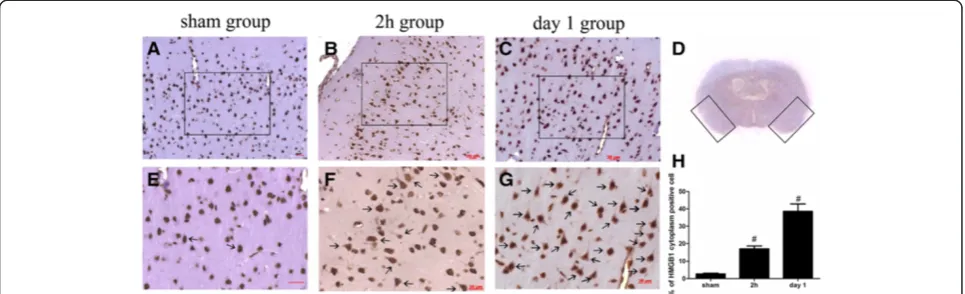
Figure 2Representative immunohistochemistry analysis of High-mobility group box 1 (HMGB1) in the brain cortex. (A)HMGB1 expression in the sham group: HMGB1 could be observed in the nucleus in the sham group, cells were seldomly stained positive for cytosolic HMGB1.(B, C)HMGB1 staining in the 2-h and day-1 groups post subarachnoid hemorrhage (SAH), respectively. The quantity of cells positive for cytosolic HMGB1 obviously increased in the SAH groups at 2 h and day 1 after SAH.(D)Black boxes show the view of observation.(E, FandG)
of the data. The measurements were subjected to one-way analysis of variance (ANOVA). Differences between experimental groups were determined by the Student
t-test. A value of P< 0.05 was considered statistically significant.
Result
General observation
In all the experimental SAH animals (n = 72), 6 of 54 (11.1%) animals injected with blood died while no ani-mals died in the sham group. All mortality occurred within 24 h of surgery. Two rats with SAH were ex-cluded from the study because of too little blood in the prechiasmatic cistern but many blood clots in the frontal lobe instead. Compared to the sham group (Figure 1A), the blood clots could easily be found on surface of the temporal lobe and around the basilar arteries (Figure 1B to E). It was also demonstrated that the blood clot in the subarachnoid space disappeared gradually with time (Figure 1B to E). No blood clots were found in the saline control group or in rHMGB1 injected groups (Figure 1F to J), and no rats in the control group died, while 3 of 45 rats died within 24 h after injection of rHMGB1.
HMGB1 expression in the sham-group brain
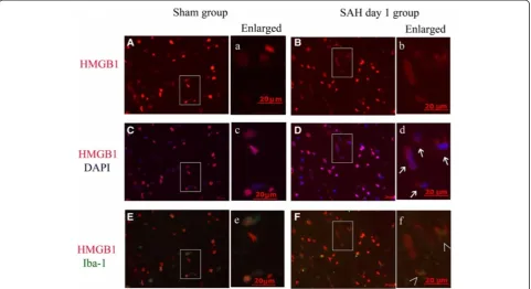
In the sham-group rat-brain coronal sections, HMGB1 was observed to be widely expressed in the nuclei of brain cells (Figure 2A), in either NeuN, GFAP, or Iba-1-positive cells (Figures 3D, 4C and 5C).
Subarachnoid hemorrhage induction induces HMGB1 translocation and release in brain cells
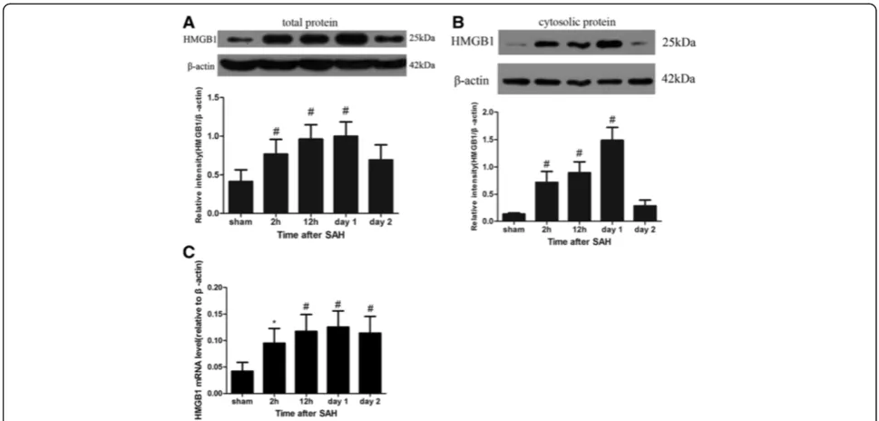
HMGB1 was reported as a late responding signal mol-ecule in sepsis [12]. Individual study indicated that HMGB1 level was increased in the late stage of SAH [13]. Little is known about the role of HMGB1 in the early stage of SAH. Thus, we examined a series of early time points in the rat SAH model to obtain a full view of HMGB1 protein level and location changes after SAH. Firstly, through western blot analysis of total tissue extracts, the level of HMGB1 protein increased signifi-cantly as early as 2 h after experimental SAH onset and peaked on day 1 post SAH when compared to the sham group(2 h, P< 0.05; 12 h,P< 0.01; day 1, P< 0.01; day 2,
P< 0.05) (Figure 6A). To identify whether the increased level of HMGB1 protein was transferred from nucleus to cytoplasm, nuclear protein fraction and cytosolic protein fraction were extracted separately (see Method and ma-terial). HMGB1 protein level in the cytosolic protein fraction was detected to significantly increase as early as 2 h after SAH induction (2 h, P< 0.01; 12 h, P< 0.01; day 1, P< 0.01) (Figure 6B). The above results showed that SAH could cause significant increased production and translocation of HMGB1 protein in the brain cortex as early as 2 h post injury.
Through quantitative real-time PCR analysis, the mRNA level of HMGB1 in SAH groups was identified to increase compared to the sham group. In detail, low level mRNA of HMGB1 could be detected in the sham group while the HMGB1 mRNA expression was significantly higher in a
time-dependent manner, similar to western blot in the SAH groups. A statistical difference was found not only between the sham and 2-h group (P< 0.05) but also between the sham group and the 12-h, day-1 groups (P< 0.01, respectively) (Figure 6C). The above results showed that SAH could cause significant active production of HMGB1 in the brain cortex as early as 2 h post injury.
HMGB1 translocation and release from brain cells was also confirmed by immohistochemistry study. The extra-nucleus or cytoplasm-positive HMGB1 staining cells were regarded as HMGB1-positive cells. As Figure 2B shows, HMGB1 translocation in the cortex occurred as early as 2 h after SAH. This result is in agreement with our west-ern blot result (Figure 6B). The amount of cytoplasm HMGB1-positive cells could be observed to increase sig-nificantly in representative cortex from the 2-h (mean = 17.03%) to day-1 group (mean = 38.57%) (Figure 2F,G) compared to the sham group (mean = 2.63%). Semi-quantitative analysis showed that there was an obvious dif-ference between the sham group and the 2-h, day-1 group (P< 0.01) (Figure 2H).
Early HMGB1 translocation induced by SAH occurred mostly in neurons in brain tissue
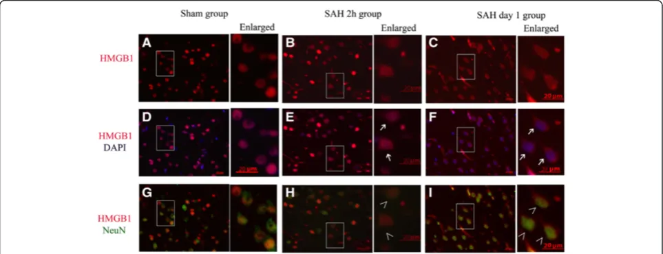
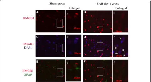
Double immunofluorescent staining was performed for HMGB1 and NeuN, Iba1 or GFAP to identify the cell types in which HMGB1 translocation occurred after SAH. We examined several time points in the SAH rat model to confirm the translocation of HMGB1 and how early it occurred after SAH. As Figure 3 shows, most cytosolic HMGB1-positive cells were also posi-tive for NeuN-staining. In comparison with the sham group, SAH also induced HMGB1 translocation in cells positive for Iba-1 (Figure 5). Few cells were found to stain positive for cytoplasmic HMGB1 and GFAP (Figure 4). These results suggest that HMGB1 trans-location mainly occurred in neurons in the injured cortex following SAH. Meanwhile a small number of Iba-1-positive cells started to secrete HMGB1 in the early phase following SAH. These findings may in part reflect the selective vulnerability of neurons to the in-jury and suggest that neurons might be one of the main sources of the released HMGB1, at least in this early phase.
Figure 4Cytosolic expression of High-mobility group box 1 (HMGB1) in Glial fibrillary acidic protein (GFAP)-positive cells of cortex from animals with subarachnoid hemorrhage (SAH). (A, B)HMGB1 immunostaining images obtained from cortex of a sham(A), and a day-1
(B)animal respectively.(C, D)Merged images of HMGB1 immunostaining (red) and 4',6-diamidino-2-phenylindole (DAPI) nuclear staining (blue).
Both passive and positive release of HMGB1 are involved in the increased level of HMGB1
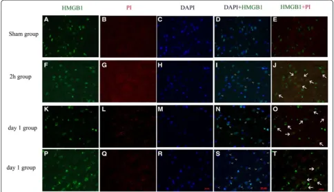
To give support to the hypothesis that both passive and positive release of HMGB1 were engaged in HMGB1 translocation. PI staining was employed to distinguish the dead cells and survival cells [21]. As Figure 7 shows, seldom cells were stained for PI in the sham group while PI-positive cells could be easily found in the 2-h and day-1 groups (Figure 7B,G,L,Q). Cells positive for PI and cytoplasmic HMGB1 were observed both in the 2-h and day-1 groups, which indicates that these cells were in an injured condition and HMGB1 might be released passively (Figure 7J,O) [9]. However, HMGB1 cytoplasm-positive cells that were not cytoplasm-positive for PI were also de-tected in another representative view of the day-1 group (Figure 7T), which means these cytoplasmic HMGB1 were actively secreted. These results were also in agreement with our real-time PCR result.
Extracellular HMGB1 induces inflammatory cytokines and cleaved caspase3 activityin vivo
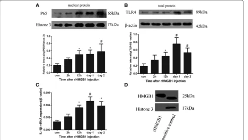
TLR4 is the important receptor of HMGB1. Western blot of total protein extraction demonstrated that rHMGB1 induced increased level of TLR4 (P< 0.05
between the control group and 12-h group; P< 0.01 be-tween the control group and day-1, day-2 groups respect-ively) (Figure 8B). This result indicates that extracellular HMGB1 could initiate TLR4 signal pathway. NF-κB activ-ity is usually measured by electrophoretic mobilactiv-ity shift assay (EMSA) [22] and nuclear translocation of its main proinflammatory subunit P65 [23,24]. Thus, the protein level of P65 in the nucleus extract is usually used to evalu-ate the activity of NF-κB in our laboratory [25,26]. The nuclear extract was prepared for western blot analysis of NF-κB(P65). As Figure 8A shows, the P65 subunit in the nucleus was significantly increased in the 12-h, day-1, day-2 groups after injection of rHMGB1 (12 h, P< 0.05; day 1,P< 0.05; day 2,P< 0.01). The downstream inflam-matory factor IL-1β, was also detected to be up-regulated by real-time PCR (12 h, P< 0.05; day 1, P< 0.01; day 2,
P< 0.05) (Figure 8C). Cleaved caspase 3 staining, a pos-sible marker of apoptosis, was used to evaluate the brain injury after rHMGB1 injection. Compared with the con-trol group, the number of cells positive for cleaved cas-pase 3 and NeuN was increased, which suggested that rHMGB1 might be a harmful molecule for brain cells, especially for neurons (Figure 9) and extracellular HMGB1 might contribute to the brain injury after SAH.
Figure 5Cytosolic expression of High-mobility group box 1 (HMGB1) in ionized calcium binding adaptor molecule 1 (Iba1)-positive cells of cortex from animals with subarachnoid hemorrhage (SAH). (A, B)HMGB1 immunostaining images obtained from cortex of a sham
Massive HMGB1 release from Hb-incubated neurons
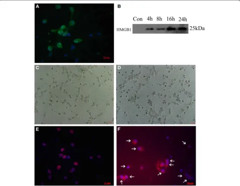
To confirm the neuronal susceptibility of early HMGB1 translocation, primary cortical neuron culture was sub-jected to Hb treatment and HMGB1 translocation was detected by immunofluorescence staining and western blot. Immunofluoresecnce staining showed more than 98% cells were positive for both NeuN (green) and DAPI (blue) which suggested the high percentage of neurons in the primary cultured cells (Figure 10A). Through western analysis, HMGB1 was undetectable in medium from untreated cells. However, HMGB1 was found to accumulate in culture medium of Hb-incubated rons (Figure 10B). A phase-contrast micrograph of neu-rons demonstrated cellular morphology of untreated (Figure 10C) and 24-h Hb (Figure 10D). Furthermore, HMGB1 was detected as nucleus-positive in The con-trol group (Figure 10E) and cytoplasm-positive in Hb-treated groups (Figure 10F). This result indicates that HMGB1 in neurons was in a process from nucleus to extracellular after Hb incubation.
Conditioned medium from Hb-treated neurons induced IL-1βin cultured mixed glial cells, which could be inhibited by HMGB1-specific inhibitor
To determine whether HMGB1 released from injured cul-tured neurons was biologically active as a pro-inflammatory mediator, we treated primary mixed glial cell cultures with
conditioned medium from Hb-treated neurons. To remove residual Hb in supernatant, neurons were cultured in fresh DMEM after being exposed to Hb (20 μmol) for 2 h. As shown in Figure 11, conditioned medium ro-bustly induced IL-1β mRNA expression in glial cells. However, IL-1βcould be inhibited after treatment with HMGB1-specific inhibitor GA (P< 0.01 between the con-trol and medium group; P< 0.05 between the medium group and medium + GA group). This result indicates that HMGB1 in the medium played an important role in acti-vation of glial cells.
Discussion
In this study, we demonstrated that (1) HMGB1 was translocated from the nucleus to the cytoplasm and re-leased from neurons as early as 2 h after SAH associated with a significant upregulation of protein and mRNA level; (2) both passive and active release of HMGB1 were involved in the process of HMGB1 translocation; (3) rHMGB1 or HMGB1 released from neurons could induce inflammatory response, and (4) extracellular HMGB1 con-tributed to the early brain injury after SAH.
Previous studies have demonstrated that HMGB1 could be released from necrotic cells passively or secreted ac-tively from immune cells or non-immune parenchymal cells, such as hepatocytes,in ischemia [27,28]. In our study, we demonstrated that HMGB1 was released from cortex
near the blood clot as early as 2 h after SAH onset accord-ing to our western blot and immunhistochemistry results. Friedrichet al. showed that cortex cell death occurred as early as 10 minutes after SAH [14]. As a consequence, passive release of HMGB1 was possibly initiated by dam-aged cellular integrity. This hypothesis was also supported in our study, which showed co-location of PI staining and cytosolic HMGB1 staining (Figure 7J). However, active se-cretion of HMGB1 was also supported by up-regulated mRNA and protein levels of HMGB1 (Figure 6A to C) in our study. Further, positive staining for cytoplasmic HMGB1 but negative for PI staining also support this the-ory (Figure 7S,T). Our research was consistent with re-search into liver ischemia, which has shown that the liver parenchymal cell, liver cells could actively release HMGB1 under ischemic conditions [27,28]. Thus, it is possible that both passive and active releases of HMGB1 are involved in the process of HMGB1 translocation. However, co-immunoprecipitation might be the best method to make sure whether cytoplasmic HMGB1 was passive or actively
released [28]. Previous study indicated that both passive and active release of HMGB1 have cytokine activity and could trigger inflammatory response [29], thus co-immunoprecipitation of HMGB1 was not performed in this study.
Extracellular HMGB1 was regarded as a member of the DAMP family [30]. HMGB1 functions as a pro-inflammatory factor through its receptors, including TLR2, TLR4 and RAGE [11]. Conceivably, up-regulated expression of HMGB1 receptors after SAH might enhance the sensitivity of brain cells to HMGB1. Both TLR4 and RAGE were reported to ascend early after SAH in recent studies [4,26,31]. Although interaction of HMGB1 with these receptors signals through different pathways, they ultimately promote the NF-κB/P65 translocation to nu-cleus and activation of NF-κB, which activates the tran-scription of downstream pro-inflammatory genes (IL-1β, TNF-α) [11]. Addition of rHMGB1 in the subarachnoid space up-regulated TLR4, P65 protein levels and down-stream inflammatory response in this study, which
Figure 8Addition of recombinant High-mobility group box 1 (rHMGB1) in the subarachnoid space triggered inflammatory response
in vivo.NF-κB was measured by the western blot of its P65 subunit in the nucleus. Toll-like receptor (TLR)4 protein level was also detected by western blot analysis. IL-1βwas measured by real-time PCR.(A)rHMGB1 up-regulated P65 subunit protein level in the nuclear protein in cortex cells;P< 0.05 between the 12-h, day-1 group and control group,P< 0.01 between the day-2 group and control group.(B)rHMGB1 increased TLR4 protein level.P< 0.05 between the 12-h and control group,P< 0.01 between the day-1, day-2 group and control group.(C)rHMGB1 upregulated IL-1βmRNA expression in cortex cells:P< 0.05 between the 12-h, day-2 group and control group,P< 0.01 between the day-1 group and control group.(D)Western blot analysis of histone 3 content in rHMGB1(left lane) and the nuclear protein extraction(right lane). The result could exclude histone 3 contamination in rHMGB1 products. Histone 3 was predominant composition of histone protein, thus, our result indicated the rHMGB1 used in the study had good purity. Bars represent the mean ± standard error (n = 6): *P< 0.05 compared with the control group;#P< 0.01 compared with the control group.
confirmed that extracellular HMGB1 could trigger the inflammatory response and the TLR4/NF-κB signal pathway might be one of the activated inflammatory pathway in HMGB1-induced inflammatory response.
Previous study has shown that the most important re-ceptors of HMGB1, TLR4 and RAGE are up-regulated 4 h and 6 h after SAH, respectively. IL-1β reaches its peak on day 1 after SAH [3,31]. In this study HMGB1 translocation was detected as early as 2 h post injury. Furthermore, in vivo, we observed a two-fold induction of IL-1β mRNA level after subarachnoid injection of rHMGB1 (Figure 8C). Previous studies also demon-strated that HMGB1 incubation in glia or neuron cells
in vitro induce 2- to 4-fold inflammatory factor mRNA
induction [10,22]. The data reported here showed the translocation of HMGB1 preceded the increase of other cytokines, which indicates that HMGB1 may act as an early upstream initiator of inflammation after SAH.
Immunohistochemical and immunofluorescence stain-ing results showed most cells positive for cytoplasmic HMGB1 were also positive for NeuN. A small number of cytoplasmic HMGB1-positive cells were found to be microglia. Neurons seem to be susceptible after brain in-jury and the main source of extracellular HMGB1. Actu-ally, not only in the SAH modelin vivoandin vitro, but also in the ischemic brain, neurons seem to be the main source of HMGB1 in the early stage of brain injury [10]. Early-released HMGB1 from neurons might be the
important early upstream factor in the following inflam-matory response after SAH. To better understand whether neuron-derived HMGB1 could signal to mixed glial cells, medium from neurons, which had been stim-ulated by Hb, was used to culture the mixed glial cells (see detailed information in Method and material). The result demonstrates that HMGB1 released from neurons could initiate the inflammatory response in mixed glial cells. Thus, it could be supposed that HMGB1 released from neurons might influence neighboring glial cells and up-regulate the inflammatory factors, which could in turn stimulate brain cells to release more HMGB1 with an up-regulated mRNA level of HMGB1 [9]. This result suggests further that HMGB1 might be an early up-stream factor in inflammation after SAH. Myeloid differ-entiation primary response protein 88 (downstream of TLRs) and RAGE were also reported to be up-regulated mainly in neurons after SAH [3,26]. Our previous study
in vitroshowed addition of rHMGB1 could also increase MyD88 expression in protein and mRNA levels in cul-tured neurons with up-regulated inflammatory factors [22]. According to these studies, it seems that relative healthy neurons nearby could also be reactive cells for extracellular HMGB1 as well as glial cells. A small num-ber of microglia was observed to release HMGB1 in the beginning. However, it was believed that more and more microglia would start to secrete HMGB1 as time devel-oped, which was observed in the late stage in a previous study [13]. Interestingly, this phenomenon also exists in
the late stage of the ischemic brain [32]. HMGB1 released from microglia might be responsible for the inflammation in the late stage.
Addition of rHMGB1 up-regulated cleaved caspase 3, a possible marker of apoptosis in the cortex, especially in neurons (Figure 9). Extracellular rHMGB1 seemed harmful for survival of brain cells. The explicit mechan-ism is not yet clear. The inflammatory response induced by rHMGB1 might be the possible reason. It is widely believed that inflammation in the early stage contributes to the brain injury and it has been confirmed that inhib-ition of NF-κB and downstream inflammatory factors confer protection in the early stage [2,8,24]. Increased inflammatory factors, such as IL-1β and TNF-α, could mediate cell apoptosis and cell damage [33,34]. Thus, it was suspected that the rHMGB1 might accelerate brain injury through up-regulated inflammation. How the rHMGB1 in the subarachnoid space influenced the cor-tex parenchymal cells is not yet clear. Maybe it shared a similar process to the intraventricular injection of lipo-polysaccharide [34] or the small molecule HMGB1 might be able to easily infiltrate the pia mater because of its small molecular weight.
Rodex modification of 106 cysteine in the HMGB1 molecule might be critical for its cytokine activity. Oxi-dization of the cysteine at position 106 in HMGB1 could suppress the pro-inflammtory activity [35,36]. However, although together with ROS, HMGB1 retains its activity during the inflammatory process in a glutamate-treated-neurons model, oxygen-glucose deprivation model [10] and an N-methyl-D-aspartic acid (NMDA)-treated model [32]. Further research using tandem mass spectrometric analysis indicated that the predominant form of HMGB1 during the inflammatory process is not oxidized. When the inflammation was resolved, the form of HMGB1 was oxidized [35]. As shown in Figure 11, although ROS could be provided by hemoglobin, HMGB1 cytokine activity still existed and triggered inflammation in glial cells. Furthermore, ROS also damages cells itself and upregu-lates HMGB1 release [37]. Thus oxidization could not silence all the HMGB1 because the treatment target of HMGB1 could alleviate the inflammatory response [38]. Early use of HMGB1 inhibitor, such as GA, might be a good choice for stopping the harmful inflammatory response.
Combining the research listed above, we could specu-late that early-released HMGB1 from neurons after SAH onset might trigger inflammation in neurons [22], and glial cells nearby with their MyD88-level up-regulated [3]. Increased levels of inflammatory factors might trigger more cells to actively secrete HMGB1 [9]. Up-regulated inflammation contributes to the BBB breakdown, brain edema, cell apoptosis and death. Extracellular HMGB1 might be the early key mediator that mediates cross-talk
Figure 11Conditioned medium from Hb-treated neurons induced IL-1βin cultured mixed glial cells, which could be inhibited by High-mobility group box 1 (HMGB1)-specific inhibitor.
between injured cells and relative healthy cells around damaged tissues [16].
The effects in clinic trials targeting a single signal in the inflammatory pathway are not as good as expected [39,40]. Inflammation is such a complex pathophysio-logical process, that treatment simply targeting a single molecule or receptor is difficult to determine. It is import-ant to find out the source of the inflammatory response. Our research points out that HMGB1 might be the early key mediator in the process of SAH and a novel potential target for treatment [41].
Summary
As mentioned above, HMGB1 is massively released from neurons early after SAH onset. Passive and active releases are involved in the translocation process. Extracellular HMGB1 represented a pro-inflammatory role and con-tributed to brain injury. HMGB1 might be the major up-stream inflammatory mediator, which might be a potential treatment target.
Abbreviations
ANOVA:analysis of variance; BBB: blood brain barrier; CNS: central nervous system; CSF: cerebrospinal fluid; DAB: diaminobenzidine; DAMP: damage-associate molecular pattern; DAPI: 4',6-diamidino-2-phenylindole; DMEM: Dulbecco's modified Eagle's medium; FBS: fetal bovine serum; EMSA: electrophoretic mobility shift assay; GFAP: Glial fibrillary acidic protein; Hb: hemoglobin; HMGB1: High-mobility group box 1; Iba1: Ionized calcium binding adaptor molecule 1; IL-1β: interleukin 1β; MyD88: Myeloid differentiation primary response gene; NeuN: Neuron-specific nuclear protein; NF-κB: Nuclear factor kappa B; NMDA: N-methyl-D-aspartic acid; PBS: phosphate-buffered saline; PCR: polymerase chain reaction;
PI: propidium iodide; RAGE: receptors for advanced glycation end-products; rHMGB1: recombinant High-mobility group box 1; ROS: reactive oxygen species; SAH: subarachnoid hemorrhage; SD: Sprague-Dawley; TLR: toll-like receptor; TNF-α: tumor necrosis factor-alpha.
Competing interests
All the authors declared that they have no competing interests.
Authors’contributions
QS and Y-CH designed the studies, carried out the surgery, physiology studies, immunohistochemical studies, and data analysis, and wrote the manuscript. WW designed the additional experiment and contributed much to the revised manuscript. HL and SL conducted the neuronal cell culture. DZ, W-DL and J-HZ contributed to the Western blotting and immunohistochemical analysis. BM and LZ contributed to the real-time PCR analysis. WL and M-LZ contributed to the design and analysis of the study. C-HH contributed to the design and analysis of the study and wrote the manuscript. All authors read and approved the final manuscript.
Authors’information
Qing Sun, Wei Wu and Yang-Chun Hu co-first authors.
Acknowledgements
The study was supported by the National Natural Science Foundation, China (81371294). The authors gratefully acknowledge the generous assistance of Dr Lin Zhu and Gen-Bao Feng.
Author details
1
Department of Neurosurgery, Jinling Hospital, School of Medicine, Nanjing University, Nanjing, Jiangsu Province, China.2Department of Neurosurgery, School of Medicine, Southern Medical University(Guangzhou), Jinling Hospital, Nanjing, Jiangsu Province, People's Republic of China.3Department of Neurosurgery, Jinling Hospital, 305 East Zhongshan Road, 210002 Nanjing, People's Republic of China.
Received: 31 December 2013 Accepted: 29 May 2014 Published: 12 June 2014
References
1. Schuette AJ, Barrow DL:Epidemiology and long-term mortality in subarachnoid hemorrhage.World Neurosurg2013,80:264–265. 2. Greenhalgh AD, Brough D, Robinson EM, Girard S, Rothwell NJ, Allan SM:
Interleukin-1 receptor antagonist is beneficial after subarachnoid haemorrhage in rat by blocking haem-driven inflammatory pathology. Dis Model Mech2012,5:823–833.
3. Sun Q, Dai Y, Zhang X, Hu YC, Zhang D, Li W, Zhang XS, Zhu JH, Zhou ML, Hang CH:Expression and cell distribution of myeloid differentiation primary response protein 88 in the cerebral cortex following experimental subarachnoid hemorrhage in rats: a pilot study.Brain Res2013,1520:134–144. 4. Zhou ML, Shi JX, Hang CH, Zhang FF, Gao J, Yin HX:Expression of Toll-like
receptor 4 in the brain in a rabbit experimental subarachnoid haemorrhage model.Inflamm Res2007,56:93–97.
5. Jiang Y, Liu DW, Han XY, Dong YN, Gao J, Du B, Meng L, Shi JG: Neuroprotective effects of anti-tumor necrosis factor-alpha antibody on apoptosis following subarachnoid hemorrhage in a rat model.J Clin Neurosci2012,19:866–872.
6. Hanafy KA, Grobelny B, Fernandez L, Kurtz P, Connolly ES, Mayer SA, Schindler C, Badjatia N:Brain interstitial fluid TNF-alpha after subarachnoid hemorrhage. J Neurol Sci2010,291:69–73.
7. Chou SH, Feske SK, Atherton J, Konigsberg RG, De Jager PL, Du R, Ogilvy CS, Lo EH, Ning M:Early elevation of serum tumor necrosis factor-alpha is associated with poor outcome in subarachnoid hemorrhage.J Investig Med2012,60:1054–1058.
8. Sozen T, Tsuchiyama R, Hasegawa Y, Suzuki H, Jadhav V, Nishizawa S, Zhang JH: Role of interleukin-1beta in early brain injury after subarachnoid
hemorrhage in mice.Stroke2009,40:2519–2525.
9. Andersson U, Tracey KJ:HMGB1 is a therapeutic target for sterile inflammation and infection.Annu Rev Immunol2011,29:139–162. 10. Qiu J, Nishimura M, Wang Y, Sims JR, Qiu S, Savitz SI, Salomone S,
Moskowitz MA:Early release of HMGB-1 from neurons after the onset of brain ischemia.J Cereb Blood Flow Metab2008,28:927–938.
11. van Beijnum JR, Buurman WA, Griffioen AW:Convergence and
amplification of toll-like receptor (TLR) and receptor for advanced glycation end products (RAGE) signaling pathways via high mobility group B1 (HMGB1).Angiogenesis2008,11:91–99.
12. Wang H, Bloom O, Zhang M, Vishnubhakat JM, Ombrellino M, Che J, Frazier A, Yang H, Ivanova S, Borovikova L, Manogue KR, Faist E, Abraham E, Andersson J, Andersson U, Molina PE, Abumrad NN, Sama A, Tracey KJ:HMG-1 as a late mediator of endotoxin lethality in mice.Science1999,285:248–251. 13. Murakami K, Koide M, Dumont TM, Russell SR, Tranmer BI, Wellman GC:
Subarachnoid hemorrhage induces gliosis and increased expression of the pro-inflammatory cytokine high mobility group box 1 protein.Transl Stroke Res2011,2:72–79.
14. Friedrich V, Flores R, Sehba FA:Cell death starts early after subarachnoid hemorrhage.Neurosci Lett2012,512:6–11.
15. Jeon H, Ai J, Sabri M, Tariq A, Macdonald RL:Learning deficits after experimental subarachnoid hemorrhage in rats.Neuroscience2010, 169:1805–1814.
16. Qiu J, Xu J, Zheng Y, Wei Y, Zhu X, Lo EH, Moskowitz MA, Sims JR: High-mobility group box 1 promotes metalloproteinase-9 upregulation through Toll-like receptor 4 after cerebral ischemia.Stroke2010, 41:2077–2082.
17. You WC, Li W, Zhuang Z, Tang Y, Lu HC, Ji XJ, Shen W, Shi JX, Zhou ML: Biphasic activation of nuclear factor-kappa B in experimental models of subarachnoid hemorrhage in vivo and in vitro.Mediators Inflamm2012, 2012:786242.
18. Girard JP:A direct inhibitor of HMGB1 cytokine.Chem Biol2007,14:345–347. 19. Mollica L, De Marchis F, Spitaleri A, Dallacosta C, Pennacchini D, Zamai M,
Agresti A, Trisciuoglio L, Musco G, Bianchi ME:Glycyrrhizin binds to high-mobility group box 1 protein and inhibits its cytokine activities. Chem Biol2007,14:431–441.
21. You Z, Savitz SI, Yang J, Degterev A, Yuan J, Cuny GD, Moskowitz MA, Whalen MJ:Necrostatin-1 reduces histopathology and improves functional outcome after controlled cortical impact in mice.J Cereb Blood Flow Metab2008,28:1564–1573.
22. Li W, Ling HP, You WC, Ji XJ, Tang Y, Zhao JB, Su XF, Hang CH: Recombinant high-mobility group box 1 protein (HMGB-1) promotes myeloid differentiation primary response protein 88 (Myd88) upregulation in mouse primary cortical neurons.Neurol Sci2013,34:847–853. 23. Bera A, Zhao S, Cao L, Chiao PJ, Freeman JW:Oncogenic K-Ras and loss of
Smad4 mediate invasion by activating an EGFR/NF-kappaB axis that induces expression of MMP9 and uPA in human pancreas progenitor cells.PLoS One2013,8:e82282.
24. Kang SR, Han DY, Park KI, Park HS, Cho YB, Lee HJ, Lee WS, Ryu CH, Ha YL, Lee DH, Kim JA, Kim GS:Suppressive effect on lipopolysaccharide-induced proinflammatory mediators by citrus aurantium L. in macrophage RAW 264.7 cells via NF-kappaB signal pathway.Evid Based Complement Alternat Med2011,2011. doi:10.1155/2011/248592. Epub 2010 Sep 21.
25. Hu YC, Sun Q, Li W, Zhang DD, Ma B, Li S, Li WD, Zhou ML, Hang CH: Biphasic activation of nuclear factor kappa B and expression of p65 and c-Rel after traumatic brain injury in rats.Inflamm Res2014,63:109–115. 26. Li H, Wu W, Sun Q, Liu M, Li W, Zhang XS, Zhou ML, Hang CH:Expression
and cell distribution of receptor for advanced glycation end-products in the rat cortex following experimental subarachnoid hemorrhage: a preliminary study.Brain Res2014,1543:315–323.
27. Tsung A, Sahai R, Tanaka H, Nakao A, Fink MP, Lotze MT, Yang H, Li J, Tracey KJ, Geller DA, Billiar TR:The nuclear factor HMGB1 mediates hepatic injury after murine liver ischemia-reperfusion.J Exp Med2005,201:1135–1143. 28. Evankovich J, Cho SW, Zhang R, Cardinal J, Dhupar R, Zhang L, Klune JR,
Zlotnicki J, Billiar T, Tsung A:High mobility group box 1 release from hepatocytes during ischemia and reperfusion injury is mediated by decreased histone deacetylase activity.J Biol Chem2010,
285:39888–39897.
29. Scaffidi P, Misteli T, Bianchi ME:Release of chromatin protein HMGB1 by necrotic cells triggers inflammation.Nature2002,418:191–195. 30. Bianchi ME, Manfredi AA:Immunology: dangers in and out.Science2009,
323:1683–1684.
31. Ma CX, Yin WN, Cai BW, Wu J, Wang JY, He M, Sun H, Ding JL, You C: Toll-like receptor 4/nuclear factor-kappa B signaling detected in brain after early subarachnoid hemorrhage.Chin Med J (Engl)2009, 122:1575–1581.
32. Kim JB, Sig CJ, Yu YM, Nam K, Piao CS, Kim SW, Lee MH, Han PL, Park JS, Lee JK:HMGB1, a novel cytokine-like mediator linking acute neuronal death and delayed neuroinflammation in the postischemic brain. J Neurosci2006,26:6413–6421.
33. van Kralingen C, Kho DT, Costa J, Angel CE, Graham ES:Exposure to inflammatory cytokines IL-1beta and TNFalpha induces compromise and death of astrocytes; implications for chronic neuroinflammation. PLoS One2013,8:e84269.
34. Hanafy KA:The role of microglia and the TLR4 pathway in neuronal apoptosis and vasospasm after subarachnoid hemorrhage. J Neuroinflammation2013,10:83.
35. Yang H, Lundback P, Ottosson L, Erlandsson-Harris H, Venereau E, Bianchi ME, Al-Abed Y, Andersson U, Tracey KJ, Antoine DJ:Redox modification of cysteine residues regulates the cytokine activity of high mobility group box-1 (HMGB1).Mol Med2012,18:250–259.
36. Kazama H, Ricci JE, Herndon JM, Hoppe G, Green DR, Ferguson TA: Induction of immunological tolerance by apoptotic cells requires caspase-dependent oxidation of high-mobility group box-1 protein. Immunity2008,29:21–32.
37. Tsung A, Klune JR, Zhang X, Jeyabalan G, Cao Z, Peng X, Stolz DB, Geller DA, Rosengart MR, Billiar TR:HMGB1 release induced by liver ischemia involves Toll-like receptor 4 dependent reactive oxygen species production and calcium-mediated signaling.J Exp Med2007,204:2913–2923.
38. Kim SW, Jin Y, Shin JH, Kim ID, Lee HK, Park S, Han PL, Lee JK:Glycyrrhizic acid affords robust neuroprotection in the postischemic brain via anti-inflammatory effect by inhibiting HMGB1 phosphorylation and secretion.Neurobiol Dis2012,46:147–156.
39. Krams M, Lees KR, Hacke W, Grieve AP, Orgogozo JM, Ford GA:Acute Stroke Therapy by Inhibition of Neutrophils (ASTIN): an adaptive dose-response study of UK-279,276 in acute ischemic stroke.Stroke2003, 34:2543–2548.
40. Enlimomab Acute Stroke Trial Investigators:Use of anti-ICAM-1 therapy in ischemic stroke: results of the Enlimomab Acute Stroke Trial.Neurology
2001,57:1428–1434.
41. Sun Q, Wang F, Li W, Li W, Hu YC, Li S, Zhu JH, Zhou M, Hang CH: Glycyrrhizic acid confers neuroprotection after subarachnoid hemorrhage via inhibition of high mobility group box-1 protein: a hypothesis for novel therapy of subarachnoid hemorrhage.Med Hypotheses2013,81:681–685.
doi:10.1186/1742-2094-11-106
Cite this article as:Sunet al.:Early release of high-mobility group box 1 (HMGB1) from neurons in experimental subarachnoid hemorrhage
in vivoandin vitro.Journal of Neuroinflammation201411:106.
Submit your next manuscript to BioMed Central and take full advantage of:
• Convenient online submission
• Thorough peer review
• No space constraints or color figure charges
• Immediate publication on acceptance
• Inclusion in PubMed, CAS, Scopus and Google Scholar
• Research which is freely available for redistribution