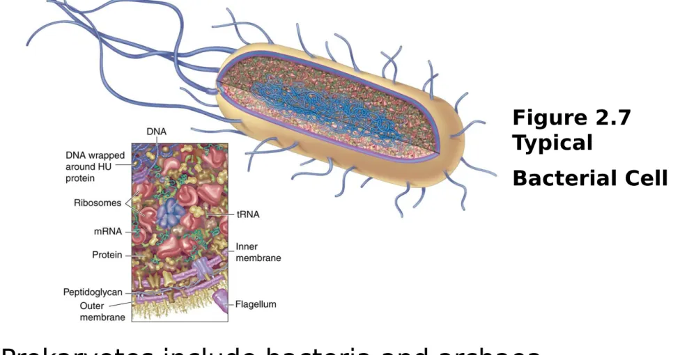
Figure 2.7
Typical
Bacterial Cell
Prokaryotes include bacteria and archaea
They have cell wall, plasma membranes, circular
DNA, and no membrane-bound organelles-
lack
a
membrane bound nucleus
Structure of Prokaryotic Cells
Figure 2.8
Bacterial Cell
Cell Wall
The prokaryotic cell wall is a complex
semi-rigid structure primarily for support
and protection
The cell wall is primarily composed of
peptidoglycan
Cell Wall and Plasma Membrane
www.torresbiocla.pbs.com
PlasmaMembrane
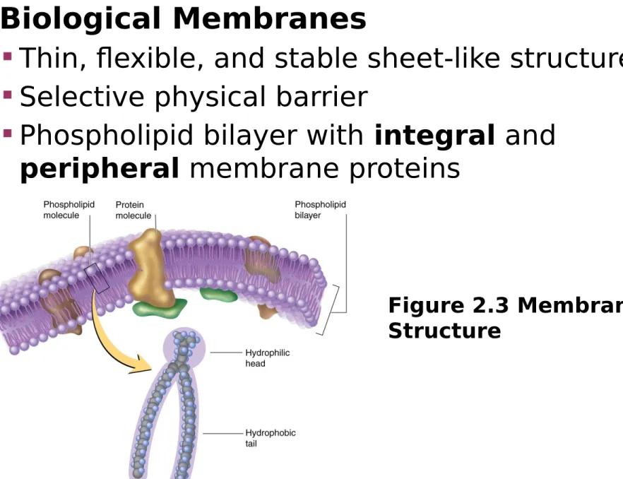
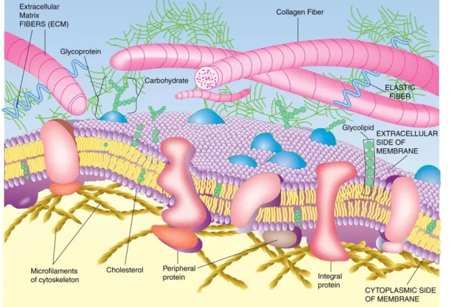
Biological Membranes
Thin, flexible, and stable sheet-like structures
Selective physical barrier
Phospholipid bilayer with
integral
and
peripheral
membrane proteins
Figure 2.3 Membrane
Structure
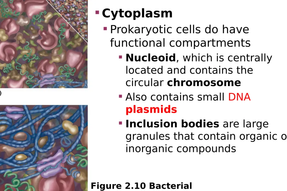
Figure 2.10 Bacterial
Cytoplasm
Cytoplasm
Prokaryotic cells do have
functional compartments
Nucleoid
, which is centrally
located and contains the
circular
chromosome
Also contains small
DNA
plasmids
Inclusion
bodies
are large
granules that contain organic or
inorganic compounds
CHROMOSOME
PLASMID
Plasmid
• Can replicate independently of chromosomes
Many bacteria have external appendages
Pili (pilus) are for attachment and conjugation
Flagella (flagellum) are used for locomotion
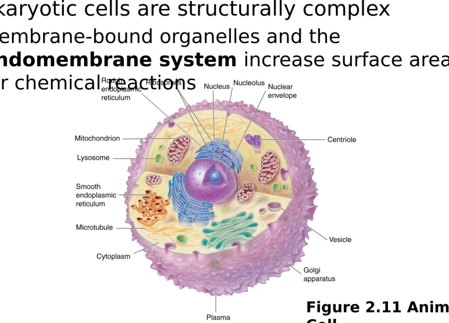
Figure 2.11 Animal
Cell
Eukaryotic cells are structurally complex
Membrane-bound organelles and the
endomembrane system
increase surface area
for chemical reactions
Animal Cell
Important structures: plasma membrane,
endoplasmic reticulum, Golgi apparatus,
nucleus, lysosomes, mitochondria,
chloroplasts, ribosomes, and the cytoskeleton
Plant Cell
Figure 2.12 Plant
Plasma Membrane
Isolates the cell and is selectively permeable
Outside the plasma membrane are the
glycocalyx
and the
extracellular matrix
Plasma Membrane
Figure 2.13
Plasma
Membrane
Endoplasmic Reticulum
The
endoplasmic reticulum
(ER)
is a series of membranous
tubules, vesicles, and flattened
sacks
Two types:
Rough ER
functions include
protein synthesis, folding, and
glycosylation
Smooth ER
functions include
lipid biosynthesis and Ca
2+storage
Endoplasmic Reticulum
Figure 2.15
Endoplasmic
Ribosomes
Figure 2.29
Ribosomes
Ribosomes
Ribosomes
are RNA/protein
complexes involved in protein
biosynthesis
Two subunits form a functional
unit
Eukaryotes and prokaryotes
have ribosomes
Eukaryotic ribosomes are larger
than prokaryotic ones although
they are similar in structure and
function
In eukaryotes RER consists
of ribosomes attached to the
ER
Golgi Apparatus
The
Golgi apparatus
is
formed of large,
flattened, sac-like
membranous vesicles
Processes, packages,
and distributes cell
products
Participates in
EXOCYTOSIS
Golgi Apparatus
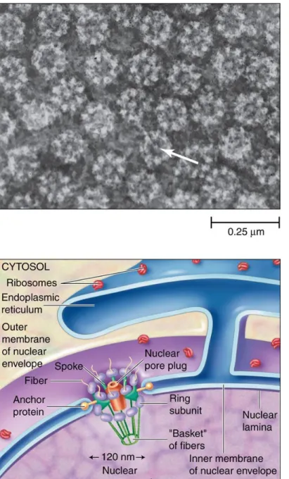
Nucleus
The
nucleus
is the most
prominent organelle
Contains the hereditary
information ( DNA)
Site of
transcription
Nuclear components:
Nucleoplasm
Chromatin
(
genome
)
Nuclear
matrix
Nucleolus
Nuclear envelope
Nucleus
Figure 2.18 Eukaryotic
The
nuclear envelope
surrounds the nucleoplasm
The nuclear envelope has
nuclear pores
referred to as
nuclear pore complexes
Structures through which
pass most of the
molecules that enter and
leave the nucleus
Nuclear Envelope
Lysosomes
Figure 2.21 Lysosomes
Vesicular Organelles
Lysosomes
are vesicles that
contain digestive enzymes
Enzymes are acid hydrolases
Degrade debris in cells and
involved in
autophagy
Mitochondria
Figure 2.23 The Mitochondrion
Mitochondria
The
mitochondria
(mitochondrion)
are
recognized as the site of
aerobic metabolism
Mitochondria are the
principle source of cellular
energy (ATP)
Have
inner
and
outer
membrane
surrounding
the
matrix
Peroxisomes
Peroxisomes
The peroxisome is a
small organelle containing
oxidative enzymes
Detoxifies peroxides
(e.g., H
2
O
2
)
Plastids
Figure 2.25
Chloroplast
Plastids
Plastids
are organelles
found only in plants, algae,
and some protists
Two types:
leucoplasts
and
chromoplasts
Chloroplasts
are
chromoplasts specialized for
Cytoskeleton
Cytoskeleton
The
cytoskeleton
is an intricate supportive
network of fibers, filaments, and associated
proteins
Three main components:
Microtubules
(largest) (contains the protein
tubulin
)
Microfilaments
(smallest) (contains the protein
actin
)
Intermediate
filaments
Main functions include cell shape, structure, and
cell movement
Cilia and Flagella are made up of cytoskeletal
structures
• Microtubules: Made up of tubulin and tubulin-binding
Proteins ( kinensin and dynein)
• Microfilaments: Made up of actin and actin-binding proteins
( myosin, troponin, tropomodulin, actinin)
Cytoskeleton
Figure 2.26 The
Cytoskeleton
Cytoskeleton