doi: http://dx.doi.org/10.18203/2319-2003.ijbcp20150879
IJBCP
International Journal of Basic & Clinical Pharmacology
Research Article
Study of evaluation of anti-inflammatory activity of macrolide
antibiotics in rats: an experimental study
Punam A. Gosavi*, Jugalkishore B. Jaju, Vishal M. Ubale, Ganesh R. Pawar,
Shrikant C. Dharmadhikari
INTRODUCTION
Inflammation is defined as series of molecular and cellular responses acquired during evolution designed to eliminate foreign agents and promote repair of damaged tissues.1
The mediators of inflammation are bradykinins, histamine, serotonin, leukotrienes, prostaglandins, lipoxins, and
lysosomal enzymes.2 In general, inflammatory response
acts to protect the host (like in localization of abscess, decreasing the bacterial, and viral load), but many times it goes unchecked with tissue destruction leading to a spectrum of inflammatory disorders, and it is under these circumstances need to resort to drug therapy to dampen or abolish the unwanted inflammatory response arises.3
The current management of inflammation involves use
of inflammatory drugs used are non-steroidal anti-inflammatory drugs (NSAIDs), glucocorticoids and disease modifying anti-rheumatic drugs (DMARDs).4
These currently available anti-inflammatory agents have different mechanisms of action to control inflammation, but they are also associated with unwanted side effects.4
It has been estimated that about 34-46% of the users of NSAIDs have sustained some gastrointestinal damage.4,5
Chronic use of NSAIDs is associated with nephrotoxicity and development of drug-induced hypertension.6 Recently,
developed selective cyclooxygenase-2 inhibitors are gastric friendly but have a potential adverse effect of prothrombotic tendency leading to myocardial infarction and death.4,5
Glucocorticoids also produce an array of side-effects on chronic administration such as hypertension, Cushing’s syndrome, hyperglycemia, osteoporosis, peptic ulceration,
ABSTRACT
Background: Inflammation is a complex and dynamic condition in which many
changes take place at the site of inflammation, as well as systemically. In general, inflammatory response acts to protect the host, but many times it goes unchecked with tissue destruction leading to a spectrum of inflammatory disorders. Anti-inflammatory drugs have long been used to treat spectrum of Anti-inflammatory conditions. Anti-inflammatory agents, in use today, though have efficacy, cause a variety of side effects causing major problems during their clinical use. Amongst newer approaches to treat inflammation, macrolides, the anti-bacterial agents, seem to be beneficial in decreasing the inflammation. Still there is much speculation about the anti-inflammatory activity of macrolide antibiotics. So, we planned this study to assess anti-inflammatory activity of macrolide antibiotics (erythromycin, roxithromycin, azithromycin, and clarithromycin) and to compare their anti-inflammatory activity with control and indomethacin (standard non-steroidal anti-inflammatory drugs).
Methods: To assess anti-inflammatory activity of macrolides, we used acute (carrageenin-induced paw edema and turpentine oil-induced arthritis), as well as chronic model of inflammation (cotton pellet induced granuloma).
Results: All the macrolides, i.e., erythromycin, roxithromycin, azithromycin and clarithromycin showed significant (p<0.05) anti-inflammatory activity in acute models of inflammation as compared to control group. However, macrolides showed insignificant activity as compared to indomethacin (acute and chronic models of inflammation) and as compared to control (chronic model of inflammation).
Conclusions: This study shows macrolide antibiotics have anti-inflammatory activity in animal models of acute inflammation.
Keywords: Anti-inflammatory, Macrolide antibiotics, Inflammation, Carrageenin Department of Pharmacology,
Government Medical College, Latur, Maharashtra, India
Received: 13 August 2015 Accepted: 10 September 2015
*Correspondence to:
Dr. Punam A. Gosavi, Email: gosavipunam@gmail.
com
DMARDs are associated with side effects such as exfoliative dermatitis, thrombocytopenia, peripheral neuritis, bone marrow depression, and anaphylaxis.8
So, constant effort is being put in to discover newer anti-inflammatory agents with improved efficacy and safety. Macrolide antibiotics have long been used as
antibacterial agents in therapeutics.9 They have once
a day administration, greater bioavailability and good
tolerability.10 It has been shown that they possess
anti-inflammatory activity in some animal models and human conditions.11 Still there is much speculation about the
anti-inflammatory effects of macrolide antibiotics. There is paucity of animal studies to evaluate the anti-inflammatory activity of macrolide antibiotics in general and of particular all four macrolide antibiotics.
So in the light of above information and evidence, we have undertaken this study to test the anti-inflammatory activity of macrolide antibiotics (erythromycin, roxithromycin, clarithromycin, and azithromycin) in various acute and chronic models of inflammation in comparison with the control, as well as standard drug indomethacin.
METHODS
Materials
Animals
Healthy adult albino rats of either sex weighing between 150 and 200 g were used in the experiment. The study was conducted after approval from the Institutional Animal Ethics Committee. The study was performed in accordance with the Committee for the Purpose of Control and Supervision of Experiments on Animals guidelines.
The animals were acclimatized for 10 days under laboratory conditions. The animals were fed with standard diet and
water ad libitum under strict hygienic conditions.
Drugs and chemicals
Drugs (erythromycin, roxithromycin, azithromycin, clarithromycin, and indomethacin) and chemicals (carrageenin, turpentine oil, formalin, and diethyl ether) were obtained from High Media, Mumbai.
a. Indomethacin: The standard solution of indomethacin was prepared by dissolving 20 mg of the pure powder form in 10 ml of 1% carboxymethylcellulose (CMC) b. Erythromycin, roxithromycin, azithromycin and
clarithromycin: The test solutions of each drug were prepared by dissolving 80 mg pure powder form in 20 ml of 1% CMC.
c. Carrageenin: It was administered as a suspension (1% in 0.9% normal saline).12
Groups
The animals were divided into six groups containing six
animals in each group.
Group I: Control: CMC in distilled water - Dose: 1 ml (p.o.). Group II: Standard drug: Indomethacin dissolved in
CMC9 - Dose: 10 mg/kg (p.o.).13
Group III: Test drug-1: Erythromycin dissolved in CMC - Dose: 40 mg/kg (p.o.).9,14
Group IV: Test drug-2: Roxithromycin dissolved in CMC - Dose: 40 mg/kg (p.o.).9
Group V: Test drug-3: Azithromycin dissolved in CMC - Dose: 40 mg/kg (p.o.).9
Group VI: Test drug-4: Clarithromycin dissolved in CMC - Dose: 40 mg/kg (p.o.).9
Experimental methods
Carrageenin induced-rat paw oedema2,5,13,15,16
Anti-inflammatory activity was evaluated by carrageenin-induced rat paw edema model, described by Winter et al.
using mercury plethysmograph.15 Paw of each animal was
marked with ink at the level of lateral malleolus. All the drugs were administered to the respective groups orally. After 1 hr of drug administration, paw edema was induced by an intradermal injection of 0.1 ml of carrageenin (1% in normal saline) into the planter surface of the right hind
paw of the rats.16
Paw volume was measured as the displacement of mercury
column on the mercury plethysmograph and was expressed in ml. The readings were taken at the baseline and at hourly interval up to 4 hrs after administering carrageenin.
The difference in paw volume was calculated by subtracting the 0 hr reading from the 4 hrs reading. Statistical analysis was done by comparing the mean difference in paw volume of control and drug-treated groups. Percentage inhibition of the edema for the test and control group was also calculated as a measure of anti-inflammatory activity by using the
following formula.5,13
Percentage anti-inflammatory activity = C T
C
- ´
100
C = Mean increase in paw volume in the control group T = Mean increase in paw volume in the drug treated group
Turpentine induced arthritis in rats13,17,18
Knee joint of each animal was marked with ink. All the drugs were administered to the respective groups orally. 1 hr later, acute inflammatory joint edema was produced by injecting 0.05 ml of turpentine oil into the right knee joint cavity of rats.17 Lateral diameter of the knee joint was
The change in lateral diameter of knee joint was calculated by subtracting the 0 hr reading from the 4 hrs reading. Statistical analysis was done by comparing the mean difference in lateral diameter of the knee joint of control and drug-treated groups. Percentage inhibition of arthritis for the test and control group was also calculated as a measure of anti-inflammatory activity by using the
following formula.13
Percentage anti-inflammatory activity = C TC- ´100
C = Mean increase in lateral diameter of knee joint in the
control group
T = Mean increase in lateral diameter of knee joint in the drug treated group
Cotton pellet granuloma in rats2,9,16,19,20
On the 1st day, drugs were administered to respective groups orally. Then rats were anesthetized lightly with ether.19 The
skin was disinfected with 70% ethanol. The small linear incisions of about 1 cm were made one near each axilla.2 Two
sterile cotton pellets weighing 10 mg each was implanted through small incisions in both the axilla.20 The wounds
were then sutured, and the animals were maintained in clean cages. Food and water were allowed throughout period of experimentation. Aseptic precautions were taken throughout the experiment. Drugs were administered to respective groups for 6 consecutive days.
On the 8th day, animals were anesthetized by diethyl ether. Cotton pellets with granulation tissue were removed. Wounds were sutured with black silk thread. Cotton pellets were cleaned of extraneous tissue and then dried in a hot air oven at 60°C for 18 hrs.16 The dry weight of the granuloma was
calculated by noting the difference in the weight of the pellets recorded before and after implantation.
Statistical analysis was done by comparing the mean dry weight of granuloma for control and drug-treated groups. Percentage inhibition of mean dry weight of granuloma for the test and control group was also calculated as a measure of anti-inflammatory activity by using the following
formula.2,9
Percentage anti-inflammatory activity = W WcW t
c
-´100
Wc = Mean dry weight of granuloma in the control group
Wt = Mean dry weight of granuloma in the drug treated group
Statistical analysis
Data was analyzed by using Graph pad prism software version 5.01. Comparison between different groups was done by one-way ANOVA followed by Tukey’s post-hoc test. The
p<0.05 was considered statistically significant.
RESULTS
Carrageenin induced rat paw edema
Mean paw volume increase is considered as a measure of inflammation and the ability to control this increase as compared to control suggests anti-inflammatory activity. In this model, at 4 hrs, all the macrolides showed statistically significant (p<0.05) curtailment of mean paw volume increase as compared to control group (Table 1). Anti-inflammatory activity shown by macrolides was not significant (p>0.05) as compared to indomethacin. Percentage inhibition of increase in mean paw volume shown by macrolides was less as compared to indomethacin (Table 1).
Turpentine oil-induced arthritis
Mean increase in lateral diameter of knee joint, is considered as a measure of inflammation and an ability to control this increase as compared to control suggests anti-inflammatory activity. Curtailment of increase in mean lateral diameter of knee joint is considered as anti-inflammatory action. In this model, at 4 hrs, all the macrolides showed statistically significant (p<0.05) curtailment of increase in mean lateral diameter of knee joint as compared to control group
(Table 2). Anti-inflammatory activity shown by macrolides was not significant (p>0.05) as compared to indomethacin. Percentage inhibition of increase in mean lateral diameter of knee joint shown by macrolides was less as compared to indomethacin (Table 2).
Cotton pellet induced granuloma
Mean dry granuloma weight is considered as a measure of inflammation and an ability to decrease this mean dry granuloma weight as compared to control suggests anti-inflammatory activity. In this model, anti-anti-inflammatory activity of macrolides as shown by decrease in mean dry granuloma weight was not significant (p>0.05) as compared
to control group (Table 3). Anti-inflammatory activity shown by macrolides was not significant (p>0.05) as compared to indomethacin. Percentage inhibition of mean dry granuloma weight shown by macrolides was very less as compared to indomethacin (Table 3).
DISCUSSION
Inflammation is the integral part of the body’s defense mechanism. It is known that the acute inflammatory response consists of three main vascular effects such as vasodilation, increased vascular permeability, and leukocytes migration to the injured tissue.21 Chronic inflammation includes
proliferation of fibroblasts, increased connective tissue, and infiltration of mononuclear cells.22 These mediators of
spectrum of inflammatory conditions. Anti-inflammatory agents, in use today, though have efficacy, cause a variety of side effects causing major problems during their clinical use.
Amongst newer approaches to treat inflammation, macrolides, the anti-bacterial agents, not only reduces infection but seem to be beneficial in decreasing the inflammation. So, we planned this study to assess anti-inflammatory activity of macrolide antibiotics using acute (carrageenin-induced paw edema and turpentine oil-induced arthritis), as well as chronic models of inflammation (cotton pellet induced granuloma).
In carrageenin-induced hind paw edema model, there is a biphasic response. The first phase is mediated through the release of histamine, serotonin and kinins, whereas the second phase is related to the release of prostaglandins and
slow reacting substances.23 In carrageenin-induced hind paw
edema model, we assessed mean paw volume. Curtailment of mean paw volume increase is considered as anti-inflammatory action. In this model, at 4 hrs, all the macrolides showed statistically significant (p<0.05) curtailment of mean paw volume increase as compared to control group (Table 1). Anti-inflammatory activity shown by macrolides was not significant (p>0.05) as compared to indomethacin. Percentage inhibition of increase in mean paw volume shown by macrolides was less as compared to indomethacin (Table 1).
Turpentine oil-induced arthritis is characterized by triphasic release of various inflammatory mediators such as histamine, serotonin, and kinin such as substance, cyclooxygenase, and lipoxygenase products. Inhibition of turpentine oil induced joint arthritis suggests the possible effect of macrolides on different phases of inflammation.24 In turpentine oil-induced
[image:4.595.51.545.102.214.2]arthritis, we assessed mean lateral diameter of knee joint. Curtailment of increase in mean lateral diameter of knee joint is considered as anti-inflammatory action. In this model, at 4 hrs, all the macrolides showed statistically significant (p<0.05) curtailment of increase in mean
Table 1: Effect of different drugs on mean paw volume (ml) at 0 hr and 4 hrs in carrageenan-induced rat paw edema model.
Groups Mean paw volume (ml) Mean difference in paw volume % inhibition
At 0 hr At 4 hrs At 4 hrs (ml) At 4 hrs
Control 0.27±0.02 0.95±0.07 0.67±0.06
Indomethacin 0.36±0.05 0.61±0.03 0.25±0.02*** 62.96
Erythromycin 0.30±0.04 0.68±0.03 0.38±0.02**# 43.21
Roxithromycin 0.28±0.01 0.67±0.07 0.39±0.06**# 41.97
Azithromycin 0.25±0.04 0.70±0.02 0.44±0.06*# 34.56
Clarithromycin 0.38±0.05 0.78±0.03 0.40±0.04*# 40.74
[image:4.595.50.545.279.404.2]Values are expressed in mean±SEM, n=6 in each group, df=5,30. *p<0.05 as compared to control. **p<0.01 as compared to control, ***p<0.001 as compared to control, #p>0.05 as compared to indomethacin. SEM: Standard error of mean
Table 2: Effect of different drugs on mean lateral diameter of knee joint (mm) at 0 hr and 4 hrs in turpentine oil induced arthritis model.
Groups Mean lateral diameter of
knee joint (mm) Mean difference in lateral diameter of knee joint % inhibition At 0 hr At 4 hrs At 4 hrs (mm) At 4 hrs
Control 7.02±0.27 8.78±0.22 1.76±0.25
Indomethacin 6.21±0.34 6.86±0.35 0.64±0.23*** 63.35
Erythromycin 7.13±0.20 7.94±0.22 0.81±0.04**# 53.82
Roxithromycin 6.95±0.30 7.77±0.31 0.82±0.03**# 53.20
Azithromycin 7.04±0.30 8.04±0.27 1.00±0.03*# 42.82
Clarithromycin 6.82±0.33 7.82±0.33 0.99±0.08*# 43.44
Values are expressed in mean±SEM, n=6 in each group, df=5,30. *p<0.05 as compared to control, **p<0.01 as compared to control, ***p<0.001 as compared to control, #p>0.05 as compared to indomethacin. SEM: Standard error of mean
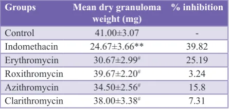
Table 3: Effect of different drugs on mean dry granuloma weight (mg) in cotton pellet induced
granuloma model. Groups Mean dry granuloma
weight (mg) % inhibition
Control 41.00±3.07
-Indomethacin 24.67±3.66** 39.82
Erythromycin 30.67±2.99# 25.19
Roxithromycin 39.67±2.20# 3.24
Azithromycin 34.50±2.56# 15.8
Clarithromycin 38.00±3.38# 7.31
Values are expressed in mean±SEM, n=6 in each group, df=5,30. **p<0.01 as compared to control, #p>0.05 as compared to control
[image:4.595.310.546.480.592.2]lateral diameter of knee joint as compared to control group
(Table 2). Anti-inflammatory activity shown by macrolides was not significant (p>0.05) as compared to indomethacin. Percentage inhibition of increase in mean lateral diameter of knee joint shown by macrolides was less as compared to indomethacin (Table 2).
Cotton pellet-induced granuloma model divided into at least three response phases, i.e., transudative phase, exudative phase and proliferative phase. The proliferative phase is measured as the increase in dry weight of the granuloma.25,26
In cotton pellet-induced granuloma model, we assessed mean dry granuloma weight. Reduction in mean dry granuloma weight is considered as anti-inflammatory action. In this model, anti-inflammatory activity of macrolides as shown by decrease in mean dry granuloma weight was not significant (p>0.05) as compared to control (Table 3). Anti-inflammatory activity shown by macrolides was not significant (p>0.05) as compared to indomethacin. Percentage inhibition of mean dry granuloma weight shown by macrolides was very less as compared to indomethacin (Table 3).
Thus, all the macrolides, i.e., erythromycin, roxithromycin, azithromycin, and clarithromycin showed significant anti-inflammatory activity in acute models of inflammation as compared to control group. However, macrolides showed insignificant activity in acute models of inflammation as compared to indomethacin. Macrolides did not show significant activity in chronic model of inflammation as compared to control group and indomethacin group.
Various mechanisms have been proposed to explain this anti-inflammatory action of macrolides. Evidences indicate that macrolides are suggested to inhibit the production of many pro-inflammatory cytokines (interleukin [IL] 1, IL 2, IL 6), nitric oxide, tumor necrosis factor-α, leukotrienes, and prostaglandins. The most probable mechanisms of acute anti-inflammatory activity of macrolide antibiotics could be due to the inhibition of synthesis of prostaglandins, serotonin, histamine and other inflammatory mediators.9,14,27 Insignificant chronic anti-inflammatory
activity of macrolides could be due to that, they do not inhibit fibroblast proliferation and synthesis of collagen in chronic inflammation.
Previous studies, such as Ianaro et al.,9 Prathima et al.,11
Khobragade et al.,14 Scaglione and Rossoni,27 and Agen
et al.,28 have also shown that macrolide antibiotics have
anti-inflammatory activity using various animal models of inflammation. Our results correlate with the findings in above studies.
So from our study, we propose that macrolide antibiotics have anti-inflammatory activity. Furthermore, these drugs have advantages such as once a day administration, greater bioavailability, and good tolerability making them a potential new anti-inflammatory drug. Since this is an animal study, these results need to be confirmed in human studies for
further establishment of the role of macrolide antibiotics in the treatment of inflammation.
CONCLUSION
From this study, we conclude that macrolides have significant anti-inflammatory activity as compared to control in animal models of acute inflammation. Further studies need to be done to establish exact role of macrolides as anti-inflammatory agents.
ACKNOWLEDGMENTS
We would like to thank, High Media Ltd., Mumbai, Maharashtra, for providing drugs in pure powder form.
Funding: No funding sources
Conflict of interest: None declared
Ethical approval: The study was approved by the Institutional Animal Ethical Committee
REFERENCES
1. Chensue SW, Ward PA. Inflammation. In: Damjanov I, Linder J, editors. Anderson’s Pathology. 10th Edition. St Louis, Missouri: Mosby Year Book; 1990: 387-413. 2. Parmar N, Rawat M, Kumar T. Evaluation of
anti-inflammatory potential of Kigella pinnata leaf extract in wistar rats. Asian J Pharm Clin Res. 2012;5(1):95-7. 3. Rang HP, Dale MM, Ritter JM. Anti-inflammatory and
immunosuppressant drugs. In: Flower RJ, editor. Rang and Dales Pharmacology. 6th Edition. China: Elsevier; 2007: 226-245.
4. Suresha RN, Brahadeesh M, Javanthi MK, Satish Am, Kalabharathi H, Pushpa VH, et al. Screening of cetrizine for its anti-inflammatory potential in albino rats. Int J Recent Sci Res. 2014;5(1):224-7.
5. Brahadeesh M, Suresha RN, Satish AM. Screening of the drug sumatriptan for its anti-inflammatory potential in albino rats. Int J Recent Trends Sci Technol. 2013;9(1):76-80. 6. Stillman MT, Schlesinger PA. Nonsteroidal
anti-inflammatory drug nephrotoxicity. Should we be concerned? Arch Intern Med. 1990;150(2):268-70.
7. Shrivastava SK. Corticosteroids. A Complete Textbook of Medical Pharmacology. 1st Edition. Sirmour, Himachal Pradesh: Avichal; 2012: 1126-7.
8. Shrivastava SK. Drug therapy of rheumatoid arthritis, gout and other types of arthritis. A Complete Textbook of Medical Pharmacology. 1st Edition. Sirmour, Himachal Pradesh: Avichal; 2012: 732-6.
9. Ianaro A, Ialenti A, Maffia P, Sautebin L, Rombolà L, Carnuccio R, et al. Anti-inflammatory activity of macrolide antibiotics. J Pharmacol Exp Ther. 2000;292(1):156-63. 10. Labro MT. Anti-inflammatory activity of macrolides:
a new therapeutic potential? J Antimicrob Chemother. 1998;41 Suppl B:37-46.
11. Prathima C, Suresha RN, Thippeswamy T. Anti-inflammatory effects of erythromycin and azithromycin in animal models. Int J Res Pharm Biomed Sci. 2012;3(3):1373-8.
Inflammation Protocols. UK: Humana Press; 2003: 115-21. 13. Suresha RN, Naidu SV, Huralikuppi J, Ashvini V. Varied
anti-inflammatory activity of indomethacin in different experimental animal models. Int J Pharm Sci Res. 2012;3(10):3993-8.
14. Khobragade AA, Patel SB, Pophale RR, Vallish BN, Kosale SP. Analgesic and anti-inflammatory activity of roxithromycin and erythromycin, alone and in combination with ibuprofen: an animal study. IOSR J Pharm. 2012;1(1):15-21.
15. Winter CA, Risley EA, Nuss GW. Carrageenin-induced edema in hind paw of the rat as an assay for antiiflammatory drugs. Proc Soc Exp Biol Med. 1962;111:544-7.
16. Ashok P, Koti BC, Thippeswamy AH, Tikare VP, Dabadi P, Viswanathaswamy AH. Evaluation of antiinflammatory activity of Centratherum anthelminticum (L) Kuntze seed. Indian J Pharm Sci. 2010;72(6):697-703.
17. Singh S, Nair V, Gupta YK. Antiarthritic activity of majoon suranjan (a polyherbal Unani formulation) in rat. Indian J Med Res. 2011;134:384-8.
18. Patwardhan MD, Muley MP, Manekar MS. Anti-inflammatory property of ranitidine, a specific H2-receptor antagonist. Indian J Physiol Pharmacol. 1986;30(3):205-9. 19. Mishra NK, Panda RK, Rajkumar V, Kumar S, Tejonidhi K,
Mishra G. Evaluation of anti-inflammatory activity and dose selection of Mollungo pentaphylla by using cotton pellet induced granuloma in rat. Int J Pharm Health Sci. 2010;1(3):155-62.
20. Choksi K, Shenoy A, Shabharaya AR, Lala M. Calcium enhances anti-inflammatory activity of aspirin. Int Res J Pharm. 2011;2(3):82-5.
21. Ching FP, Omogbai EK, Okpo SO, Ozolua RI. Antiinflammatory Activity of Aqueous Extract of
Stereospermum kunthianum (Cham, Sandrine Petit) stem
bark in rats. Indian J Pharm Sci. 2009;71(1):106-10. 22. Rappl G, Kapsokefalou A, Heuser C, Robler M, Ugurel S,
Tilgen W, et al. Dermal fibroblasts sustain proliferation of activated T cells via membrane-bound interleukin-15 upon long-term stimulation with tumor necrosis factor-alpha. J Invest Dermatol. 2012;116(1):102-9.
23. Brahmbhatt MR, Patel JM, Patel VB, Saluja AK. Analgesic and anti-inflammatory activity of leaves of rivea hypocrateriformis. J Pharm Phytother. 2010;1(1):001-3. 24. Gundamaraju R, Sheeba DS, Ramesh C. Evaluation of
anti-arthritic effects of Lantana camara var linn. Using acute model on albino rats. Int J Adv Pharm Sci. 2012;3(5):272-7. 25. Sireeratawong S, Itharat A, Lerdvuthisopon N, Piyabhan P,
Khonsung P, Boonraeng S, et al. Anti-inflammatory, analgesic, and antipyretic activities of the ethanol extract of Piper interruptum Opiz. and Piper chaba Linn. ISRN Pharmacol. 2012;2012:480265.
26. Swingle KF, Shideman FE. Phases of the inflammatory response to subcutaneous implantation of a cotton pellet and their modification by certain anti-inflammatory agents. J Pharmacol Exp Ther. 1972;183(1):226-34.
27. Scaglione F, Rossoni G. Comparative anti-inflammatory effects of roxithromycin, azithromycin and clarithromycin. J Antimicrob Chemother. 1998;41 Suppl B:47-50.
28. Agen C, Danesi R, Blandizzi C, Costa M, Stacchini B, Favini P, et al. Macrolide antibiotics as antiinflammatory agents: roxithromycin in an unexpected role. Agents Actions. 1993;38(1-2):85-90.