R E V I E W
Open Access
Restricting retrotransposons: a review
John L. Goodier
Abstract
Retrotransposons have generated about 40 % of the human genome. This review examines the strategies the cell has
evolved to coexist with these genomic
“
parasites
”
, focussing on the non-long terminal repeat retrotransposons of humans
and mice. Some of the restriction factors for retrotransposition, including the APOBECs, MOV10, RNASEL, SAMHD1, TREX1,
and ZAP, also limit replication of retroviruses, including HIV, and are part of the intrinsic immune system of the cell. Many
of these proteins act in the cytoplasm to degrade retroelement RNA or inhibit its translation. Some factors act in the
nucleus and involve DNA repair enzymes or epigenetic processes of DNA methylation and histone modification. RISC and
piRNA pathway proteins protect the germline. Retrotransposon control is relaxed in some cell types, such as neurons in
the brain, stem cells, and in certain types of disease and cancer, with implications for human health and disease. This
review also considers potential pitfalls in interpreting retrotransposon-related data, as well as issues to consider for future
research.
Keywords:
Alu, Autoimmunity, Epigenetics, LINE-1, Methylation, Restriction, Retrovirus, RNAi, SINE, SVA
Background
Sixty-five years on from Barbara McClintock’s seminal
discovery of mobile DNA [1] we now understand that
genomes are dynamic and changeable, with transposable
elements (TEs) being major contributors to their fluidity.
We recognize that TEs, sometimes called
“junk DNA”,
are major players in genome evolution and have helped
shape the form and function of many genes [2].
Never-theless, TEs are foremost parasitic DNA, and parasites
must be controlled or they will destroy a host. There is
far more junk than treasure in mobilomes.
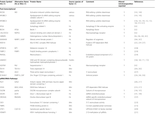
DNA transposons comprise about 3 % of the human
genome and most move by a
“cut and paste”
mechanism
involving excising an element and reinserting it elsewhere
(Fig. 1 [3]). With the exception of at least one family of
piggyBac elements in little brown bats [4], no active DNA
transposons are known in mammals. There are two
clas-ses of retrotransposon. Both move by a
“copy and paste”
mechanism, involving reverse transcription of an RNA
intermediate and insertion of its cDNA copy at a new site
in the genome. LTR retrotransposons are named for the
long terminal repeats that flank their sequences (reviewed
in [5–7]). Endogenous retroviruses (ERVs) are relics of
past germline viral infections and for the most part are
highly mutated. However, some intracisternal A-particle
(IAP) and Etn/MusD family LTR elements remain
inser-tionally active in mice [8], and formation of infective virions
by recombination or phenotypic mixing of intact proteins
from different ERV proviruses has been reported [9–12].
Among the 31 human endogenous retrovirus subfamilies
extant in the human genome, no replication-competent
HERVs are known, although their existence has not been
ruled out [13, 14], and recently an unfixed fully intact
HERV-K (HML2) provirus was identified in some
individ-uals [15]. Many HERVs, and their lone LTRs that populate
the genome as a consequence of non-homologous
recom-bination, remain capable of expression and may act as
tran-scriptional regulatory elements for genes (reviewed in [16]).
Non-LTR retrotransposons are as old as the earliest
multi-cellular organisms and their 28 clades have origins
in the Precambrian Era of 600 million years ago [17, 18].
Long Interspersed Elements (LINEs) and Short
Inter-spersed Elements (SINEs) comprise most of this group
in mammals. LINE-1s (L1s), the only currently active
autonomous mobile DNA in humans, have been
evolv-ing durevolv-ing at least 150 million years of mammalian
radi-ation. Multiple active L1 lineages coexisted in ancestral
primates, but for the past 40 Myr there has been a single
unbroken lineage of subfamilies [19, 20]. Expansion of L1s
was massive, and roughly 500,000 copies now occupy about
17 % of the human genome. Remnant copies of extinct L2
Correspondence:[email protected]
McKusick-Nathans Institute for Genetic Medicine, Johns Hopkins University School of Medicine, Baltimore, MD, USA212051
© 2016 The Author(s).Open AccessThis article is distributed under the terms of the Creative Commons Attribution 4.0 International License (http://creativecommons.org/licenses/by/4.0/), which permits unrestricted use, distribution, and reproduction in any medium, provided you give appropriate credit to the original author(s) and the source, provide a link to the Creative Commons license, and indicate if changes were made. The Creative Commons Public Domain Dedication waiver (http://creativecommons.org/publicdomain/zero/1.0/) applies to the data made available in this article, unless otherwise stated. GoodierMobile DNA (2016) 7:16
and L3 family elements comprise an additional 4 %
[21]. L1s have also been responsible for genomic
inser-tion of 8000 processed pseudogenes and over a million
non-autonomous SINEs [22]. B1s and Alus, the
pre-dominant SINEs of mice and men, respectively, originate
from the 7SL RNA component of the signal recognition
particle. Alus are about 300 base pairs in length with a
dimeric structure; B1s are monomeric (reviewed in [23]).
SVAs are hominid-specific SINEs, and the youngest family
of active human retrotransposons. Their name is an
acro-nym reflecting their composite nature: a
HERV-K(HML-2)-derived SINE-R, variable-number-of-tandem-repeats
(VNTR), and an Alu-like region. There are roughly 2700
SVA copies per human genome, most of which are
full-length and about 50 of which may be active [24–31].
SVA-like variants have been described, including a
human-specific subfamily generated by fusion of the first intron of
the MAST2 gene with an SVA [29, 32, 33], and the LAVA,
PVA and FVA elements of non-human primates [34–36].
From 12 Myr ago, the primate LINE-1 expansion
slo-wed, and most insertions are molecular fossils, truncated,
rearranged, or mutated [20]. However, although most L1s
no longer
“jump”, at least 100 remain potentially mobile
in any individual diploid human genome [37, 38]. Many
more L1s are transcribed. Interestingly, only a small
num-ber of the active L1s are
”hot”
for retrotransposition and
Fig. 1Types of transposable elements in mammals. Abbreviations: DR, direct repeat; ITR, inverted terminal repeat; Gag, group-specific antigen; Prt, protease; Pol, polymerase; Env, envelope; RT, reverse transcriptase domain; INT, integrase domain; TSD, target site duplication; LTR, long terminal repeat; EN, endonuclease domain; C, zinc knuckle domain; An, poly (A); A/B, A- and B-box Pol III promoter; SVA, SINE-R, VNTR, Alu element; VNTR, variable number tandem repeats (reproduced from [3]; Elsevier license number 3803340576977)
these have accounted for most
de novo
insertions.
How-ever, when several of these
“hot”
Ta-1 L1s were examined
across diverse human populations, considerable individual
allelic variation affected their ability to retrotranspose
[39]. Up to 5 % of newborn children have a new
retro-transposon insertion, and to date there are 124 known
human disease-causing germline insertions of L1s, Alus,
and SVAs [40–42]. The current residual activity of human
retrotransposons is the background that escapes a variety
of mechanisms that have evolved to limit replication of
mobile DNA. This review focuses on mammalian
non-LTR retrotransposons and how the cell controls them.
Non-LTR retrotransposons are mobilized by a
mechan-ism very different from that used by retroviruses and LTR
retrotransposons. Extensive biochemical analyses of insect
R1 and R2 elements, together with genomic sequence
ana-lyses, indicate that L1s likely retrotranspose by a process
known as target-primed reverse transcription (TPRT) that
occurs at the site of DNA insertion. According to this
model, L1-encoded endonuclease nicks the bottom strand
of target DNA exposing a 3'-hydoxyl that primes reverse
transcription of bound L1 RNA. Second-strand DNA
syn-thesis follows and the integrant is resolved in a manner still
poorly understood [43]. Short target site duplications
(TSDs) of variable length, and occasionally deletions, are
generated at new L1 insertion sites.
The 6 kilobase bicistronic L1 has a 5' untranslated region
(UTR) that functions as an internal promoter, a 3' UTR that
ends in a poly (A) signal and tail, and two open reading
frames (ORF1 and ORF2) on the sense strand. A weak
pro-moter on the antisense strand of the human 5' UTR [44]
lies upstream of a recently identified 216-nt
translation-competent ORF0 [45]. Unlike human L1s, mouse L1s have
a 5′
UTR consisting of tandemly repeated
∼
200 bp
sequences called monomers [46]. ORF2 encodes a 150 kD
protein with endonuclease and reverse transcriptase (RT)
activities. While the 40 kD ORF1p RNA-binding protein is
essential for LINE-1 retrotransposition, its precise function
remains unclear, although it possesses chaperone activity
in
vitro
[47, 48]. Early L1 investigations showed ORF1p to be
predominantly cytoplasmic where it forms large aggregates,
subsequently identified as stress granules (SGs) and
pro-cessing bodies (PBs) [49
–
51]. Endogenous L1 RNA has also
been detected in PBs [52]. SGs are discrete cytoplasmic
ag-gregates which can be induced by a range of stress
condi-tions, including heat shock, osmotic shock, oxidative stress,
viral infection, and overexpression of some proteins. PBs
are dynamic cytoplasmic compartments containing
mole-cules involved in mRNA decay and translation inhibition
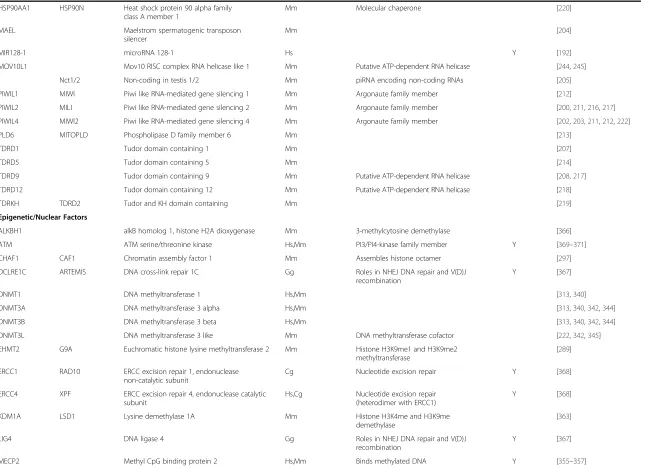
(reviewed in [53, 54]). ORF1p can also concentrate at the
perinucleus, is detected faintly in the nucleus, and is seen
in nucleoli of a small fraction of cells [55
–
57] (Fig. 2).
Expressed from a full-length L1 construct, ORF1p is
present in SGs as a ribonucleoprotein (RNP) complex
together with L1 RNA, ORF2p, and many other
RNA-binding proteins [58, 59]. Recently, endogenous ORF1p and
ORF2p have been reported to also colocalize in nuclear foci
of cancer cells [60].
How retrotransposons impact the mammalian cell
and genome has been the subject of many other reviews
[3, 41, 42, 61–67]. These effects extend beyond simple
mutation by genomic insertion. L1 RNA and protein
overexpression has been linked with apoptosis, DNA
damage and repair, tumor progression, cellular
plasti-city, and stress response [68–72]. Consequently, the cell
has evolved a battery of defenses to protect against the
dangers of unfettered retrotransposition. It is not
surpris-ing that many of the known anti-retrotransposon
restric-tion factors are also anti-retroviral. Phylogenetic analyses
suggest that eukaryote non-LTR retrotransposons predate
LTR retrotransposons, which in turn gave rise to
retrovi-ruses through the acquisition of an envelope (env) gene
[73–76]. Indeed, some restriction factors may have first
evolved to control ancient endogenous retroelements and
were later recruited to the fight against exogenous
in-vaders. It is reasonable to presume that from the study of
factors controlling endogenous retrotransposition new
in-sights into the control of viral infections will emerge.
Until recently, our knowledge of the cellular factors that
interact with mammalian retrotransposons to facilitate or
frustrate their activity lagged behind our understanding of
such factors in yeast and flies [77–80]. Nevertheless, in
recent years, with the aid of mouse transgenic models,
improved antibodies, efficient strategies for
immunopreci-pitating retrotransposon RNP complexes from cells, new
high-throughput (HT) DNA resequencing strategies, and
cell culture retrotransposition assays, we have significantly
increased our understanding of how the mammalian cell
at-tempts to coexist with a molecular parasite whose
un-checked activity could be bad news indeed.
Of all the tools in the toolbox of mammalian
retrotrans-poson research, after twenty years the cell culture assay
for retrotransposition remains the most important (Fig. 3;
reviewed in [81, 82]). It built upon earlier assays that
tracked Ty1 LTR retrotransposition in budding yeast [83].
A reporter gene cassette, interrupted by a backwards
in-tron and inserted in opposite transcriptional orientation
into the 3' UTR of a retrotransposition-competent L1, is
expressed only when the L1 transcript is spliced,
reverse-transcribed, its cDNA inserted in the genome, and the
reporter gene expressed from its own promoter. The
ori-ginal neomycin phosphotransferase gene reporter [84–86]
was later joined by enhanced green fluorescent protein,
blasticidin S-resistance, firefly luciferase, and secreting
gaussia luciferase gene constructs [51, 87–89] (Fig. 3).
Alu, SVA, and mouse SINE non-LTR, and IAP and HERV
LTR retrotransposition assays have also been established
[10, 90–96]. While immensely effective in revealing
cis
and
trans
-acting factors of retrotransposition, the degree
to which these plasmid-based assays truly reflect
endogen-ous levels of retrotransposition is often uncertain.
Fortu-nately, cell culture results can now be confirmed by HT
genome sequencing [97].
Lines of defense
To a significant degree, non-LTR retrotransposon sequence
itself and the nature of TPRT mitigate genomic insertions.
Most L1s die at the time of TPRT, undergoing 5'
trunca-tions or inversions, or internal deletrunca-tions. Most of the
inser-tions that remain intact ultimately lose their ability to
remobilize due to DNA recombination or mutation [98]. It
has also been suggested that the adenosine richness of the
L1 template retards processivity of transcription and limits
retrotransposition [99]. Mutations to binding sites for
tran-scription/enhancer factors, including E2F1/RB1, ETS, p53,
RUNX3, SOX2, SP1, TCF-LEF, and YY1 for L1s and AHR,
CTCF, RAR and SLUG for mouse SINEs, modulate TE
expression and in some cases retrotransposition [100–116]
(All factors and their full names are listed in Table 1).
Cryp-tic splice sites in L1 RNA transcripts induce a complex
pattern of splicing that may remove portions of the ORFs
or the 5' UTR [117, 118]. Alus also contain cryptic splice
sites, and when resident in genes are frequently exonized
into mRNA transcripts and are occasionally translated
[119–122]. Interestingly, heterogeneous nuclear
ribonucleo-protein C (HNRNPC) protects the cell from Alu-mediated
aberrant exonization by competing with splicing factor
U2AF2 for binding at Alu splice sites [123]. Alu lacks its
own Pol III transcription termination signal, requiring
termination at signals in downstream flanking DNA with
possible loss of retrotransposition efficiency. The L1
pos-sesses a poly (A) termination signal that is inherently weak
and permits occasional read-through of L1 transcripts,
necessitating their termination at signals downstream.
Interestingly,
in silico
studies show that approximately
15 % of L1s have transduced 3’
flanking DNA to a new
genomic location, in the process generating between 19
and 30.5 Mb of new DNA or as much as 1 % of the
hu-man genome [124–127]. Cryptic polyadenylation signals
are also scattered along the A-rich length of the L1, and
consequently a majority of L1 RNAs are prematurely
trun-cated and incapable of forming functional RNPs [99, 128].
Post-translational protein modifications, including
phos-phorylation of ORF1p [129, 130], may also modulate
retrotransposition.
The cell has also evolved a phalanx of trans-acting
re-striction factors that function as an early defense against
both viral infection and endogenous retroelements. Many
of these proteins are involved in nucleic acid metabolism
and may be constitutively expressed or induced, often by
type I interferons. Typically they form a rapid response to
infection, and act in the cytoplasm. Early examples were
found by comparing cell lines that were permissive or
re-strictive for viral infection.
Apolipoprotein B mRNA editing enzyme, catalytic
polypeptide-like (APOBEC)/ Activation-induced cytidine
deaminase (AID) proteins
The first anti-retrotransposon restriction factors identified
were AID/APOBEC proteins, an evolutionarily conserved,
vertebrate-specific family of cytidine deaminases. While
rodents have a single APOBEC3 family member, humans
have seven APOBEC3s (A3A-D, A3F, A3G and A3H). It
was discovered that A3G is packaged into virions of
Vif-deficient HIV-1, where during reverse transcription it
deaminates cytosines to uracils in the nascent first-strand
HIV cDNA. Uracils in the cDNA cause dG > dA
hypermu-tations during second strand synthesis, limiting viability of
Fig. 2Subcellular distribution of LINE-1 ORF1 protein.a. Endogenous ORF1p detected in human embryonal carcinoma 2102Ep cells by a monoclonal antibody [57]. ORF1p is mostly cytoplasmic where it concentrates in SGs and PBs and occasionally at the nuclear membrane. It is faintly detectable in some nuclei and concentrates in nucleoli of a small percentage of cells. Expression of GFP-tagged TDP43 in nuclei but not in nucleoli is shown as a marker.b. Exogenously expressed GFP-tagged ORF1p strongly concentrates at the nuclear membrane and in perinucleolar foci of 5 % or fewer human embryonic kidney (HEK) 293T cells, with attendant reduction in size and number of cytoplasmic granules (left panel). Construct ORF1-EGFP L1-RP contains a CMV promoter, ORF1 C-terminally tagged with EGFP, followed by intact downstream L1 sequence. Nucleoli are marked byα-C23 (nucleolin) antibody (Santa Cruz) and nuclei are stained with Hoechst (right panel)
Fig. 3Cell culture retrotransposition assay reporter gene cassettes come in a variety of flavors.a. LINE-1 assays. A retrotransposition-competent L1 and reporter cassette is cloned in pCEP4 (Invitrogen)-based vectors, which encode EBNA-1 and OriP and so replicate in primate cells. Variants of the vectors also contain or lack an exogenous promoter upstream of the L1, and encode resistance to hygromycin or puromycin permitting antibiotic selection of transfected cells.mneoIandmblastIreporter cassettes confer drug resistance to cells having a retrotransposition event. These cells are expanded in culture to form colonies, fixed, stained, and the number of colonies scored. ThemEGFPIcassette fluorescently marks cells with retrotransposon insertions and allows their numbers to be counted by flow cytomentry. Firefly luciferase genemFlucIreporter vectors may be cotranfected with pGL4.73 (Promega) or other vector which constitutively expresses renilla luciferase from transfected cells. Following cell lysis, retrotransposition levels, indicated by firefly luciferase, are adjusted to renilla expression to control for differences in transfection efficiency. The
mGlucIcassette expresses Gaussia luciferase which when secreted into the media serves as an effective read-out of accumulated retrotransposition events. Levels of Gluc may be normalized to those of Cypridina luciferase (which is also secreted and does not cross-react with Gluc) constiitutively expressed from the cotransfected pSV40-CLuc vector (NEB). Simply by sampling small aliquots of cell culture media, retrotransposition may be assessed in a single well at multiple time points without cell lysis. Luciferase-based reporter cassettes are amenable to HT retrotransposition screening.b. The Alu assay. An active Ya5 Alu and neoTETcassette interrupted by aTetrahymena thermophilaself-splicing 23S rRNA Group I intron is cloned between the 7SL pol III enhancer and terminator. When this construct is co-expressed with L1 ORF2 alone or a full-length retrotransposition-competent L1, Alu RNAs are reverse transcribed along with the splicednptgene and integrated into the genome to confer neomycin resistance. Abbreviations: 7SL enh, 7SL enhancer; 7SL TTTT, 7SL transcription terminator; ampR, ampicillin resistance gene; bsd, blasticidin S deaminase gene; CMV, cytomegalovirus promoter; EBNA-1, Epstein-Barr nuclear antigen 1; EGFP, enhanced green fluorescent protein; L mon, left monomer; mini, chimeric mini-intron of the plasmid psiCHECK-2 (Promega); npt, neomycin phosphotransferase gene; oriP, latent origin of replication; pCI: synthetic intron from pCI (Promega); R mon, right monomer; SA, splice acceptor; SD, splice donor; SV40, simian virus 40 early enhancer/promoter; TET,T. thermophilaself-splicing intron; TK, herpes simplex virus thymidine kinase poly(A) signal
Table 1
Cellular factors associated with mammalian non-LTR retrotransposon activity
Protein Symbol (Hs) (1)
Alternative Name (Hs or Mm) (1)
Protein Name Source species of factor (1)
Comment Altered
retrotransposition reported
References
Post-transcriptional
AICDA AID Activation-induced cytidine deaminase various RNA-editing cytidine deaminase Y [142,144]
APOBEC1 Apolipoprotein B mRNA editing enzyme
catalytic subunit 1
various RNA-editing cytidine deaminase Y [143,145]
APOBEC3 Apolipoprotein B mRNA editing enzyme
catalytic subunit 3
Hs RNA-editing cytidine deaminase Y [136,145,149,152,153,
160,515–521]
ATG5 Autophagy related 5 Hs Autophagy E1-like activating enzyme Y [52]
BECN1 ATG6 Beclin 1 Mm Autophagy factor Y [52]
CALCOCO2 NDP52 Calcium binding and coiled-coil domain 2 Hs Macroautophagy receptor Y [52]
HNRNPL Heterogeneous nuclear ribonucleoprotein L Hs Y [56,236,240,261]
KIAA0430 MARF1, LKAP Meiosis arrest female protein 1 Hs Regulator of oogenesis [266,267]
MOV10 Mov10 RISC complex RNA helicase Hs Putative ATP-dependent RNA
helicase
Y [232,234–237]
MTNR1A MT1 Melatonin receptor 1A Hs Y [258]
PABPC1 PABP1 Poly(A) binding protein cytoplasmic 1 Hs Y [259]
RNASEL Ribonuclease L Hs Endoribonuclease/component of
2-5A system
Y [254]
SAMHD1 SAM and HD domain containing deoxynucleoside
triphosphate triphosphohydrolase 1
Hs,Mm Y [168,169,171,172]
SQSTM1 P62 Sequestosome 1 Hs Macroautophagy receptor Y [52]
TEX19 TEX19.1 Testis expressed 19 Mm [264,265]
TREX1 AGS1 Three prime repair exonuclease 1 Mm 3' exonuclease Y [257]
ZC3HAV1 PARP13, ZAP Zinc finger CCCH-type containing, antiviral 1 Hs Antiviral protein Y [236,240,250]
piRNA/RNAi-Pathways
ASZ1 GASZ Ankyrin repeat, SAM and basic leucine zipper domain containing 1
Mm [206,217]
DDX4 MVH, VASA DEAD-box helicase 4 Mm ATP-dependent RNA helicase [210,217]
DGCR8 pasha DGCR8 microprocessor complex subunit Hs Subunit of microprocessor Y [196,197]
DICER1 DCR1 Dicer 1, ribonuclease type III Hs dsRNA endoribonuclease Y [191,194]
DROSHA RNASEN Drosha ribonuclease type III Hs dsRNA-specific endoribonuclease/
subunit of microprocessor
Y [197]
EXD1 Exonuclease 3'-5' domain containing 1 Mm 3'-5' exonuclease activity [223]
FKBP6 FK506 binding protein 6 Mm Cis-trans peptidyl-prolyl isomerase [215]
GTSF1 CUE110 Gametocyte specific factor 1 Mm UPF0224 (FAM112) family member [209]
HENMT1 HEN1 methyltransferase homolog 1 Mm 2'-O-methylation pf piRNAs [221]
Goodier
Mobile
DNA
(2016) 7:16
Page
6
of
Table 1
Cellular factors associated with mammalian non-LTR retrotransposon activity
(Continued)
HSP90AA1 HSP90N Heat shock protein 90 alpha family class A member 1
Mm Molecular chaperone [220]
MAEL Maelstrom spermatogenic transposon
silencer
Mm [204]
MIR128-1 microRNA 128-1 Hs Y [192]
MOV10L1 Mov10 RISC complex RNA helicase like 1 Mm Putative ATP-dependent RNA helicase [244,245]
Nct1/2 Non-coding in testis 1/2 Mm piRNA encoding non-coding RNAs [205]
PIWIL1 MIWI Piwi like RNA-mediated gene silencing 1 Mm Argonaute family member [212]
PIWIL2 MILI Piwi like RNA-mediated gene silencing 2 Mm Argonaute family member [200,211,216,217]
PIWIL4 MIWI2 Piwi like RNA-mediated gene silencing 4 Mm Argonaute family member [202,203,211,212,222]
PLD6 MITOPLD Phospholipase D family member 6 Mm [213]
TDRD1 Tudor domain containing 1 Mm [207]
TDRD5 Tudor domain containing 5 Mm [214]
TDRD9 Tudor domain containing 9 Mm Putative ATP-dependent RNA helicase [208,217]
TDRD12 Tudor domain containing 12 Mm Putative ATP-dependent RNA helicase [218]
TDRKH TDRD2 Tudor and KH domain containing Mm [219]
Epigenetic/Nuclear Factors
ALKBH1 alkB homolog 1, histone H2A dioxygenase Mm 3-methylcytosine demethylase [366]
ATM ATM serine/threonine kinase Hs,Mm PI3/PI4-kinase family member Y [369–371]
CHAF1 CAF1 Chromatin assembly factor 1 Mm Assembles histone octamer [297]
DCLRE1C ARTEMIS DNA cross-link repair 1C Gg Roles in NHEJ DNA repair and V(D)J
recombination
Y [367]
DNMT1 DNA methyltransferase 1 Hs,Mm [313,340]
DNMT3A DNA methyltransferase 3 alpha Hs,Mm [313,340,342,344]
DNMT3B DNA methyltransferase 3 beta Hs,Mm [313,340,342,344]
DNMT3L DNA methyltransferase 3 like Mm DNA methyltransferase cofactor [222,342,345]
EHMT2 G9A Euchromatic histone lysine methyltransferase 2 Mm Histone H3K9me1 and H3K9me2
methyltransferase
[289]
ERCC1 RAD10 ERCC excision repair 1, endonuclease non-catalytic subunit
Cg Nucleotide excision repair Y [368]
ERCC4 XPF ERCC excision repair 4, endonuclease catalytic subunit
Hs,Cg Nucleotide excision repair (heterodimer with ERCC1)
Y [368]
KDM1A LSD1 Lysine demethylase 1A Mm Histone H3K4me and H3K9me
demethylase
[363]
LIG4 DNA ligase 4 Gg Roles in NHEJ DNA repair and V(D)J
recombination
Y [367]
MECP2 Methyl CpG binding protein 2 Hs,Mm Binds methylated DNA Y [355–357]
Goodier
Mobile
DNA
(2016) 7:16
Page
7
of
Table 1
Cellular factors associated with mammalian non-LTR retrotransposon activity
(Continued)
MORC1 MORC family CW-type zinc finger 1 Mm Role in early spermatogenesis [352]
PRKDC XRCC7,
DNA-PKcs
Protein kinase, DNA-activated, catalytic polypeptide
Cg NHEJ DNA double-strand break
repair
Y [68,72]
SIRT6 Sirtuin 6 Mm NAD-dependent protein deacetylase Y [473]
SUV39H Suppressor of variegation 3–9 homolog 1 Hs,Mm Histone H3-K9 methyltransferase 1 [286,288,291]
TRIM28 KAP1 Tripartite motif containing 28 Hs,Mm Nuclear corepressor for KRAB-ZFPs [313]
UHRF1 NP95, ICBP90 Ubiquitin-like with PHD and ring finger domains 1
Mm RING-finger type E3 ubiquitin ligases [351]
XRCC4 X-ray repair cross complementing 4 Cg DNA single-strand break repair Y [68,72]
XRCC6 KU70 X-ray repair cross complementing 6 Gg ssDNA-dependent ATP-dependent
helicase
Y [367]
Krüppel-associated box domain-containing zinc finger proteins (KRAB-ZFPs)
GM6871 Predicted gene 6871 Mm [313]
ZBTB16 PLZF Zinc finger and BTB domain containing 16 Hs,Mm Y [317]
ZFP819 Zinc finger protein 819 Mm [314]
ZNF91 Zinc finger protein 91 Hominidae [315]
ZNF93 Zinc finger protein 93 Hominidae Y [315]
Other transcription factors
AHR Aryl hydrocarbon receptor Mm,Dr Ligand-activated transcription factor [109,114]
CTCF CCCTC-binding factor various BORIS + CTCF gene family member [108,116]
ETS1 ETS proto-oncogene 1, transcription factor Hs ETS transcription family member [102]
RAR Retinoic acid receptor Hs Thyroid-steroid hormone receptor
superfamily member
[107,115]
RB/E2F1 RB transcriptional corepressor proteins/E2F transcription factor 1
Hs,Mm Transcription repressor complex [112,298]
RUNX3 Runt-related transcription factor 3 Hs Runt domain-containing transcription
family member
Y [104]
SNAI2 SLUG Snail family transcriptional repressor 2 Mm,Dr Snail C2H2-type zinc finger
transcription family member
[109,114]
SOX2 SRY-box 2 Hs,Mm SRY-related HMG-box (SOX)
transcription family member
[103,106,111]
SP1 Sp1 transcription factor Hs zinc finger transcription factor [102]
TCF-LEF T-cell factor/lymphoid enhancer factor Rn Wnt transcription factors [111]
TP53 p53 tumor protein p53 Dr,Hs,Mm tumor suppressor protein Y [110,299,300]
YY1 YY1 transcription factor Hs,Mm GLI-Kruppel zinc finger transcription
family member
[101,105,113]
(1) Cg,Cricetulus griseus; Cl,Canis lupus; Dr,Danio rerio; Gg,Gallus gallus; Hs,Homo sapiens; Mm,Mus musculus, Rn,Rattus norvegicus
Goodier
Mobile
DNA
(2016) 7:16
Page
8
of
the viral progeny [131–133]. In the past 10 years, A3B and
A3F have also been shown to have antiretroviral activity,
and some APOBEC3 proteins are effective against other
classes of virus (reviewed in [134]).
The discoveries that AID/APOBEC proteins restrict
not only infecting viruses but also LTR and non-LTR
retrotransposons have been summarized in previous
re-views [135–141]. All APOBEC3 proteins inhibit LINE-1
retrotransposition to varying degrees, with A3A and
A3B being most effective. AID and APOBEC1 proteins
both inhibit cell culture L1 and LTR element
retrotranspo-sition [142–145]. AID may also promote methylation of
TEs in the nuclei of primordial germ cells (PGCs) [146].
Interestingly, Khatua et al. [147] revealed a way in which
restriction may be transferred from one cell to another,
showing that extruded exosome vesicles can encapsulate
A3F and A3G mRNAs and be taken up by other cells to
inhibit their ability to support Alu and L1
retrotransposi-tion. Tumor-derived microvesicles are also enriched in
LINE-1, Alu, and especially HERV RNAs [148]. Apart
from being a potential conduit for moving TEs between
cells, these tumor-derived microvesicles may make useful
cancer biomarkers if they can be confidently detected in
human blood or sera.
Unexpectedly, catalytically inactive APOBEC3s still
inhibit non-LTR retrotransposons, and several
investiga-tions found scant genomic evidence for L1 editing by
cyti-dine deamination [149–151]. Deamination-independent
mechanisms of APOBEC action were therefore proposed,
including sequestration of retrotransposon RNPs in high
molecular weight cytoplasmic complexes and their
target-ing to SGs and PBs for possible degradation by RNAi
silencing [152–158]. However,
in silico
analyses by Carmi et
al. [159] confirmed extensive editing of LTR
retrotranspo-sons and found strong evidence for editing of SVAs (20 %)
and mouse L1 elements (0.74 %), but minimal editing of
human
L1s
(which
occurred
mostly
within
older
subfamilies). Richardson et al. [160] then proposed that
annealed L1 RNA normally protects first-strand cDNA
from deamination, but that transiently exposed
single-stranded (ss) cDNA occurring during TPRT becomes
accessible to deamination by A3A. Normally the cell repairs
U mutations, but by inhibiting uracil DNA glycosylase in
cell culture, these authors detected A3A-induced L1
muta-tions. Moreover, overexpression of both A3A and RNase H,
which degrades RNA:DNA hybrids, increased L1 cDNA
mutation in an
in vitro
RT assay [161]. HIV, unlike L1,
encodes RNase H activity, which may make its cDNA more
susceptible to APOBEC3-mediated deamination.
SAM domain and HD domain 1 (SAMHD1)
Another important member of the anti-retroviral arsenal is
SAMHD1, a dGTP-activated deoxynucleoside triphosphate
triphosphohydrolase. It has been proposed that SAMHD1
degrades the dNTP pool in non-dividing cells to levels
below that necessary for reverse transcription of
retrovi-ruses and replication of some DNA viretrovi-ruses [162–166]. Loss
of SAMHD1 has been linked to Aicardi-Goutières
syn-drome (AGS), an early-onset inflammatory disorder
affect-ing particularly the brain [167].
Overexpression of SAMHD1 inhibits, while coexpression
of SIV-encoded accessory protein viral protein X (Vpx) or
depletion of endogenous SAMHD1 increases non-LTR
ret-rotransposition in cell culture. Seven of eight AGS-related
mutations in SAMHD1 reduced inhibition of cell culture
LINE-1 retrotransposition by 40 % or more [168]. On the
other hand, nine naturally occurring polymorphisms failed
to alter SAMHD1 inhibition of retrotransposition [169].
One might expect patients with mutant SAMHD1 alleles
to show increased retrotransposition; however, sequencing
of bulk tissue and single neurons from the brain of one
AGS patient revealed no increase of L1 insertions
com-pared with controls [170].
Although SAMHD1 restricts HIV and SIV in
non-dividing cells only, non-LTR retrotransposition is reduced
in dividing cells where dNTPs are constantly replenished.
Furthermore, SAMHD1 proteins with mutations in the
NTPase catalytic domain or at a residue whose
phosphor-ylation is important for retroviral restriction still inhibit
cell culture retrotransposition [168] (although Hu et al.
[171] reported an NTPase mutant that failed to inhibit
retrotransposition). Tetramer formation by SAMHD1 is
required for both dNTPase activity and regulation of
HIV-1 and LINE-HIV-1s [HIV-172].
These data predict a mechanism other than dNTPase
activity for restricting L1s. SAMHD1 also possesses
ribonuclease activity, which even in the absence of
functional dNTPase inhibits HIV-1 replication [173]: its
effect on retrotransposons remains to be tested. Zhao
et al. [168] reported that SAMHD1 reduced L1 reverse
transcription by inhibiting ORF2p but not ORF1p. Hu
et al. [171] proposed a novel mechanism whereby
SAMHD1 enhances assembly of cytoplasmic stress
granules that then sequester L1 RNPs and prevent their
retrotransposition. Depletion of SG proteins G3BP1
(which binds the L1 RNP) or TIA1 prevented SG
for-mation and reduced SAMHD1 inhibition of LINE-1s.
While LINE-1 proteins and RNA concentrate in SGs
and PBs along with factors linked with their restriction,
a direct role for cytoplasmic granules in modulating
retrotransposition remains unclear. Previous
experi-ments investigated PBs and LTR retrotransposons only,
and results were conflicted. PBs were required for yeast
Ty1 and Ty3 virus-like particle (VLP) assembly and
ret-rotransposition [174–176], but PBs inhibited mouse
IAPs [157]. It remains to be determined if cytoplasmic
aggregates are a retrotransposition dead-end or an
inte-gral part of the L1 life cycle.
RNA-induced Silencing Complex (RISC) and Piwi-interacting
RNA (piRNA) pathway proteins
Small interfering RNA (siRNA)-mediated post-transcriptional
gene silencing is an ancient strategy for limiting the
spread of mobile genetic elements. RNA interference
(RNAi) can act at the post-transcriptional level by
causing RNA degradation and loss of translation, or
at the transcriptional level by inducing epigenetic
modifications. Several lines of evidence suggest a
dir-ect role for small RNAs in mammalian
retrotrans-poson silencing (reviewed in [177–183]. A large
number of endogenous retrotransposon-related small
RNAs of a size consistent with siRNAs, miRNAs and
piRNAs have been detected in cells [179, 184–188]
(reviewed in [189]). Treating cells with
in vitro
diced
L1 siRNAs hindered cell culture retrotransposition [190],
and L1-related endo-siRNAs decreased retrotransposon
activity, apparently by promoter hypermethylation [191].
Recently, a specific microRNA, mir-128, was found to bind
L1 RNA and repress its integration in HeLa and induced
pluripotent stem cells (iPSCs) [192]. Indeed, it has been
proposed that miRNAs originally evolved from TEs [189].
The question remains, however, as to whether RNAi
path-ways evolved to silence TEs themselves or gene transcripts
that happened to contain target TE sequences [193].
In the nucleus, DGCR8 binds DROSHA, an RNase
III-type enzyme, to form the Microprocessor complex.
Microprocessor cleaves primary miRNAs (pri-miRNAs),
which are then further processed in the cytoplasm to
mature miRNAs by DICER and loaded into Argonaute
(AGO)-containing RISCs. Knockdown of DICER1 (which
also processes siRNAs from dsRNAs) or AGO2 causes an
increase in the rate of retrotransposition of tagged L1s in
cell culture [191, 194]. Elevated transcription of murine
L1 and IAP elements has been observed in embryonic
stem cells (ESCs) of
Dicer
-null mice [195]. Interestingly,
DGCR8 also directly binds L1- and SINE-derived RNAs,
presumably at hairpin structures, which are apparently
cleaved by Microprocessor in a manner independent of
DICER and miRNAs. Both DROSHA and DGCR8 affect
cell culture retrotransposition [196–198]. Non-LTR
retro-transposon RNAs that escape Microprocessor surveillance
in the nucleus may be captured in the cytoplasm for
further processing by DICER and RISC loading.
piRNAs are small RNAs slightly longer than siRNAs
(24
–
30 nt) that are processed independently of DICER
and silence TEs specifically in the germline. They
medi-ate both PIWI protein endonuclease-slicer activity [199]
and
de novo
methylation of TE sequences (discussed
below). A large proportion of mouse prepachytene piRNAs
derives from retrotransposon sequences [200–202], and the
importance of piRNA pathway proteins in repressing
retro-transposons in prenatal gonad development and
spermato-genesis has repeatedly been demonstrated in mutant mouse
lines. Loss of EXD1, FKBP6, GASZ/ASZ1, GTSF1,
HENMT1, HSP90α, MAEL, MILI/PIWIL2, MIWI/PIWIL1,
MIWI2/PIWIL4, MVH/DDX4, PLD6/MITOPLD, TDRD1,
TDRD5, TDRD9, TDRD12, or TDRKH/TDRD2 protein, or
the piRNA-encoding non-coding RNAs Nct1/2 is
accom-panied by derepression of LINE-1 and IAP
retrotranspo-sons [201–223]. These studies generated much discussion
in the RNAi and retrotransposon fields. However, they
failed to provide a crucial piece of information: do the
ob-served accumulation of retrotransposon RNAs and proteins
mean increased numbers of endogenous insertion events?
Also, it remains to be determined if increased
retrotranspo-sition contibutes to the male germline defects and sterility
observed in many of these knockout (KO) mice. With the
advance of HT genome sequencing, this information can
now be obtained.
Moloney leukemia virus 10, homolog (mouse) (MOV10)/
Moloney leukemia virus 10-like 1, homolog (mouse)
(MOV10L1)
MOV10 is a member of the UPF1-like superfamily1
of ATP-dependent RNA helicases and was first
identi-fied as a protein that prevents infection of mice by
Moloney leukemia virus [224, 225]. It is a homolog of
SDE3, a helicase for RNAi in
Arabidopsis
, and
Armi-tage, a protein involved in RISC assembly and piRNA
control of RNA viruses and endogenous retroelements
in
Drosophila
[226, 227]. In humans, MOV10
associ-ates with APOBEC3 proteins and components of
RISC in SGs and PBs [156, 228]. Several groups
ex-amined the role of MOV10 in limiting HIV-1
replica-tion but results were conflicted [229–233]. However,
MOV10 strongly inhibits all human non-LTR
retro-transposons in cell culture, consistent with its
subcel-lular colocalization with L1 ORF1p in cytoplasmic
granules, co-immunoprecipitation (co-IP) with the L1
RNP, and binding of L1 transcripts [232, 234–236]. Li
et al. [237] showed that overexpression of MOV10
strongly reduced levels of exogenously expressed IAP and
L1 RNAs at a post-transcriptional step, while inhibition of
endogenous MOV10 increased RNA levels of transfected
L1s. On the other hand, Lu et al. [238] found that MOV10
decreased IAP RT products but not IAP RNA or protein.
The exact mode of MOV10 restriction remains uncertain.
MOV10 binds mRNA surveillance protein UPF1 and
pro-motes UPF1-induced nonsense-mediated decay, possibly
by unwinding mRNA secondary structure and displacing
proteins from 5' UTRs [235]. UPF1 itself binds both L1
ORF1p and ORF2p RNPs, and conceivably could recruit
MOV10 to the L1 RNP. Paradoxically, however, while
de-pletion of endogenous UPF1 increases L1 expression, cell
culture retrotransposition is reduced [239].
Overexpres-sion of UPF1 has no effect on retrotransposition [240].
MOV10L1, a MOV10 paralog, is expressed specifically
in the mouse male germline and is required for both
fertil-ity and meiosis. Its RNA helicase activfertil-ity is necessary for
the proper biogenesis of pre-pachytene and pachytene
piRNAs [241, 242] (reviewed in [243]). Loss of MOV10L
in mice leads to depletion of MILI- and
MIWI2-associated piRNAs, DNA demethylation in the testes,
se-vere DNA damage in spermatids, and elevated expression
of LINE-1 and IAP retrotransposons [244, 245].
Zinc finger CCCH-type, antiviral 1 (ZC3HAV1/ZAP/PARP13)
ZAP is a member of the poly (ADP-ribose) polymerase
(PARP) family of proteins. Human ZAP is a
predomin-antly cytoplasmic protein that exists in two alternatively
spliced isoforms, the shorter form being inducible by
interferon (IFN) [246]. The longer isoform possesses a
de-fective C-terminal PARP-like domain incapable of
poly-ADP ribosylation. An N-terminal CCCH-type zinc finger
domain binds and induces the degradation of transcripts
from several positive and negative-strand RNA viruses,
possibly by recruiting the RNA processing exosome and
targeting viral RNA to cytoplasmic granules [247–249].
Both ZAP isoforms potently restrict cell culture insertion
of non-LTR and mouse IAP retrotransposons through loss
of retroelement RNA. ZAP closely colocalizes with L1
ORF1p and RNA in SGs, and binds the L1 RNP [236, 250].
While it is likely that ZAP recruits RNA degradation
pro-teins to retrotransposon transcripts, inhibition of
transla-tion by ZAP has been reported for some viruses and
cannot be excluded for L1s [251, 252]. Any roles for the
exosome and SGs in ZAP-mediated retrotransposon
re-striction remain to be determined. Observations that both
ZAP and MOV10 co-IP, overlap in cytoplasmic granules
to-gether with the L1 RNP, and promote loss of L1 RNA and
proteins suggest the two proteins may act in the same
path-way [250].
Ribonuclease L (2',5'-oligoisoadenylate
synthetase-dependent ribonuclease) (RNASEL)
RNaseL is an IFN-inducible endoribonuclease that binds
and cleaves single-stranded regions of viral and cellular
RNAs, and upon prolonged activation induces autophagy
and apoptosis and the death of virus-containing cells.
Viral double-strand (ds) RNAs activate oligoadenylate
syn-thetase (OAS), which uses ATP to synthesize 2',5'-linked
oligoadenylates (2-5As). 2-5A molecules bind latent
RNA-SEL inducing its active dimer form (reviewed in [253]).
RNASEL restricts retrotransposition of both IAP and L1
elements in cultured human cells, and causes loss of L1
RNA. Zhang et al. [254] hypothesized that RNASEL is
activated by double-stranded regions existing within L1
RNA or that are formed by annealing of
complemen-tary transcripts generated by the sense and antisense
promoters of the L1 5' UTR,
Three prime repair exonuclease 1 (TREX1)
TREX1, the most abundant 3′–5′
DNA exonuclease in
mammalian cells, targets reverse-transcribed retroviral
cDNAs to prevent their accumulation in the cytosol.
Para-doxically, TREX1 has also been identified as a cofactor for
HIV-1 replication, and it has been proposed that HIV in
part evades host innate immunity by exploiting TREX1 to
clear its non-pre-integration complex cDNAs to levels
unable to trigger cytosolic DNA receptors [255, 256].
Stetson et al. [257], showed that overexpression of
TREX1 dramatically reduced retrotransposition of L1
and IAP elements in cell culture, and that ssDNA
fragments from endogenous retroelements, including
LINE-1s, SINEs and ERVs, accumulate in heart cells of
Trex1
KO mice, demonstrating that TREX1
metabo-lizes reverse transcribed cDNA.
Others
Other cellular proteins strongly inhibit retrotransposition,
mostly by unknown mechanisms. For example, melatonin,
the hormonal regulator of circadian rhythms and sleep, and
its MT1 receptor suppress L1 expression in an in vivo
can-cer model and dramatically decrease retrotransposition in
cultured cells [258]. Poly-A binding protein C1 (PABPC1)
is important for L1 RNP formation, and perturbing its
levels alters cell culture retrotransposition and subcellular
localization of ORF1p [259]. An affinity capture screen of
factors that bind the internal ribosome entry site (IRES) of
mouse L1 RNA revealed HNRNPL and nucleolin, whose
depletion, respectively, increased and decreased mouse L1
cell culture retrotransposition 10-fold [260, 261]. Evidence
suggested that while nucleolin functions as an
IRES-dependent trans-acting factor for mouse ORF2 translation,
HNRNPL behaves like a host restriction factor by
decreas-ing levels of L1 RNA and protein. In separate studies,
human HNRNPL bound the L1 RNP and strongly reduced
cell culture retrotransposition [56, 236, 240].
TEX19.1 is a mammalian-specific protein of unknown
function whose expression is limited to germ and
pluri-potent stem cells, and the placenta [262]. Mouse
TEX19.1 is important for normal placenta development
and spermatogenesis. It also represses expression of
transposable elements, including MMERVK10C LTR
ele-ments in the male germline and LINE-1 in embryonic
stem cells (ESCs) and hypomethylated
trophectoderm-derived cells of the placenta [263–265]. Although the
mouse
Tex19.1
KO phenotype resembles those of
Miwi2
and
Mili
mutants, there are indications that TEX19.1
pro-tein may inhibit retroelements at a post-transcriptional
step, distinct from the piRNA pathway [263].
MARF1 is an essential regulator of mouse oogenesis, and
loss of function causes infertility in females only. LINE-1
and IAP retrotransposon expression is upregulated in
mutant Marf1 oocytes coincident with an increase in
dsDNA breaks. While its mechanism of inhibition is
un-known, structural similarities have been noted between
MARF sequence and RNase-like and RNA binding-motifs
of PIWI and TDRD5/7 proteins, respectively [266, 267].
Limkain B, the human orthologue of MARF1, is a
compo-nent of P-bodies [268].
Macroautophagy traps cellular components in
double-walled vesicles called autophagosomes and delivers them
to lysosomes for degradation. Autophagy also plays a
role in the metabolism of Alu and L1 transcripts, which
colocalize and copurify with autophagosomes.
Knock-down of autophagy receptor proteins increased Alu and
L1 cell culture retrotransposition, and qPCR analyses
showed LINE-1 insertions to increase in mice lacking
the autophagy regulatory protein Beclin1 (BECN1/
ATG6) [52]. Autophagic control of retrotransposition is a
strategy also conserved in
Saccharomyces cerevisiae
, which
targets Ty1 VLPs to autophagosomes via interaction with
Atg19p [269].
Adenosine deaminase acting on RNA (ADAR) proteins
bind dsRNAs and convert adenosines to inosines. Both
antiviral and proviral roles have been reported for
ADARs (reviewed in [270]). In humans A-I RNA editing
occurs primarily in Alus present in the non-coding
re-gions of pre-mRNA transcripts [271–274]. Alus with
inverted orientations and proximal to each other, and
there are many in the human genome, form dsRNA
stem loop structures that are preferred templates for
ADAR editing. Editing of Alus has been linked with
al-ternative splicing, gene silencing, and altered RNA
trans-port (reviewed in [275–277]). While roles for ADAR
editing in the evolution of Alu subfamilies and the
sup-pression of their retrotransposition by mutation has not
yet been determined, it is logical to assume they exist.
Yeast Two Hybrid assays and recent affinity-capture and
co-IP experiments have identified many other
predomin-antly RNA-binding proteins that bind and colocalize with
L1 RNP complexes. Some of these proteins strongly repress
cell culture retrotransposition when overexpressed and are
obvious candidates for future investigation [51, 56, 236,
239]. CSDA, DDX39, HNRNPA1, HNRNPU, MX2, PURA,
SRSF1, and YB1 form a partial list. Roles for many of these
proteins in viral replication are known.
The nuclear option
Most of the restriction factors described so far function
largely in the cytoplasm, limiting retrotransposition by
post-transcriptional mechanisms. Other factors function
in the nucleus, suppressing transcription at the first step
of retrotransposition, or interfering with DNA integration
at the last (reviewed in [278, 279]).
In plants much crosstalk exists between DNA
methy-lation, histone modification, and RNA interference, each
of which has been implicated in transcriptional silencing
of retrotransposons [280, 281]. Our understanding of
their united effects in mammals is less developed and
derives mainly from studies in mouse ESCs and embryos
and extensive work on the regulation of ERVs.
Repres-sion of IAP retrotransposons in early mouse
embryogen-esis is maintained primarily by histone methylation, but
in post-mitotic germ and other differentiated cells DNA
methylation assumes importance [222, 282–284]. SINEs,
LINEs and SVAs typically bear histone H3 methylated at
Lys9 (H3K9me2/me3) repressed chromatin marks, and
H3K9 methyltransferases EHMT2/G9A and SUV39H
have been implicated in their repression [285–289]
(al-though Dong et al. [290] failed to detect increased
expression of LINE-1s in a
G9A
−/−cell line despite their
hypomethylation). Inhibition of SUV39H In human cells
reduces H3K9 histone trimethylation and stimulates
recruitment of polymerase III together with increased
expression of some subfamilies of Alu [291]. Loss of
ESET/SETDB1 methyltransferase in mouse PGCs is
marked by a decrease of H3K9me3 and H3K27me3
marks on LTRs and LINE-1s, with widespread
transcrip-tional derepression of ERVs but not L1s [284, 292].
Other repressive histone marks may be enriched on
non-LTR retrotransposons, although the predominant
mark may vary with cell type and species, and
discrepan-cies between study results exist [285, 286, 293–295].
Fadloun et al. [296], for example, found that repression
of L1s during preimplantation follows loss of active
chromatin marks such as H3K4me3 rather than gain of
repressed H3K9me3 marks.
Additional chromatin-associated proteins have been
im-plicated in repression of non-LTR retrotransposons. Loss of
histone chaperone chromatin assembly factor 1 (CAF-1)
leads to significant up-regulation of L1s, B2 SINES, and
IAPs in morula-stage mouse embryos, together with
in-creased histone H2AX phosphorylation and developmental
arrest. Treatment with RT inhibitors rescues some of these
embryos and so implicates retrotransposon activation
in their arrest [297]. Mouse embryonic fibroblasts
(MEFs) deficient for all retinoblastoma susceptibility
protein family members show upregulation of L1
expression and diminished HDAC1, HDAC2 and NuRD
(nucleosomal and remodeling deacetylase) corepressor
complex
recruitment
with
consequent
epigenetic
changes at the L1 promoter [112, 298].
Retrotransposi-tion has salted the human genome with p53
transcrip-tion factor binding sites present in the L1 5' UTR, with
potentially significant effects on the expression of
neighboring genes [110]. Functional p53 represses
DNA damage-induced SINE transcription [299]. Loss
of p53 increases activity of
Drosophila
non-LTR
retro-transposons, and a human L1 introduced into
tp53-mutant zebrafish showed increased retrotransposition
and loss of H3K9me3 marks on the 5'UTR. Elevated
LINE-1 expression is also a feature of p53 mutant
can-cer cell lines [300].
KRAB-associated protein 1 (KAP1/TRIM28) is a
tran-scriptional corepressor essential for normal development
and cell differentiation. KAP1 mediates the recruitment
of chromatin remodeling complexes to DNA by binding
Krüppel-associated box domain-containing zinc finger
proteins (KRAB-ZFPs) and other DNA-binding proteins
(reviewed in [301, 302]). Roles for KAP1 and its
KRAB-ZFPs in the control of both exogenous and endogenous
retroviruses of mice are well established [303–307]
(reviewed in [31, 308]). In mouse ESCs, silencing of
many ERV elements is maintained by SETDB1-mediated
H3K9me3 methylation, and KAP1 is required for their
repression [309–312]. In human ESCs, KAP1 is recruited
to older L1PA6 to L1PA3 subfamilies, but is largely absent
from young human-specific L1Hs elements. Its binding is
associated with H3K9me3 enrichment and its depletion
with expression of these older elements [313]. Some
non-LTR retrotransposons are bound by species-specific
KRAB-ZFPs, including mouse ZFP819, which inactivates LINES
and SINES [314], mouse GM6871, which weakly suppresses
two relatively young but retrotransposition-incompetent
mouse L1 subfamilies (L1MdF2 and L1MdF3) [313], and
ZNF91 and ZNF93, for which there is evolutionary
evi-dence for suppression of now inactive human SVA and L1
subfamilies [315] (reviewed in [316]). ZBTB16/PLZF, a
regulator of cell growth and differentiation, also binds L1
DNA, altering local chromatin acetylation and methylation,
and repressing L1 expression in germ and progenitor cells
and retrotransposition in the cell culture assay [317].
Methylation regulation of retrotransposons is complex
and controlled by interacting factors whose activity has
been linked to mammalian germline development. Half
the CpGs in the human genome reside in repeats, 25 % of
them in Alus and 12 % in LINEs [318, 319]. While most
CpG islands in gene promoters are undermethylated if the
genes are expressed, an island in the L1 5' UTR is typically
heavily methylated in somatic cells and L1 expression is
suppressed [320–323]. Indeed, it has been proposed that
DNA methylation of CpGs evolved primarily as a host
defense mechanism against TEs [324]. Garcia-Perez et al.
[325] made the interesting observation that in human
em-bryonic carcinoma-derived cell lines an L1 reporter was
strongly silenced by methylation during or shortly after
retrotransposition and could be desilenced with histone
deacetylase inhibitors; a control reporter gene inserted
alone into the genome was not. This suggested that
epigenetic silencing specifically targets the TPRT event.
Significantly, methylation and other repressive chromatin
marks may spread beyond LTR and non-LTR insertions
into flanking DNA, and effects on the expression of
nearby genes are possible [326–330].
In adult mice, IAP elements are heavily methylated in
mature eggs and sperm, but L1s are undermethylated in
eggs compared with sperm and somatic cells [331, 332].
Successive waves of demethylation occur in the developing
mouse embryo. The first wave is shortly after fertilization
until the morula stage and involves LINES, SINES and
LTRs. Demethylation occurs again around E8.5 in
post-implantation primordial germ cells that are entering the
hindgut endoderm, and continues through to E12.5 to
E13.5 when PGCs have colonized the genital ridges.
LINE-1 methylation is largely erased in PGCs, but IAP CpGs
remain more resistant (summarized in [181, 333–337]).
Some retrotransposons evade the remethylation that
occurs post-E13.5 and are subject to piRNA
pathway-mediated methylation from E16.5 [202, 338]. However,
substantial demethylation of L1s is not always mirrored by
transcriptional activation [337].
DNMT1, the most abundant DNA methyltransferase
(MTase) in mammals, preferentially methylates
hemi-methylated DNA (maintenance methylation); DNMT3
MTases are more involved in
de novo
methylation of
unmethylated CpGs. However, the three MTases
func-tion cooperatively. Both IAPs and L1s are demethylated
in
Dnmt1
−/−mouse ES cells [339–344]. B1 SINEs are
methylated by DNMT3A, and IAP and LINE-1 elements
are methylated by both DNMT3A and DNMT3B.
DNMT3L regulatory factor lacks catalytic activity, but
recruits DNMT3A and DNMT3B to their targets. In
nondividing
prospermatogonia,
DNMT3L
functions
mainly in establishing methylation of retrotransposons,
including L1s and IAPs. Deletion of
Dnmt3a
or
Dnmt3l
results in uncontrolled transposon expression in the
mouse male germline with spermatogenesis failure and
sterility [342, 345]. Zamudio et al. [222] found that DNA
methylation is dispensable for TE silencing prior to male
germ cell meiosis. With the onset of meiosis and
pro-grammed loss of chromatin repression, L1s were
acti-vated in
Dnmt3l
−/−mice, accompanied by precocious
loss of H3K9me2 marks and gain of H3K4me3 marks
and SPO11-induced dsDNA breaks in TEs. However, no
attendant increase in TE genomic copy number was
detected by qPCR.
In addition to MTases, several cofactors are involved in
DNA methylation and suppression of TEs.
Lymphoid-specific helicase (LSH/HELLS) belongs to the SNF2
heli-case family of chromatin remodeling proteins and may
recruit DNMT3B to chromatin [346]. LSH is essential for
normal embryonic development, and its loss in mouse
embryos or the female germline is accompanied by DNA
demethylation, altered histone acetylation, abnormal
het-erochromatinization, and hypomethyation of
pericentro-meric satellite repeats and IAP elements [347, 348]. In the
absence of LSH, IAPs are upregulated, but LINE-1
sequences remain repressed despite being hypomethylated
[349]. UHRF1 (also known as NP95 in mice and ICBP90
in humans) is another cofactor that recruits DNMT1 to
hemimethylated CpGs. Its loss leads to hypomethylation
and upregulation of IAPs, L1s, and SINEs in mouse
em-bryos [350, 351]. In the mouse male germ line, MORC1, a
member of the Microrchidia (Morc) family of GHKL
ATPases, regulates repression of various families of
retro-transposons, including L1s. Although KO mice suffer
germ cell loss and infertility similar to animals defective
for piRNA proteins, MORC1 appears to act independently
of the piRNA pathway and may facilitate DNA
methyla-tion of TEs [352]. Morc family homologs in
Arabidopsis
also repress transposons [353].
DNMT3A and DNMT3B also mediate
methylation-independent gene repression through their association
with heterochromatin protein 1 (HP1),
methyl-CpG-binding proteins (MeCP), and histone MTase activity
[354]. MECP2 binds methylated CpG dinucleotides and
forms complexes with DNA MTases or histone
deacety-lases. In cell culture, MECP2 binds the L1 5' UTR to
limit expression and retrotransposition [355].
Retro-transposon expression is increased, and, as shown by
qPCR, L1 copy numbers are elevated in brains of
Mecp2
KO mice as well as in patients with Rett syndrome, a
rare neurodevelopmental disorder caused by mutations
in
MECP2
[356, 357].
The piRNA pathway not only degrades RNAs
post-transcriptionally but may induce gene silencing by
promot-ing DNA and histone methylation (reviewed in [358]).
PIWI proteins play important roles in
de novo
DNA
methylation. The fact that
de novo
CpG DNA methylation
and transcriptional silencing of transposable elements is
re-duced during fetal spermatogenesis of
Mili
and
Miwi2
KO
mice suggests that piRNA RISC recruits methylation
pro-teins to TE loci, including the L1 5' UTR [200–203, 359].
The piRNA pathway is also important in mouse germ cells
for deposition of H3K9me3 marks on young active L1s
[329], as well as on TEs in flies [360, 361].
The enzymes that catalyze DNA and histone methylation
are well characterized; those that remove methyl-groups
much less so. Deficiency of maternal LSD1/KDM1A
histone H3 mono- and dimethyl K4 demethylase during
early mouse development leads to desilencing of
muERV-L/MERVL elements and LINE-1s, and an
in-crease in L1 ORF1p expression that is most obvious in
cell nuclei [362, 363]. TET1 (ten-eleven translocation)
enzyme oxidizes 5mC to 5-hydroxymethylcytosine
(5hmC), an intermediate for the removal of 5mC [364].
5hmC is enriched at the promoters of L1s in murine ESCs
[365]. Recently the notion that DNA methylation in
mam-mals occurs only as 5-methylcytosines was challenged
with the discovery of N6-methyladenine modification in
mouse ES cells. Knockout of the demethylase gene
Alkbh1
caused increased N6-mA deposition on the 5' ends of
young but not old L1s that correlated with an increase in
other repressive marks on the L1s and transcriptional
si-lencing of nearby genes and enhancers [366]. Discoveries
such as these add new layers of complexity to the
regula-tion of retrotransposons by DNA methylaregula-tion
DNA repair proteins have been implicated in
modula-tion of retrotransposimodula-tion, although their roles in TPRT
are unclear. Endonuclease-independent
retrotransposi-tion is strongly elevated in Chinese hamster ovary cells
lacking non-homologous end-joining (NHEJ) repair
proteins [68, 72]. Chicken DT40 cell lines defective for
NHEJ genes, including
DCLRE1C
(artemis),
LIG4
, and
XRCC6/KU70
, show restricted retrotransposition of
transfected human L1 and zebrafish ZfL2-2 LINE2
ele-ments [367]. The ERCC1/XPF heterodimer is involved
in nucleotide excision, recombination, and inter-strand
crosslink repair, and limits non-LTR retrotransposition
in cell culture [368]. Ataxia telangiectasia mutated
(ATM), a serine/threonine protein kinase activated by
dsDNA breaks, has been linked with retrotransposition,
although results are contradictory. Cell lines mutated
for
ATM
, or with ATM protein levels reduced by
ex-pression of human papillomavirus E6 oncoprotein, had
attenuated L1 activity, implying a supportive role for
ATM in retrotransposition [369, 370]. On the other
hand, ATM-deficient neuronal precursor cells and the
brains of
Atm
KO mice showed elevated activity of an
L1-mEGFPI reporter transgene (Fig. 3), and ataxia
tel-angiectasia patients had increased L1 copy numbers as
detected by PCR [371]. Thomas et al. [372] suggested
that use of G418 antibiotic to select the L1-
mneoI
reporter construct used by Gasior et al. [369] may have
caused cell toxicity and affected results (although,
GFP-induced cytotoxicity has also been reported [373]).
Despite the numerous cellular proteins implicated in the
control of mammalian non-LTR retrotransposon
integra-tion, beyond first strand synthesis, no comprehensive
model of integrant resolution and repair exists. This failure
has been in part due to the lack of an effective
in vitro
assay
that recapitulates later steps of the TPRT reaction. Such
as-says have been instrumental in detailing the mechanisms of
genome insertion by bacterial and yeast Group II introns
and insect R1 and R2 retrotransposons [374, 375].
Al-though Cost et al. [376] reconstituted the initial stages of
L1 element transposition
in vitro
, the field has failed to take
up the challenge to refine this assay and apply it to
mech-anistic investigations.
An arms race
It has been proposed that cells are engaged in a genetic
“arms race”
with infecting retroviruses and endogenous
ret-rotransposons, and must constantly evolve new strategies
to fight infection or transposition. This places selective
pressure on the parasitic element, which contrives to evolve
measures to evade repression, which in turn may be
coun-tered by new changes within a host restriction factor [377].
One signature of the struggle between host and pathogen is
positive (diversifying) selection for alleles that confer fitness
benefit. For examples, the C-terminal PARP-like domain of
ZAP-L displays recurrent positive selection and enhanced
ZAP-mediated anti-viral and anti-retrotransposon activites
[250, 378]. APOBEC3A has undergone diversifying
selec-tion in response to a changing repertoire of viral pathogens,
while maintaining the ability to inhibit L1s through 40
mil-lion years of primate evolution [379]. A recent study by
Jacobs et al. [315] provided two examples of dynamic
coevolution between KRAB-ZFPs and their retrotransposon
targets. Eight to 12 million years ago, a series of structural
modifications enabled ZNF91 to bind and repress SVA
ele-ments. In turn, until about 12.5 million years ago, ZNF93
suppressed early primate L1s until there arose the younger
L1PA3 subfamily that lacked the ZNF93 target sequence. It
has been proposed that KRAB-ZFP genes reflect a classic
arms race between retroelements and their hosts, with ZFP
repressors evolving novel DNA binding specificities that
target retrotransposon subfamilies as they became newly
active in the genome [380]. Indeed, of 18 KRAB-ZFPs
tested, 16 bound specific classes of endogenous
retroele-ments [381]. Moreover, the predicted ages of the ZFPs
and the retroelements they bound were correlated with
only two exceptions, ZNF33A, which primarily bound
SVAs, and ZNF382, which associated with younger L1Hs
elements.
LINE-1
type
Transposase
Domain-containing
1
(L1TD1/ECAT11), the sole known example of functional
domestication of LINE-1-derived protein sequence,
con-tains two ORF1-like domains. L1TD1 is associated with
self-renewal and the maintenance of pluripotency in
em-bryonic stem cell culture (although not in KO mice) [382,
383]. Its loss or pseudogenization in multiple mammalian
lineages, together with evidence for diversifying selection,
prompted McLaughlin et al. [384] to propose that L1TD1
originally evolved as a host restriction factor against
retro-transposons that was later coopted as a pluripotency
fac-tor. Like L1 ORF1p, endogenous L1TD1 is detected in
PBs [382], although any effect on L1 activity is so far
un-known. Conceivably, expression of L1TD1 protein might
exert a dominant-negative effect on ORF1p function.
There is some evidence for positive selection within
and in the near vicinity of L1s. Using genome-wide
ana-lyses, Kuhn et al. [385] detected extended haplotype
homozygosity around some L1 insertions with evidence
for recent positive selection; this predicts potential
phenotypic effects of the L1s, although, no supporting
functional studies were attempted. Since the mammalian
radiation, a single lineage of L1s has been active in both
mice and humans, each subfamily losing activity due to
mutations, to be then supplanted by the next, until today
there remains one active subfamily in humans (L1PA1)
and three in mice (A, T
F, and G
F) [386, 387]. Positive
se-lection is evident in the coiled-coil domain of human L1
ORF1p; coiled-coils mediate protein-protein interactions
[19, 20, 388]. Although the coiled-coil domain of mouse
ORF1 fails to show positive selection, there has been
considerable structural instability in this region. It has
been suggested that the diversity of 5' UTRs and novel
ORF1 sequence variants that distinguish mouse L1
sub-families arose from recombination and may reflect an
evolutionary drive for the L1 to adapt to cellular host
factors [46, 47, 389].
Some lentiviruses have evolved small accessory proteins
that both modify cellular functions and mute the cell’s
antiviral response. Vif and Vpx, for example, target
APO-BEC3G and SAMHD1 for ubiquitination and degradation,
and BST-2 is neutralized by HIV-1 Vpu, SIV Nef, and
HIV-2 Env (reviewed in [390–392]). It is therefore
reason-able to consider that by disrupting host restriction factors,
HIV infection might stimulate retrotransposition, Indeed,
this effect was observed [393], and expression of Vif or
Vpr was necessary for maximal induction of HIV-infected
Jurkat cell culture retrotransposition. An increase in L1
and Alu DNA copy numbers was also detected by qPCR
of DNA from infected CD4
+T cells, but could not be
con-firmed to represent new insertions [393]. Vpr is a
multi-functional accessory protein that regulates nuclear import
of the HIV-1 preinitiation complex; it is not known if it
targets a host restriction factor [394]. Recombinant Vpr
protein added to cell culture increased tagged L1
retro-transposition, and when injected into transgenic mice
caused an increase in genomic L1 copy number as
deter-mined by qPCR [395, 396].
At this point, a note of caution may be in order.
Com-mencing with investigations of retrotransposition in brain
tissue samples [356, 371, 397, 398], the use of sensitive
qPCR strategies to assess variation in the copy number of
L1 genomic insertions is becoming
de rigeur
in the field.
This trend is likely to increase with the development of
more sensitive digital droplet PCR protocols [399].
Appar-ent changes in retrotransposon copy number are never
confirmed by downstream genome sequencing to detect
new insertions. Previously, I proposed a possible source of
bias for such PCR-based studies [400]. Cellular conditions
that stimulate expression of L1s or HERVs, and therefore
their encoded reverse transcriptases, might also induce
promiscuous reverse transcription of retrotransposon
RNAs not engaged in TPRT at the site of chromatin
inte-gration. The cDNAs so generated would be amenable to
qPCR amplification, biasing upwards estimates of genomic
L1 copy numbers. Although an unverified concern, recent
studies suggest that it is not an unreasonable one. For
example, elevated levels of Alu- and LINE-1-containing
hybrid RNA/DNA molecules have been detected in cancer
cell lines and are lost upon treatment with RT inhibitor
[401]. cDNA complementary to infecting viruses and
cel-lular mRNA is generated independent of genomic
integra-tion in the presence of LINE-1 ORF2p [402, 403].
Protocols that isolate only high molecular weight DNA
(such as gel purification) and that apply RNase H (to
de-grade RNA/DNA substrates) and ssDNA nucleases prior
to qPCR could remove contaminating molecules that
might confound data interpretation.
Since the 1980s, extrachomosomal small polydisper
![Fig. 1 Types of transposable elements in mammals. Abbreviations: DR, direct repeat; ITR, inverted terminal repeat; Gag, group-specific antigen; Prt,protease; Pol, polymerase; Env, envelope; RT, reverse transcriptase domain; INT, integrase domain; TSD, target site duplication; LTR, long terminalrepeat; EN, endonuclease domain; C, zinc knuckle domain; An, poly (A); A/B, A- and B-box Pol III promoter; SVA, SINE-R, VNTR, Alu element; VNTR,variable number tandem repeats (reproduced from [3]; Elsevier license number 3803340576977)](https://thumb-us.123doks.com/thumbv2/123dok_us/637856.2063867/2.595.58.539.85.510/transposable-abbreviations-polymerase-transcriptase-duplication-terminalrepeat-endonuclease-reproduced.webp)
![Fig. 2 Subcellular distribution of LINE-1 ORF1 protein. a. Endogenous ORF1p detected in human embryonal carcinoma 2102Ep cells by a monoclonalantibody [57]](https://thumb-us.123doks.com/thumbv2/123dok_us/637856.2063867/4.595.58.541.88.221/subcellular-distribution-protein-endogenous-detected-embryonal-carcinoma-monoclonalantibody.webp)