UNIVERSITY OF TRENTO
International PhD Program in Biomolecular Sciences
XXVI Cycle
Mechanisms of post-transcriptional regulation of genes
involved in FTDP-17
Tutor:
Prof. Michela Alessandra Denti
Advisor
: Dr.Margherita Grasso
Laboratory of RNA Biology and Biotechnology
Centre for Integrative Biology (CIBIO)
Ph.D. Thesis of
Francesca Fontana
Laboratory of RNA Biology and Biotechnology
Centre for Integrative Biology (CIBIO)
I
Declaration of authorship
I confirm that this is my own work and the use of all material from other sources has
been properly and fully acknowledged.
II
III
Acknowledgements
Firstly I would like to thank Professor Michela Alessandra Denti for the opportunity to start this new experience that deeply changed my way of thinking and my perspective to observe the molecular world. I would like to thank her for all the precious advices and new ideas that she gave me during the project development, allowing me to try my own independent research.
A particular thank to my advisor, Dr. Margherita Grasso and Dr. Valerio del Vescovo for their support and all the provided suggestions. I thank Dr. Giuseppina Covello, all the lab members and visiting students that contribute to the project.
My great gratitude goes to Professor Catherine M. Greene and all her laboratory members that host me during the period abroad. I would really like to thank Professor Catherine M. Greene for the possibility to change and improve the project results, through the use of the miR-CATCH technique. In particular I thank Dr. Sebastian Vencken for teach me useful protocols and important tips for the experimental procedures. Their supervision and support was really important to complete the project.
A special thanks to Dr. Paola Zuccotti for her very kind help with polysomal analysis and western blot, her experience and knowledge was extremely important for the project. I would like to really thank Valentina Adami, staff scientist from High Throughput Screening facility, Dr. Viktoryia Sidarovich and Dr. Valentina Greco for their contribution and their important suggestions with cell analysis.
I want to thanks also my external advisor Dr. Marco Venturin for his support, Professor Annamaria Confaloni and Dr. Paola Piscopo for the opportunity to collaborate and work together.
My special gratitude goes to Kavitha Siva and Niccolò Bacchi. I shared this experience with them, starting the PhD during the same year. Thanks a lot for the enjoyable working experience filled with true friendship and routine experimental help.
IV
Abstract
MicroRNAs (miRNAs) are small non coding RNAs of 18-25 nt, capable of regulating mRNA
translation and gene expression at post-transcriptional level. Alteration of miRNAs
expression is often associated with human diseases, such as cancers and neurodegenerative
pathologies. The main objective of this study is an analysis of the post-transcriptional
regulation played by miRNAs of two important genes, MAPT and GRN, involved in
Frontotemporal Dementia with Parkinsonism linked to chromosome 17 (FTDP-17). This is
one of the major degenerative dementia syndromes, characterized by atrophy of the
prefrontal and anterior temporal lobes. Several studies identified 44 pathogenic mutations
in MAPT, which encodes for microtubule associated tau protein. In the brain tau has
important functions in the assembly and stability of microtubules, that are fundamental for
neuronal integrity and function. Disruption of tau role due to tau aggregations cause
devastating effects that trigger neurodegeneration. To date 69 different mutations were
found in GRN in the presence of FTD. GRN encodes a secreted precursor protein called
progranulin that is expressed in neurons, microglia and represents an important growth
factor involved in the regulation of multiple processes. The study identified miRNAs that can
bind to GRN and MAPT mRNAs, affecting the translation efficiency of these transcripts, with
a consequently reduction of the protein production. The project combined standard
bioinformatic tools with a novel capture affinity assay, called miR-CATCH, in order to
identify the best putative miRNAs. The assay was performed with specific biotinylated
anti-sense oligonucleotides for the pull-down of GRN and MAPT mRNAs in different
neuroblastoma cell lines. The binding of selected miRNAs on the 3’UTR regions of these
genes, was validated using over-expressing vectors and reporter constructs to perform
luciferase assays. Regulation of selected miRNAs over-expression on progranulin and tau
proteins was investigated in a neuroblastoma cell line, using western blot. This effect was
further analyzed with Real Time PCR reactions and polysomal analysis to understand if the
selected miRNAs control progranulin and tau expression through a mechanism of
translational repression or transcripts degradation. Two of the analyzed candidates,
miR-659-3p and miR-608 were found important for the regulation of both genes, GRN and MAPT
implicated in the disease, with a subsequent control on the endogenous progranulin and tau
V
involved in the regulation of GRN expression. In addition we identified several SNPs located
on miRNAs binding sites that are predicted to increase or decrease miRNAs binding. Since
neurological disorders are strongly influenced by common genetic variability, SNPs or
variations that overlapped miRNA-binding sites, could represent potential and important
VI
List of Abbreviations
FTD Frontotemporal dementia
FTLD Frontotemporal lobar degeneration
MAPT Microtubule associated protein tau gene
VCP Valosin-containing protein
CHMP2B Charged multivesicular body protein 2B
C9ORF72 Chromosome 9 open reading frame 72
TARDBP TAR DNA binding protein
FUS Fused in sarcoma
ALS Amyotrophic lateral sclerosis
i-CLIP Individual-nucleotide resolution cross-linking and
immunoprecipitation
RNA Ribonucleic acid
miRNA microRNA
IBMPFD Inclusion Body Myopathy with Paget’s disease of the bone and
frontotemporal dementia
ESCRT-III Endosomal sorting complex required for transport-III
VPS4A Vacuolar protein sorting 4
RAN Repeat-associated non-ATG translation
ER Endoplasmic reticulum
SORT1 Sortilin1
TNFR Tumor necrosis factor receptor
CNS Central nervous system
CSF Cerebrospinal fluid
NFT Neurofibrillary tangles
RISC RNA-induced silencing complex
UTR Untranslated region
RT-PCR Reverse-transcriptase polymerase chain reaction
cDNA Complementary DNA
VII
FBS Fetal bovine serum
EMEM Eagle’s minimum essential medium
DMEM Dulbecco’s modified eagle’s medium
ELISA Enzyme-linked immunosorbent assay
miR-CATCH MiRNA Capture Affinity Technology
RIPA Radio immuno precipitation assay
SDS-PAGE SDS-polyacrylamide gel electrophoresis
HPRT1 Hypoxanthine phosphoribosyltransferase
SDHA Succinate dehydrogenase complex subunit A
GAPDH Glyceraldehydes-3-phosphate dehydrogenase
qRT-PCR Quantitative Real Time PCR
sRNU Small nuclear RNA
AD Alzheimer’s disease
ROS Reactive oxygen species
HIF Hypoxia inducible factor
HREs Hypoxia responsive elements
VIII
Table of Contents
Declaration of authorship………I Acknowledgements………III Abstract………..IV List of Abbreviations………..VI Table of Contents………VIII
CHAPTER ONE: INTRODUCTION
1.1 Frontotemporal Dementia (FTD): General Introduction………1
1.1.1 The Molecular Genetics of FTLD………2
1.1.2 Frontotemporal Dementia in an RNA Perspective………6
1.2 GRN and Progranulin Structure……….9
1.2.1 Progranulin Localization and Function………10
1.2.2 GRN Mutations Causing FTLD-TDP……….11
1.3 MAPT and Tau Structure……….13
1.3.1 Tau Localization and Function………..14
1.3.2 MAPT Mutations in FTLD……….15
1.4 miRNAs Biogenesis and Function……….17
1.4.1 Non-Canonical Biogenesis………19
1.4.2 miRNAs in Neurodegenerative Diseases……….……….…….21
CHAPTER TWO: MATERIALS AND METHODS 2.1 Cloning of Reporter Vectors……….23
2.2 Cloning of Over-Expressing Plasmids……….26
2.3 Cell Cultures and Transfection Experiments……….27
2.4 miR-CATCH Experiments……….28
2.4.1 Formaldehyde Cross-Linking and Cell Lysis………..28
2.4.2 Streptavidin Bead Preparation and miRNA:GRN or MAPT mRNAs Pulldown...29
2.5 Luciferase assay………30
IX
2.7 Quantitative Real Time-PCR……….32
2.8 Polysomal analysis………..33
CHAPTER THREE: IDENTIFICATION OF miRNAs INVOLVED IN THE POST-TRANSCRIPTIONAL REGULATION OF GRN EXPRESSION 3.1 Introduction………...35
3.2 Results……….37
3.2.1 GRN mRNA as a direct target of putative microRNAs……….37
3.2.2 GRN 3’UTR Analysis of SNPs Located on miRNAs Binding Sites and Sequencing of GRN 3’UTR in Different Cell Lines ………..38
3.2.3 Expression of GRN mRNA in different neuroblastoma cell lines………43
3.2.4 miRNAs Basal Expression in different cell lines……….44
3.2.5 GRN microRNAs Capture Affinity Technology (miR-CATCH)……….45
3.2.5.1 Biotinylated DNA Oligonucleotides Design……….46
3.2.5.2 Validation of GRN mRNA:miRNA Isolation………..49
3.2.5.3 GRN mRNA Enrichment in KELLY, SK-N-BE and SH-SY5Y cell lines…….51
3.2.5.4 Selected miRNAs Profiling on Capture Samples in KELLY, SH-SY5Y and SK-N-BE cell lines………..52
3.2.6.1 Luciferase Assays using reporter vectors of the full GRN 3’UTR in HeLa cell line………..55
3.2.6.1.1 Analysis of the miRNAs Overexpression in HeLa cell line…...55
3.2.6.1.2 Validation of mirton-939 over-expression………...56
3.2.6.2 Luciferase Assays using reporter vectors containing different regions of GRN 3’UTR in HeLa cell line………..61
3.2.7 Effect of Selected miRNAs Over-expression on Progranulin level in KELLY cell line………..65
3.2.8 Effect of miR-939-5p Inhibition on Progranulin Expression in HeLa cell line……67
X
3.2.10 Effect of miRNAs Over-expression on GRN mRNA in polysomal and
sub-polysomal compartments in KELLY cell line………70
CHAPTER FOUR: PROGRANULIN AND HYPOXIA 4.1 Introduction……….73
4.2 Results……….76
4.2.1 Functional assays to validate miR-659-3p binding on the GRN 3’UTR…………....76
4.2.2 Effect of miR-659-3p over-expression on progranulin protein, analyzed with ELISA and western blot………...77
4.2.3 Validation of the physical interaction between miR-659-3p and GRN mRNA….78 4.2.4 GRN 3’UTR sequencing in HeLa and SK-N-BE cell lines………79
4.2.5 Negative correlation between miR-659-3p expression and GRN mRNA levels in SK-N-BE cell line under hypoxic condition………..80
4.2.6 Negative correlation between GRN mRNA and miR-659-3p in vivo………81
CHAPTER FIVE: IDENTIFICATION OF miRNAs INVOLVED IN THE POST-TRANSCRIPTIONAL REGULATION OF MAPT EXPRESSION 5.1 Introduction………83
5.2 Results……….85
5.2.1 MAPT mRNA as a direct target of putative microRNAs………85
5.2.2 MAPT 3’UTR Fragment V Analysis of SNPs Located on miRNAs Binding Sites………92
5.2.3 MicroRNA Capture Affinity Technology (miR-CATCH)………..95
5.2.3.1 MAPT Biotinylated DNA Oligonucleotide Design………96
5.2.3.2 Validation of MAPTmRNA:miRNA Isolation………99
XI
5.2.3.4 Selected miRNAs enrichment on MAPT Capture Samples in KELLY,
SH-SY5Y and SK-N-BE cell lines……….101
5.2.4 Luciferase Assays using reporter vectors of the MAPT 3’UTR in HeLa cells……104
5.2.5 Effect of Selected miRNAs Overexpression on Tau protein level in KELLY cell line...107
5.2.6 Effect of Selected miRNAs Overexpression on MAPT mRNAs level in KELLY cell line……….108
5.2.7 Effect of miRNAs Over-expression on MAPT mRNAs in polysomal and sub-polysomal compartments in KELLY cell line………109
CHAPTER SIX: DISCUSSION AND FUTURE PERSPECTIVES 6.1 PROGRANULIN AND HYPOXIA……….111
6.2 IDENTIFICATION OF miRNAs INVOLVED IN THE POST-TRANSCRIPTIONAL REGULATION OF GRN EXPRESSION………..114
6.3 IDENTIFICATION OF miRNAs INVOLVED IN THE POST-TRANSCRIPTIONAL REGULATION OF MAPT EXPRESSION………117
6.4 FUTURE PROSPECTIVES……….118
References……….………121
List of my Contributions…………..……….145
1
CHAPTER ONE: INTRODUCTION
1.1 Frontotemporal Dementia (FTD): General Introduction
Frontotemporal dementia (FTD) is a neurodegenerative disease, characterized by
atrophy of the frontal and anterior temporal lobe that determines behavior and personality changes with a gradual deterioration of languages skills. All the pathological manifestations associated with FTD are collectively termed frontotemporal lobar degeneration (FTLD) (Nicholson A.M. et al. 2011). After Alzheimer’s disease, the second most common cause of early-onset dementia is Frontotemporal lobar degeneration (FTLD) (Graff-Radford N.R and Woodruff B.K. 2007). Indeed FTLD affects mainly individuals under the age of 65 (Nicholson A.M. et al. 2011). The 30-50% of FTLD cases have a familial component (Lashley T. et al.
2011), although sporadic forms of FTLD are also known. In 10-23% of cases the disorder segregates with an autosomal dominant inheritance pattern (Goldman J.S. et al. 2005 and 2007). Molecular genetic studies have identified so far seven genes that cause FTLD in presence of mutations: the microtubule associated protein tau gene (MAPT), progranulin gene (GRN), the gene encoding valosin-containing protein (VCP), the charged multivesicular body protein 2B (CHMP2B), abnormal expansion of a hexanucleotide repeat in C9orf72, TARDBP and FUS genes (Cruts M. et al. 2012). The last three genes can also cause a clinical picture of amyotrophic lateral sclerosis (ALS), or a combination of FTD and ALS (ALS/FTD) (Pearson J.P. et al. 2011; Neumann N. et al. 2006, Yan et al. 2010). Frontotemporal dementia appears to be a complex pathology caused by an heterogeneous group of different genes that lead to different neuropathological characteristics (Cairns NJ, et al.
2
binding protein (TDP-43) (Arai T et al. 2006). Subsequently a third subgroup was identified with inclusions immunoreactive for FUS termed (FTLD-FUS) (Neumann N et al. 2009).
Table 1. Molecular classification of FTLD with the genes involved in the pathology and the clinical phenotypes. * Pathological subtype indicates the pattern of pathology (Adapted from Riedl et al. 2014).
1.1.1 The Molecular Genetics of FTLD
A large proportion of FTD patients have a family history, the first two genes that were identified associated with FTD were MAPT and GRN, located on chromosome 17 (Baker et al. 2006). These genes will be further discussed in the following chapters (Chapter three and Chapter four). TDP-43, encoded by TARDBP (TAR DNA Binding Protein) gene is a ubiquitous protein of 43 KDa and 414 amino acids (Wang et al. 2008; Neumann et al. 2006). This protein is mainly localized in the nucleus, however a cytosolic expression was observed in large motor neurons, with a role in mRNA transport and dendritic branching (Ayala et al.
3
2011). TDP-43 is not only involved in mRNA transport, stability and maturation but increasing evidences reveals its role on splicing process, influencing the splice site selection through a binding on exon-intron junctions and intronic regions (Tollervey et al. 2011).
TDP-43 was also found involved in miRNA biogenesis, since it is able to associate with the Drosha microprocessor and directly interact with Drosha at different levels, influencing
miRNAs production (Di Carlo et al. 2013). i-CLIP experiments demonstrated that TDP-43 could interact and bind to long non coding RNAs (lncRNAs) such as the nuclear-enriched autosomal transcript 1 (NEAT1) and the metastasis lung adenocarcinoma transcript 1 (MALAT1) (Tollervey et al. 2011). Even though the binding and regulation of non coding RNAs (ncRNAs) and miRNAs on TDP-43 expression need to be further investigate, TDP-43 can autoregulate its own expression through the activation of a cryptic polyA site and a nuclear retention mechanism (Avendaño-Vázquez et al. 2012). So far 34 different TDP-43 mutations have been found in different FTD or ALS/FTD families (Cruts et al. 2012), indicating that an altered role of TDP-43 can lead to neurodegeneration and frontotemporal dementia. Another RNA and DNA binding protein that present similar localization and functions is FUS, a multifunctional protein, often associated with TDP-43. Indeed FUS is actively implicated in mRNA transport, stability and translation, shuttling between the nucleus and the cytoplasm through a nuclear localization signal, as reported for TDP-43 (Iko
4
synaptogenesis, is controlled by FUS (Morlando et al. 2012). FUS can also bind ncRNAs such as CCND1, inducing an allosteric change of FUS and allowing the interaction between FUS and CBP/p300 that lead to the inhibition of histone acetyltransferase function of CBP/p300 (Wang X. et al. 2008). FUS was also found to interact with NEAT1 ncRNA, which assembles and organizes the core proteins of paraspeckles (Wang X. et al. 2008; Hoell et al. 2011;
Lagier-Tourenne et al. 2012). Paraspeckles are present in almost all the cultured cells and in tissues with high levels of NEAT1 RNA, representing a nuclear retention of long hyperedited transcripts or a storage for the rapid release of RNAs during stress conditions (Prasanth et al. 2005; Chen and Carmichael, 2009). Reported mutations and aberrations localized mainly in the glycine rich region and C-terminal part of FUS are linked to the pathogenesis of FTD, ALS or ALS/FTD (Kwiatkowski et al. 2009; Vance et al. 2009; Lagier-Tourenne et al. 2010).
So far 18 different VCP mutations have been found in families with ALS/FTD, inclusion body myopathy and Paget disease (Cruts et al. 2012). A mutation identified on the VCP gene in a French family leaded to the Inclusion Body Myopathy with Paget’s disease of the bone and frontotemporal dementia (IBMPFD), with a psychiatric onset of front temporal dementia and aggregation of TDP-43 (Jacquin et al. 2013).
VCP is one of the most abundant cytosolic protein in mammals and member of the AAA-ATPase gene superfamily (AAA-ATPase Associated with diverse cellular Activities) (Woodman, 2003; Wang et al. 2004). The protein is formed by 806 amino acids, with a N-terminal domain implicated in ubiquitin binding function and C-terminal domain with a strong ATPase activity (Wang et al. 2004). VCP is involved in the extraction of polyubiquitin chains from ubiquitinated proteins to facilitate the delivery to the proteasome (Meyer et al. 2012), especially in ER-associated degradation (Stolz et al. 2011). The first role that was associated with VCP was the regulation of the membrane dynamics, since VCP affects the reformation of the Golgi apparatus after mitosis and the ER network maintenance (Uchiyama and Kondo, 2005). Recently, evidences connect VCP with other membrane-mediate processes: endosomal trafficking and autophagy (Ramanathan and Ye, 2011; Ritz et al. 2011; Zehner et al. 2011). Another protein with a similar function in these two processes and implicated in the pathogenesis of frontotemporal dementia is the charged multivesicular body protein (CHMP2B).
5
endocytosed protein trafficking from endosome to lysosomes, acting at the sorting phase of protein and regulating their efficient lysosomal degradation (Urwin et al. 2010). Endosomal trafficking represents a fundamental process for the internalization, transport and secretion of signaling molecules or neuronal growth factors and an alteration of this mechanism could have heavy effects on communication between cells (Bronfman et al. 2007). In the cytosol,
the C-terminal domain interacts with the N-terminal part, creating an autoinhibitory structure that require an activation done by VPS4, through a binding on the C-terminal domain of CHMP2B (Whitley et al. 2003; Shim et al. 2007). VPS4 is indeed a member of the AAA-ATPase family, with a role in catalyzing the dissociation of ESCRT complexes from endosomes and its activity is essential for the correct function of the endosomal sorting (Katzmann et al. 2002; Obita et al. 2007). CHMP2B is expressed in all the major parts of the brain, with an enhanced expression in the hippocampus, in the frontal, temporal lobes and in the cerebellum observed through an in situ hybridization of mouse brain (Skibinski et al. 2005). Mutations found in the CHMP2B gene are reported in frontotemporal dementia linked to chromosome 3 (FTD-3), an autosomal dominant dementia with an onset of 48-67 years, usually characterized by TDP-43 negative, ubiquitin positive inclusions (FTLD-U) (Urwin et al. 2010). Different missense mutations in the CHMP2B gene are connected with FTD-3 (Skibinski et al. 2005), with familial or sporadic cases of ALS (Urwin et al. 2010, Parkinson et al. 2006), familial frontotemporal lobar degeneration (FTLD) (Ghanim et al. 2010) or cortico-basal degeneration (CBD) (Van der Zee et al. 2008), however only few studies analyzed the pathogenic features of these mutations.
Recently a large expansion of the hexanucleotides non-coding repeat GGGGCC in the C9ORF72 was demonstrated to cause ALS and FTD (De Jesus-Hernandez et al. 2011; Renton
6
intracellular trafficking processes of the endosomal and autophagy-lysosomal compartments (Farg et al. 2014). Similarly to what reported for CHMP2B, in neuronal cell lines and primary cortical neurons, C9ORF72 colocalizes with four Rab proteins involved in endosomal trafficking, with a higher colocalization with Rab7 that is implicated in the biogenesis of lysosomes, the transport of endosomes and the maturation of
autophagosomes (Gutierrez et al. 2004). So far the pathological mechanism involved in the presence of the hexanucleotides repeats is not known, however some studies found that the repeats transcripts tend to accumulate and create structure in the nucleus called RNA foci, which can produce neurodegeneration through a toxic effect (De Jesus-Hernandez et al. 2011). The repeat-associated non-ATG (RAN) translation can aberrantly express these transcripts with all the possible reading frame, producing poly(GP), poly(GA) and poly(GR) proteins. Inclusions in neurons composed by RAN proteins are considered an hallmark of the disease (Mori et al. 2013b; Ash et al. 2013) RNA foci could also sequester important proteins, causing alterations or dysfunctions inside the cells (Mori et al. 2013a; Reddy et al. 2013; Xu et al. 2013; Lee et al. 2013; Gendron et al. 2013). A recent work described a molecular mechanism for ALS/FTD with hexanucleotide repeats in C9ORF72. The mechanism consists in the alteration of RNA transcription, with transcriptional pausing and abortion due to the DNA and RNA-DNA structure formed in the repeat expansion regions. The accumulation of abortive transcripts with hexanucleotides repeats, creates G-quadruplexes and hairpins structures that lead to the binding of essential proteins, causing nuclear stress and further defects (Haeusler et al. 2014).
1.1.2 Frontotemporal Dementia in an RNA Perspective
7
and regulations played by RNA. The review provides a broad summary of the recent literature in order to identify a common network of combined regulations, with an RNA perspective. RNA and its processing could have a key role for the unique integration of all the known genes involved in FTD and for the possible explanations of their pathological role or dysfunction mechanisms. The long non coding RNA regulations is still at the beginning
phase and further work has to be done, however a global picture of interactions could help to open new questions, different experimental approaches to better decipher the signs of neurodegeneration. Please refer to the attached review “A network of RNA and protein interactions in Fronto Temporal Dementia” pubblished in Frontiers in Molecular Neuroscience in March 2015.
8
9 1.2 GRN and Progranulin Structure
10
Figure 2. Schematic representation of progranulin structure. GRN gene encodes for a protein of 593 amino acids composed by intra-linked granulin peptides that can be cleaved by protease such as elastase or protect in the full-length PGRN conformation through the presence of protease inhibitor such as the secretory leukocyte protease inhibitor (SLPI) (Gass et al. 2012).
1.2.1 Progranulin Localization and Function
Progranulin and its fragments are both expressed in different tissues of the periphery, as well as in the central nervous system (Nicholson et al. 2011), especially in cerebral cortex, hippocampus and cerebellum (Daniel et al. 2000; Ahmed et al. 2007). In the brain, progranulin is expressed in different levels in neurons and microglia. In neurons the expression of progranulin increases with age, while in glial cells the level of progranulin depends on cells state of activation. Indeed an upregulation of proganulin is part of the proliferative and inflammatory response (Petkau et al. 2010). The subcellular location of progranulin seems to be the endoplasmic reticulum (ER) and Golgi, where it is particularly abundant in mouse cortical neurons and microglia (Almeida et al. 2011).
11
several groups have concentrated their attention to search for potential progranulin receptors. Sortilin (SORT1), the neuronal growth factor receptor (Jansen et al. 2007; Teng et al. 2005) has been identified as a receptor of progranulin by two different studies (Carrasquillo et al. 2010; Hu et al. 2010). Indeed a knockdown of SORT1 in HeLa cells increased the extracellular levels of progranulin, while overexpression of SORT1 led to a
reduction of progranulin levels (Carrasquillo et al. 2010). Progranulin is able to directly interact with SORT1 on the surface of neuronal cells. Moreover it was demonstrated that SORT1 could mediate progranulin levels in mouse brain (Hu et al. 2010). However another potential identified progranulin receptor is the tumor necrosis factor receptor (TNFR), suggesting a role for progranulin as TNFα antagonist (Tang et al. 2011).
Progranulin is implicated in a wide range of biological processes such as embryogenesis (Diaz-Cueto et al. 2000; Daniel et al. 2003; Bateman and Bennet, 2009), cell survival and cell growth (Plowman et al. 1992; He and Bateman, 1999), inflammation and wound repair (Zhu
et al. 2002; Kessenbrock and Froehlich, 2008; Yin et al. 2010), transcriptional repression (Hoque et al. 2003; Hoque et al. 2010) and several reports suggest its role in neuronal development (Van Damme et al. 2008). Interestingly, progranulin and the proteolytically cleaved granulins can have coherent functions, such as in the regulation of neurite outgrowth (Van Damme et al. 2008), or they can have contrasting roles, since granulin were found to have both agonistic and antagonistic role in cellular proliferation (Nicholson et al. 2011) and a pro-inflammatory function in inflammation processes (He and Bateman, 2003).
1.2.2 GRN Mutations Causing FTLD-TDP
12
decrease of progranulin and a loss of its function (Mukherjee and Pastor, 2006; Mukherjee
et al. 2008; Shankaran et al. 2008; Wang et al. 2010), silent and intronic mutations with unknown pathology can also occur. Usually all these type of mutations lead to a decrease of GRN through nonsense-mediated mRNA decay and subsequent haploinsufficiency (Baker et al. 2006; Cruts et al. 2006). However GRN mutations can cause a reduction of cellular
progranulin levels through additional mechanisms, such as a decrease observed in the amount of secreted protein due to the presence of mutations that affect the signal peptide sequences (Gass et al. 2006; Le Ber et al. 2008) or a loss of GRN mRNA translation due to mutations located in the Kozak sequence of the GRN gene (Baker et al. 2006; Cruts et al. 2006; Gass et al. 2006; Le Ber et al. 2008). In any case the result is a reduction of progranulin levels that can also be observed in either serum or cerebrospinal fluid (CSF) of GRN mutation patients, where progranulin is ~30-50% of the normal amount (Van Damme
et al. 2008). A decrease in progranulin levels was also measured in plasma of GRN mutation patients (Finch et al. 2009) and a reduced GRN mRNA level was detected in patient whole blood samples (Coppola et al. 2008).
13 1.3 MAPT and Tau Structure
The microtubule associated protein tau, termed MAPT gene is located on the chromosome 17 at the locus q21.3. The gene contains 16 exons, out of which 11 are expressed in CNS (Wolfe et al. 2012). The primary transcript contains 13 exons with eight
constitutive exons: 1, 4, 5, 7, 9, 11, 12, 13, three alternative spliced exons: 2, 3, and 10 and two transcribed but not translated exons: -1 and 14 (Luna-Munoz et al. 2013). The extensive alternative splicing process gives rise to six different mRNAs, translated in six different isoforms present in the CNS. MAPT gene encodes for tau protein, whose largest isoform is composed by of 441 amino acids. The isoforms differ for 29 amino acids encoded by exon 2 or exon 3 and the presence of three (3R) or four (4R) C-terminal repeat-regions, according to the presence or the absence of exon 10 that encodes for the additional microtubule binding domain. Indeed as reported in Figure 3, the tau protein structure contains in the C-terminal part several repeated microtubule binding domains of 18 residues that regulate the rate of microtubules polymerization. Whereas the N-terminal projection domain includes an acid and proline rich regions which interacts with cytoskeletal elements (Luna-Munoz et al. 2013).
14 1.3.1 Tau Localization and Function
Tau is expressed in different tissues and highly enriched in neurons in the CNS. Indeed considering its association and interaction with microtubules that are particularly important for neuronal structure and function, tau is primarily found in axons and also in dendrites (Binder et al. 1985; Ittner et al. 2010). All the six isoforms are expressed in adults and the shorter tau isoform, containing three microtubule binding domains and the skipping of exons 2 and 3 is expressed also in fetal brain (Goedert et al. 1989; Goedert and Jakes 1990). It has been reported that tau can be secreted unconventionally in naked form (Chai et al. 2012) or associated to exosomes (Saman et al. 2012) and/or to other membrane vesicles (Simón et al. 2012). Detectable level of tau was also measured in the CSF of human tau transgenic mice (Andorfer et al. 2003; Yamada et al. 2011) and comparable levels was found in the CSF of healthy individuals (Hampel et al. 2010), leading to the hypothesis that tau release occurs under physiological conditions in vitro and in vivo. It is still unclear if the secretion of tau could have a physiological or pathological role. However the unconventional secretion of tau species could represent the way required for spreading and transmission of tauopathy observed in mice brain (Clavaguera et al. 2009; Chai et al. 2012).
15
tau in vitro, however the process in vivo seems quite unclear (Mandelkow et al. 2004; Doble and Woodgett, 2003). Phosphatases such as PP1 and PP2A can dephosphorylate tau and the balance between kinase or phosphatase activity will determine tau functionality.
1.3.2 MAPT Mutations in FTLD
In frontotemporal dementia (FTLD-Tau), patients show inclusions in neurons and glia of hyperphosphorylated tau called neurofibrillary tangles (NFT) (Lee et al. 2005). However if the hyperphosphorylation of tau precede or is a consequence of the filament assembly and which site is relevant for the pathology is still not clear (Wolfe et al. 2012). It was reported that through its tandem repeats, with the amino-terminal and the carboxy-terminal part, tau can assemble into filaments (Wischik et al. 1988; Wegmann et al. 2013). These filaments can also be assembled from non-phosphorylated full length recombinant tau with the interaction of negatively charged compounds such as free fatty acids and RNA (Kampers et al. 1996; Wilson et al. 1997). However the tangles found in patient neurons consist of hyperphosphorylated aggregates of insoluble tau. A direct toxic effect of these inclusions and/or a loss of the axonal functionalities of tau have been considered to contribute to the disease. So far 44 different MAPT mutations have been discovered in FTD population (Cruts
16
17 1.4 miRNAs Biogenesis and Function
microRNAs (miRNAs) are a group of small non-coding RNAs with important regulatory roles on the post-transcriptional expression of target mRNAs (Bartel, 2009; Ghildiyal and Zamore, 2009), they are identified in animals, plants, green algae and viruses
18
between the miRNA-RISC complex and the target mRNA (Helwak et al. 2013). Frequently these sequences are located in the 3’untranslated region (UTR) of target mRNAs, however they were subsequently localized in the coding region and the 5’UTR of a mRNA (Rigoutsos, 2009; Orom et al. 2008). The RISC complex induces mRNA downregulation through two different functional pathways: either the mRNA cleavage if there is a perfect
complementarity between the sequences of the miRNA and its target mRNA, or translation inhibition in case of imperfect binding (Bartel, 2009). In animals translational repression is the most frequent way of action for miRNAs, however in case of perfect complementarity, Ago2 is the protein involved in the cleavage of the target mRNA in humans. (Liu et al. 2004). The whole process is summarized in Figure 4. The mechanisms for translational repression played by miRNAs are still unclear, however different works suggested the promotion of target mRNA deadenylation, sequestration of miRNAs and target mRNAs by stress granules and P bodies, disruption of translation initiation or degradation caused by RISC after translation (Tang et al. 2008). For this reasons the translational repression induced by miRNAs seems to be a complex process that lead to a fine tuning downregulation effect at the protein level. miRNAs modulate through their binding the transcriptome of cells (Guo H.
19
Figure 4. Scheme of miRNAs biogenesis. The primary transcript (pri-miRNA) is cleaved by Drosha and DGCR8, generating a miRNA precursor (pre-miRNA) that is exported to the cytoplasm through Exportin5 and Ran-GTP61. Dicer and TRBP process the pre-miRNA to produce the mature miRNA, that is incorporated to the RISC and subsequently binds to the 3’UTR of a target mRNA leading to either mRNA degradation or translational repression. (Adapted from Sapadaro and Bredy, 2012)
1.4.1 Non-Canonical Biogenesis
20
incorporation into the silencing complex (Okamura et al. 2007; Berezikov et al. 2007). Originally mirtrons were identified in flies and worms, however similar loci were recognized in rodents and primates (Berezikov et al. 2007; Babiarz et al. 2008). These loci give rise to shorter precursor stem lengths, lacking the lower stem which usually recruits Drosha/DGCR8 for the cleavage (Han et al. 2006). As depicted in Figure 5, in all the spliced
21
Figure 5. Representation of the canonical miRNAs and mirtrons biogenesis pathways. A) Canonical miRNAs biogenesis, through Drosha cleavage of pri-miRNA transcripts. B) Lariat debranching enzyme (Ldbr) debranch the spliced introns to produce pre-miRNA hairpins. C) Tailed mirtrons are spliced, debranched and the tails is trimmed by an RNA exosome in case of 3’tails. 5’ trimming is performed by unknown enzymes. The pre-miRNA hairpins generated from these alternative pathways are export to the cytoplasm where they undergo the usual process for the production of a mature and active miRNAs (Westholm and Lai, 2011).
1.4.2 miRNAs in Neurodegenerative Diseases
22
Damiani et al. 2008; Davis et al. 2008; Haramati et al. 2010) and in glial cells (Tao et al. 2011; Shin et al. 2009; Wu et al. 2012). Several studies showed that the DGCR8 haploinsufficiency leads to a decreased production of miRNAs, with neuronal alteration as result (Stark et al.
2008; Fenelon et al. 2011; Schofield et al. 2011). Hebert and colleagues found an altered phosphorylation pattern of tau after the deletion of Dicer, suggesting the presence of
miRNAs’ control on specific aspects of the neurodegeneration process (Hebert et al. 2010). Microarray analyses demonstrated a specific brain expression of different miRNAs (Lim
et al. 2005; Manakov et al. 2009), which is particularly relevant during mouse or zebrafish brain development (Miska et al. 2004; Kapsimali et al. 2007), such as miR-17-5p, miR-199a, miR-92 found important and higher expressed during the embryonic and post-natal days, while miR-29b, miR-138 were more expressed in the adult stages of mouse development (Miska et al. 2004). Sequencing data lead to the development of a mammalian miRNAs expression atlas in different cell types (Landgraf et al. 2007). In particular the neuronal specific miRNAs clusters miR-9 and miR-124 or the miRNAs clusters miR-15a and miR-126 were found abundant in the adult frontal cortex, cerebellum, hippocampus regions and higher expressed in neuroblastoma cell lines such as SH-SY5Y (Landgraf et al. 2007). Recent studies showed an interesting correlation between the expression pattern of miRNAs in brains of different primates and human development (Somel et al. 2011 and 2010; Hu HY et al. 2011) suggesting that changes in miRNAs profiles induced significant differences at mRNA and protein levels between human and other primates’ brain. Therefore, not only the global loss of miRNAs can cause neurodegeneration, but in some cases a specific alteration of a single miRNA pattern in the brain could be linked to a particular disease. For this reasons in the last years different research groups focused their efforts on exploring circulating miRNAs as possible biomarkers for neurodegenarative disorders.
23
CHAPTER TWO: MATERIALS AND METHODS
2.1 Cloning of Reporter Vectors
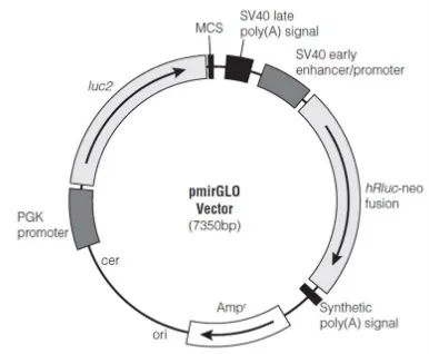
GRN 3’UTR was amplified through RT-PCR, starting from RNA extracted from HeLa cells and retro-transcribed into cDNA. The full length GRN 3’UTR of 304 bp was amplified using GRN Forward and GRN Reverse Primers containing restriction sites for XbaI enzyme (refer to table 2.1a). The amplified product of 304 bp was purified using PCR purification kit from Qiagen and cloned inside pmirGLO plasmid vector, in the multiple cloning site, located at the 3’ of firefly luciferase (Figure 2.1). The cloning was performed through the sticky ends produced with XbaI digestion. pmirGLO plasmid (from Promega) is a dual-luciferase plasmid containing both firefly luciferase gene (luc2), under the control of PGK promoter, and Renilla luciferase gene, under the control of SV40 promoter. The vector is designed to measure miRNAs activity, through the inclusion of the putative miRNA binding sites or the entire 3’UTR of interest at the 3’ of luc2. Firefly luciferase assay is used to analyze mRNA regulation, while Renilla luciferase is a control reporter for normalization.
In order to investigate the effect of miRNAs on different parts of GRN 3’UTR, the GRN 3’UTR full lenght was firstly divided in two parts, using the restriction site for SacI, located after 114 bp. The first part of GRN 3’UTR of 114 bp was called GRNIPart, while the second part of 190 bp was called GRNIIPart. Following the digestion with SacI, the products were run on agarose gel and extracted through gel extraction kit from Qiagen. The samples were then purified using PCR purification kit from Qiagen and used for the cloning procedure. GRNIPart and GRNIIPart were both cloned inside pmirGLO plasmid vector at the multiple cloning site, using sticky ends derived from the digestion with XbaI and SacI respectively.
24
MAPT 3’UTR was amplified from human genomic DNA using MAPT Forward and MAPT Reverse Primers, containing SacI and NheI restriction sites (refer to the table 2.1b for primer sequences). The amplified product of 4167 bp was purified using PCR purification kit from Qiagen and cloned inside pmirGLO plasmid vector, in the multiple cloning site, located at
the 3’ of luc2.
MAPT 3’UTR was further divided into 8 different partially overlapped fragments of 700 bp. Amplification of the fragments was obtained with specific primers reported in table 2.1b.
The different fragments were cloned inside pmirGLO vector, using SacI and NheI or XbaI and SalI restriction sites.
25 Table 2.1a
GRN PRIMERS SEQUENCES
GRN Forward Primer 5’- AAATCTAGAGGGACAGTACTGAAG -3’
GRN Reverse Primer 5’- ATCTAGAGAAAGTGTACAAACTTTATTG-3’
RevIGRNa 5’- AAAGTCGACAGGCCTGAGCAGA- 3’
ForIIGRNb 5’-AAATCTAGACCTCCCTAGCACCTCC-3’
RevIIGRNb 5’- AAAGTCGACGTGATGGGGAGCTCA- 3’
Table 2.1b
MAPT PRIMERS SEQUENCES
MAPT Forward primer 5'-AAAGAGCTCTTTGTGATCAGGCCC-3'
MAPT Reverse primer 5’-AAAGCTAGCAATCAGAGTAATAACTTTATTTCC-3’
MAPT Reverse I 5'-ATAGCTAGCACTGACCCACAGCAG-3'
MAPT Forward II 5'-ATAGAGCTCTGAGTGTGACGGGG-3'
MAPT Reverse II 5'-ATAGCTAGCTGGGAATTCGGGACA-3'
MAPT Forward III 5'-ATAGAGCTCCTAACCAGTTCTCTT-3'
MAPT Reverse III 5'-ATAGCTAGCCTGATACTATGCATG-3'
MAPT Forward IV 5'-ATAGAGCTCGAGAGAGCCCTTTCC-3'
MAPT Reverse IV 5'-ATAGCTAGCACCTAGTCTGTGCCC-3'
MAPT Forward V 5'-ATAGAGCTCACGAGGTGTCTCTCA-3'
MAPT Reverse V 5'-ATAGCTAGCCAAACCAGAAGTGGC-3'
MAPT Forward VI 5'-GGATCTGCTC TAG AGGCCCAAG-3'
MAPT Reverse VI 5'-ATAGTCGACGGGATTGTCCTCATTT-3'
MAPT Forward VII 5'-ATAGAGCTCGGTTCCTCTTCCTGA-3'
MAPT Reverse VII 5'-ATAGCTAGCGCTTAGAGGGAAGGA-3'
26 2.2 Cloning of Over-Expressing Plasmids
27
Figure 2.2 Map of psiUX vector used for the production of miRNAs over-expressing plasmid. This vector was produced and published by Prof. Michela Denti in 2004 (Denti et al. 2004).
Table 2.2
miRNAs PRIMERS SEQUENCES
Pre-miR-939 Forward 5'-ACTGCTCGAGGGTCCTCCAGAACGTGTTCA-3'
Pre-miR-939 Reverse 5'-ACTGAGATCTCGCTACCAGGAGGAAAGCAA-3'
Pre-miR-659 Forward 5'-ACTGCTCGAGCACTGTCATTATTTTCTCAC-3'
Pre-miR-659 Reverse 5'-ACTGAGATCTGCGTTCTTGTTTTGTGTTTC-3'
Pre-miR-615 Forward 5'-ACTGAGATCTCGACGACATAATTGGATCAT-3'
Pre-miR-615 Reverse 5'-ACTGGGTACCAGGAAGGGGTGAATAGCTTG-3'
Pre-miR-608 Forward 5'-ACTGAGATCTGGTAATGGCTCCATCTGGAG-3'
Pre-miR-608 Reverse 5'-ACTGCTCGAGTTGCAGACTCTTGGGCCCTT-3'
2.3 Cell Cultures and Transfection Experiments
28
F12 Medium (Gibco®, Life Technologies) supplemented with 2 mM L-Glutamine, Penicillin/Streptomycin and 10% Fetal Bovine Serum (FBS). All cell cultures were maintained at 37°C in a humidified atmosphere of 5% CO2.
Cells were seeded on 24-well plate (Luciferase Assay) and 6-well plate (Western Blotting and ELISA assay). Cells were transfected at 80% confluence with Lipofectamine LTX and Plus
reagent (Life Technologies) for Luciferase and ELISA Assays and with with Lipofectamine 3000 (Life Technologies) for Western Blot analysis. Whereas cells were plated on culture dishes and transfected with Lipofectamine 3000 (Life Technologies) for polysomes analysis.
2.4 miR-CATCH Experiments
2.4.1 Formaldehyde Cross-Linking and Cell Lysis
29
2.4.2 Streptavidin Bead Preparation and miRNA:GRN or MAPT mRNAs Pulldown
Dynabeads MyOne Streptavidin C1 magnetic beads (Life Technologies, Grand Island, USA) were prepared to the manufacturer’s instructions. An excess of 800 pmol of biotinylated capture oligonucleotides was immobilised to the magnetic beads in 1X B&W
30
Figure 2.4 miR-CATCH strategy. a) synthesis of a biotin oligonucleotide targeting a single strand region of a target mRNA. b) Formaldehyde cross-linking step. c) oligonucleotide immobilization to magnetic streptavidine beads. d) cell lysis. e) the target mRNA is captured and purified. f) Formaldehyde cross-links are reversed (Vencken et al. 2015).
2.5 Luciferase assay
31
Dual-GLO Luciferase Assay System (Promega) with Infinite® M200 (Tecan) plate reader. The statistical significance was determined using Student’s t-test comparison. Data were considered significant when p<0,05.
2.6 Western blotting and ELISA assay
HeLa cells were seeded in a 6-well plate and transfected using Lipofectamine LTX and Plus reagent (Life Technologies) or Lipofectamine 3000 with 2,2 μg of miRNA-overexpressing plasmids. At 48h after transfection, proteins were extracted with Radioimmunoprecipitation Assay Buffer (RIPA) supplemented with a protease inhibitor cocktail (Sigma-Aldrich) and analyzed by Western Blotting and ELISA assays. Proteins (20 µg) were separated on 10% SDS-polyacrylamide gel electrophoresis (SDS-PAGE), and transferred to a nitrocellulose membrane by using iBlot® Dry Blotting System (Life Technologies®) at 20V for 7 minutes. Blots were first blocked with 5% non-fat powdered milk in PBS/Tween 0.1%, then probed overnight at 4 °C with mouse monoclonal anti-GRN (Abcam® no. ab55167, 1:500), rabbit polyclonal anti-Tau Dako (Agilent Technologies® A0024 , 1:1000) and rabbit polyclonal anti-HPRT (FL-218) (Santa Cruz Biotechnology® no. sc-20975, 1:500). Membranes were subsequently washed and incubated with anti-Mouse IgG and Anti-Rabbit IgG secondary antibodies (1:5000). Membranes were processed by ECL plus detection kit (Amersham®) according to manufacturer’s instructions. Densitometric analysis was performed using ImageJ program.
32 2.7 Quantitative Real Time-PCR
Total RNA was extracted from transfected or non transfected cells using TRIzol reagent (Invitrogen®), according to the manufacturer’s instruction. For the quantification of GRN and MAPT transcripts from total RNA or polysomal/subpolysomal fractions, cDNA was
synthesized by retrotranscription using RevertAid First Strand cDNA Synthesis Kit (Thermo Scientific®) with oligo (dT) primers, according to the manufacturer’s protocol. Real-Time PCR (RT-qPCR) was performed using Kapa SYBR fast qPCR master mix (Kapa Biosystem®) and specific primers reported in table 2.7. PCR reaction (10 µl) contains 0,3 µM of each primer, 5 µl of master mix and 10 ng of cDNA. The expression of GRN and MAPT mRNAs was normalized with expression of different reference genes: glyceraldehydes-3-phosphate dehydrogenase (GAPDH), hypoxanthine phosphoribosyltransferase (HPRT1), succinate dehydrogenase complex subunit A (SDHA).
33 Table 2.7
Real Time PCR PRIMERS SEQUENCES
Primer Forward GRN 5'-TTCTGGACAAATGGCCCAC-3'
Primer Reverse GRN 5'-ACCCACGGAGTTGTTACCTG-3'
MAPT Forward 5'-ACATCCATCATAAACCAGGAGGT-3'
MAPT Reverse 5'-TGTCTTGGCTTTGGCGTTCT-3'
HPRT I Forward 5'-TGACACTGGCAAAACAATGCA-3'
HPRT I Reverse 5'-GGTCCTTTTCACCAGCAAGCT-3'
SDHA Forward 5'-TGGGAACAAGAGGGCATCTG-3'
SDHA Reverse 5'-CCACCACTGCATCAAATTCATG-3'
GAPDH Forward 5'-TCTCCTCTGACTTCAACAGC-3
GAPDH Reverse 5'-CGTTGTCATACCAGGAAATGA-3'
2.8 Polysomal analysis
35
CHAPTER THREE: IDENTIFICATION OF miRNAs INVOLVED IN THE
POST-TRANSCRIPTIONAL REGULATION OF
GRN
EXPRESSION
3.1 Introduction
36
(Wang-Xia et al. 2010). Later on it was demonstrated that this miRNA family regulates insulin sensitivity and its silencing leads to an improved glucose homeostasis (Trajkovski et al. 2011).
Another scenario in which the decrease of GRN expression below a critical threshold can cause the development of FTD is due to the presence of SNPs (Rademakers et al. 2008). The
37 3.2 Results
3.2.1 GRN mRNA as a direct target of putative microRNAs
In order to identify the key miRNAs that could regulate GRN mRNA expression, three different bionformatic software programs were used to predict the potential miRNAs binding on the GRN 3’UTR sequence. Predictive results derived from PITA (http://genie.weizmann.ac.il/), Targetscan (http://www.targetscan.org/) and Targetprofiler (http://mirna.imbb.forth.gr/Targetprofiler.html) were integrated to increase the specificity. Each database predicted binding sites for hundreds of miRNAs, including one for miR-939-5p and one for miR-615-miR-939-5p on GRN 3’UTR that was predicted by all three programs. The results were further screened and miR-608, miR-939-5p, miR-615-5p and miR-659-3p were selected according to the best score in terms of free binding energy, site accessibility and seed type (Figure 3.2.1.1). A multiple alignment using GRN 3’UTR sequences of three different species (mouse, rat and human) demonstrated a relevant level of conserved regions that might represent or indicate possible regulatory sequences (Figure 3.2.1.2). As described in Figure3.2.1.1, miR-659-3p presents only one binding site, whereas miR-939-5p and miR-615-5p show three recognition elements with different specificity and binding energy. miR-608 shows two binding sites that are partially overlapping with the binding recognized by miR-939-5p, since they have a similar seed region. Overall the less conserved regions on GRN 3’UTR seem to be regions without putative miRNA binding sites.
38
Figure 3.2.1.2 Conservation analysis of GRN 3’UTR between human, mouse and rat.
Multiple alignment of GRN 3’UTR sequence between human, mouse and rat species performed through ClustalW and graphically represented with Jalview. Black squares below the alignment and dark blue color on the sequences indicate the 100% conservation between human, mouse and rat.
3.2.2 GRN 3’UTR Analysis of SNPs Located on miRNAs Binding Sites and Sequencing of
GRN 3’UTR in Different Cell Lines
39
the ∆∆G values: (∆∆G>0 kcal/mol) SNPs that increase miRNA binding, (∆∆G<0 kcal/mol) SNPs that reduce miRNA binding and (∆∆G=0 kcal/mol) neutral SNPs that have no effect on miRNA binding (Saba et al. 2014). In the Figure 3.2.2a are reported the comparisons between the wild type and the variant miRNA-GRN 3’UTR interactions and the computed ∆∆G value for each binding. Only two SNPs caused an enhanced miRNA binding, one is
41
Figure 3.2.2a. SNPs overlapping miRNA binding sites analysis for GRN 3’UTR
Representation of the miRNA-GRN mRNA functional characterization technique following the method reported by Saba and colleagues (Saba et al. 2014).
42
Figure 3.2.2b GRN 3’UTR sequencing in HeLa, KELLY, SH-SY5Y and SK-N-BE cell line.
43
3.2.3 Expression of GRN mRNA in different neuroblastoma cell lines
Neuroblastoma cell lines were used as a neuronal-like system to study the regulation of our genes of interest, GRN and MAPT in normal cell condition. RT-PCR was used to better analyze GRN mRNA expression in different neuroblastoma cell lines. Amplification of a 200 bp product, located on GRN mRNA between exon 1 and 4, was obtained with specific primers recognizing regions of exons junctions. In particular, forward primer binds to a region located between exon 1 and 2, while reverse primer recognizes a sequence located on exons 3 and 4.
The level of progranulin expression as mRNA was quite similar in all cell lines analyzed: IMR32, LA-N-1, LA-N-2, SK-N-MC, SK-N-AS, SK-N-DZ, SK-N-SH, SK-N-BE, CHP134, CHP212, SIMA, KELLY, NB-69, except for SIMA and IMR32 cell lines, where GRN mRNA seems to be expressed at a lower level. In order to investigate GRN mRNA post-transcriptional regulation, KELLY, SH-SY5Y (derived from SK-N-SH) and SK-NBE cell lines were selected since they show a meaningful level of GRN mRNA expression.
44 3.2.4 miRNAs Basal Expression in different cell lines
Using TaqMan quantitative RT-PCR (qRT-PCR), the endogenous expression of miR-939-5p, miR-608, miR-659-3p and miR-615-5p was analyzed in four cell lines: HeLa, KELLY, SK-N-BE and SH-SY5Y. All CT levels were normalized against snRNU6 to calculate the values 2 -∆Ct. In Figure 3.2.4 we can observe that miR-939-5p, in all the different cell lines, is expressed
at higher level compared to the other selected miRNAs. Indeed, miR-659-3p is expressed at low level, mainly in SH-SY5Y and SK-NBE cell lines, whereas miR-615-5p and miR-608 are present at minimal level or totally absent in the analyzed cells.
Figure 3.2.4. miRNAs basal expression levels in HeLa, KELLY, SH-SY5Y and SK-NBE cell lines.
45
3.2.5 GRN microRNAs Capture Affinity Technology (miR-CATCH)
miRNA targets have been predicted through different bioinformatic programs that are based on the free binding energy, site accessibility, seed type and seed sequence conservation. However, the miRNAs target recognition mechanism remains challenging, due to the high false-positive rates and to the fact that computational analysis relies on different algorithms that yield different results. For this reason it became clear the necessity to use a biochemical strategy based on physiological interactions between miRNPs and their specific target mRNAs to reliably identify subset of miRNAs targeting GRN mRNA. The microRNA Capture Affinity Technology (miR-CATCH) developed by Prof. Catherine M. Greene and her team (Hassan et al. 2013, Vencken et al. 2015), allows to selectively identify miRNAs binding to a specific mRNA under different conditions, eliminating false positive mRNA:miRNA interactions that can be predicted in silico. This technique offers the possibility to identify miRNAs that bind to the entire mRNA transcripts, therefore it is not limited to the 3’UTR region. It is a novel way to bypass or complement the initial use of bioinformatic predictions. I moved to the laboratory of Prof. Catherine M. Greene, where I worked for four months to learn and applied this technique in collaboration with Dr. Sebastian Vencken*. Firstly, we used KELLY cells for the validation of the isolation method and the efficacy of the miR-CATCH protocol specificity. Then, the miR-CATCH strategy was used to investigate the regulation played by miRNAs of GRN mRNA in the selected neuroblastoma cell lines: KELLY, SH-SY5Y and SK-N-BE.
46 3.2.5.1 Biotinylated DNA Oligonucleotides Design
47
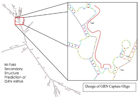
Figure 3.2.5.1 Design of GRN mRNA Capture Oligonucleotide. Secondary structure of GRN mRNA was elaborated through M-Fold. The exposed single strand region located between bases 1276 and 1303 of the transcript was used as a target.
SINGLE STRAND REGION : 5’-AGCCTGCCAGACCCACAAGCCTTGAAGA-3’ REVERSE AND COMPLEMENT SEQUENCE: ………. 5’-TCTTCAAGGCTTGTGGGTCTGGCAGGCT-3’ OLIGO SEQUENCE: Biotin 5’-TCT TCA AGG CTT GTG GGT CTG GCA GG-3’ LENGTH: 26
GC CONTENT: 57,7% MELTING TEMPERATURE: 79,8°C
HAIRPINS TM: 36,5°C 34,5°C 29,4°C 31,6°C 31,5°C HOMO DIMER ANALYSIS: ≥-5,5 Kcal/mole
Table 3.2.5.1a Sequence and characteristics of the designed GRN mRNA Capture Oligonucleotide
48
OLIGO SEQUENCE: 5’-TCT TCA AGG CTT GTG GGT CTG GCA GG-3’ MISMATCH OLIGO SEQUENCE: Biotin 5’-TCT TCA AGG CTT ACA GGT CTG GCAGG-3’
LENGTH 26
GC CONTENT 53,8%
MELT TEMP 76,5°C
Hairpins Tm: 39,4-36,5-29,9-31,6-30,6
49 3.2.5.2 Validation of GRN mRNA:miRNA Isolation
50
Figure 3.2.5.2a GRN mRNA enrichment. Representation of the real time PCR results showing the fold enrichment level (2-∆∆Ct) obtained with GRN capture oligonucleotide compared to the total RNA that did not undergo the miR-CATCH protocol. Expression of GRN mRNA was normalized against GAPDH expression. Representative data of one biological experiment and three technical replicates.
51
3.2.5.3 GRN mRNA Enrichment in KELLY, SK-N-BE and SH-SY5Y cell lines
Following the validation of the miR-CATCH protocol in the KELLY cell line, the pull-down method was performed in biological triplicates in selected neuroblastoma cell lines. “Scrambled” samples were obtained from the same cell lysate of each biological experiment, using a scrambled capture oligonucleotide biotinylated at the 5’end for the miR-CATCH strategy. Using qRT-PCR, the level of GRN mRNA enrichment was assayed individually in each biological experiment and compared to the scrambled control through a
2-∆∆Ct method. In particular 47 fold enrichment of GRN mRNA was obtained in the KELLY cell
line, whereas 66 fold enrichment was found in SH-SY5Y cell line and 23 fold enrichment for SK-N-BE cell line, as reported in the Figure 3.2.5.3. Data represents the mean of three biological replicates.
Figure 3.2.5.3 GRN mRNA Enrichment in different neuroblastoma cell lines.
52
3.2.5.4 Selected miRNA Profiling on Capture Samples in KELLY, SH-SY5Y and SK-N-BE cell
lines
53
Figure 3.2.5.4.a. Selected miRNA enrichment in KELLY cell line. Representation of miR-659-3p A) and miR-939-5p B) enrichment in GRN capture samples compared to scrambled controls. Graphs on the right part of the figure C) represent the negative control miR-323 that shows no enrichment in the capture samples. miRNAs expression was represented as fold enrichment value (2-∆Ct).
A) and B) Mean±SEM of three different biological experiments (*P<0,05; *** P<0,0001). C)
54
Figure 3.2.5.4.b Selected miRNAs enrichment in SH-SY5Y and SK-NBE cell lines. A)
Representation of miR-659-3p and miR-939-5p enrichment found in capture samples compared to scrambled controls in SH-SY5Y samples. B) Graphs representing the enrichment of miR-659-3p and miR-939-5p compared to the scrambled controls in SK-NBE cell line. miRNA expression was represented as fold enrichment value (2-∆Ct).
55
3.2.6.1 Luciferase Assays using reporter vectors of the full GRN 3’UTR in HeLa cell line
Following the results obtained with miR-CATCH strategy, the putative miRNA’s regulation on GRN 3’UTR was further investigated using luciferase activity assays in HeLa cell line. HeLa cell line is a reliable cell system, easy to handle and useful to explore with high efficiency the regulation of our miRNAs of interest by co-transfection experiments. According to our bioinformatic prediction, miR-181a does not have binding sites localized on GRN 3’UTR and for this reason it was selected as a negative control. HeLa cells were co-transfected with reporter vectors containing the full length or different regions of GRN 3’UTR and miRNA over-expressing plasmids.
3.2.6.1.1 Analysis of the miRNAs Overexpression in HeLa cell line
Firstly, was checked the specific miRNA’s over-expression, during the luciferase analysis performed at 24h and 48h after transfection, the RNA was extracted and miRNAs quantified by qRT-PCR based on TaqMan assay. All the CT levels were normalized to RNU6, to calculate the fold change with the 2-∆Ct method. In Figure 3.2.6.1.1a the basal endogenous level of expression of selected miRNAs (light green) is compared to the level of miRNAs over-expressed from plasmids (dark green). Moreover the ∆∆Ct values were calculated using as normalizers the miRNA levels measured after transfection of backbone empty plasmid, utilized for miRNA cloning. These fold changes are reported in the Figure 3.2.6.1.1b.
Figure 3.2.6.1.1a Basal and Over-expression levels of selected miRNAs in HeLa cell line.
56
from the luciferase analysis at 24h and 48h. Ct levels are normalized against RNU6 to calculate the fold change with the 2-∆Ct method. Data are obtained from biological duplicates and technical triplicates (Mean SEM).
Figure 3.2.6.1.1b Over-expression levels of selected miRNAs in HeLa cell line.
Representation of the 2-∆∆Ct values obtained through the comparison between the over-expression of miRNAs and the endogenous basal level of over-expression in HeLa cell line. Data represent the summary of biological duplicates and technical triplicates (Mean SEM).
3.2.6.1.2 Validation of mirtron-939 over-expression
57
results, showing a stronger production of the unspliced product of 465 bp compared to the spliced product of 272 bp represented by a faint band. This result demonstrated a major recruitment of the microprocessor complex on the unspliced precursor region of miR-939-5p. The production of miR-939-5p seems to be preferred compared to the splicing of that region that would lead otherwise to the disruption of the precursor sequence for
miR-939-5p.
Figure 3.2.6.1.2a Ensembl Genome Browser representation of miR-939-5p precursor
region.
58
procedure and for the RT-PCR reaction. Red square indicates the precursor region of miR-939-5p overlapping the 5’splice site.
Figure 3.2.6.1.2c RT-PCR reaction of miR-939-5p precursor region. Figure of the 2% agarose gel where samples derived from the amplification of the precursor region of miR-939-5p were loaded. RT-PCR reactions performed with the RNA extracted from cells that were transfected with miR-939-5p over-expressing plasmid. Sample 1 and 2 are showing the amplification of the unspliced region cloned inside the over-expressing plasmid for miR-939-5p (465 bp band) and the spliced product of the same region (272 bp band). Blank represents the specific control of the RT-PCR reaction that does not contain the cDNA sample.
59
60
61
3.2.6.2 Luciferase Assays using reporter vectors containing different regions of GRN 3’UTR
in HeLa cell line
62
64
65
3.2.7 Effect of Selected miRNAs Over-expression on Progranulin level in KELLY cell line
The level of progranulin protein was measured using western blot analysis after miRNAs over-expression. KELLY cell line was transfected with the over-expressing plasmid for putative miRNAs entire panel and the backbone empty plasmid, used for miRNA cloning, as negative control. The efficiency of miRNAs over-expression was analyzed with TaqMan qRT-PCR, using RNA extracted from parallel samples, that underwent the same transfection experiments. In Figure 3.2.7a the basal endogenous level of expression of selected miRNAs (light grey) is compared to the level of miRNAs over-expressed plasmids (dark grey). Moreover the ∆∆Ct values were calculated using as normalizers the miRNA levels measured after transfection of backbone empty plasmid, utilized for miRNA cloning. These fold changes are reported in the Figure 3.2.7b.
66
Figure 3.2.7a Basal and Over-expression levels of selected miRNAs in KELLY cell line.
TaqMan real time PCR was performed on the RNA extracted after 48h from cells transfected with over-expressing miRNA plasmids. CT levels are normalized against RNU6 to calculate the fold change with the 2-∆Ct method. Data are obtained from biological duplicates and technical triplicates (Mean SD).


![Sun Solaris =10 rpc ypupdated Remote Root Exploit [superdk] pdf](data:image/gif;base64,R0lGODlhAQABAIAAAP///wAAACH5BAEAAAAALAAAAAABAAEAAAICRAEAOw==)