Pollen Image Management: Using Digital Images to Teach
Recognition Skills and Build Reference Collections
Peter Shimeld, Feli Hopf and Stuart Pearson
Geomorphology and Quaternary Science Research Unit, School of Geosciences, The University of Newcastle, Australia
http://www.newcastle.edu.au/department/gg/pol/0intro.html
Introduction
A reference collection is a necessary outgrowth of many research projects, including some undergraduate PBL. The need for modern morphological information at appropriate taxonomic levels seems to drive the early stages of many projects. In palynology, the development of these collections is often a fundamental part of researcher training. It is a time consuming and
materials-expensive task that includes: the gathering and identification of herbarium specimens; the destructive processing of the specimen; the storage of reference vials of material; and the recording of the images for later cross-referencing. The microscope slides and processed materials have a limited (undefined) shelf-life. Retrieving reference material for routine
comparison with unknown materials can be frustratingly slow using reference microscope slides. Consequently, students have traditionally used a combination of annotated sketches and
photographic print film to compile a reference collection. Most pollen laboratories are festooned with hardcopies of pollen images for the enjoyment of those in the laboratory and the collections tend to 'go with' the collector. This does not result in accumulation of taxonomic information.
The 'siliciophobic' response of teachers and researchers described by Attwood (2000) has an additional variety called 'pixilophobia'. Digital-image technology and the Web are now widely available and accepted in most applications except in the collection and sharing of reference materials. For example, the field of palynology has its professional heart in image recognition, yet the systematic collection and sharing of images over the Web or on CD-ROM is relatively recent. This paper seeks to promote this recent development. Although this paper relates to a pollen collection, however, the principles and techniques used have universal application to people working with image management. We believe the model described here is a useful development, and although the endpoint may be LucID (Norton et al. 2000), we hope this low-budget model encourages others in the sharing of reference materials in digital forms.
Greater efficacy in image management
To achieve greater effectiveness we have developed a simple system that students can use to build high quality databases of reference and unknown materials with little training or
Figure 1. Examples of digital images, Acacia terminalis and Eucalyptus gummifera
The system uses digital images that replace: the delays between taking a photograph and having it added to the reference collection; the delay of duplication for sharing images with others; and the costs and delays of photographic procedures. Digital images also allow easy duplication without any loss of resolution. Training of undergraduate and research students is more efficient because images and unknown grains can be routinely compared on the screen. With a few minutes training, the same equipment can be used for 'capturing' images of unknown grains for later identification or comparison. Preliminary results with honours students suggests it reduces the microscope work time by approximately 50% and concentrates the time needed for experts to identify unknown grains.
Why a digital collection from the Geomorphology and Quaternary Science
Research Unit?
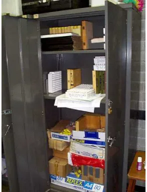
Figure 2. A traditional format reference collection
In response to multi-user demands for reference materials, and falling costs per unit of computer processing and storage, we investigated top-of-the range image grabbing and processing
configurations. The set-up costs of these systems on existing scopes were in excess of $ 20 000 and required specialised operators and software. During our recoil from this discovery we identified a 'diminishing return' in performance improvements beyond a few thousand dollars. In other words, to get small increases in performance we were expected to spend large amounts of money on a single unit.
With the impatient enthusiasm of postgraduate researchers driving the project, the occasional injection of small amounts of money and even an 'experiment' on a stolen Unix machine, the image grabbing capability was built onto two existing microscopes linked to a computer.
grabbing system at the Southern Connections Congress at Lincoln University, New Zealand, 17-22 January 2000 and a poster and display at the Quaternary Studies Meeting at the Australian National University, Canberra, 7-9 February 2000. There have been requests for copies of the CD-ROM, offers of additional collections and information on how to set-up similar hardware and software systems. The step-by-step protocol for developing images for the collection are available from the authors who are also happy to discuss set-ups for new applications.
The collection system
The collection is designed for use on standard PCs running software such as Netscape Navigator or Internet Explorer. The JPEG image file format was selected for small file size and
compatibility across software. At the moment the collection contains about 450 taxa, with another 900 taxa in progress. The CD-ROM can hold images representing 2000 taxa.
The software is structured to use the brain's remarkable ability to recognise spatial pattern and make visual comparisons. Unlike conventional databases searches are image-driven rather than text driven, using selectable sketches and thumbnails to move through the collection and select best matches. The hierarchical complexity is minimised - researchers are never more than two mouse clicks from an image. The system is computer mouse-controlled and does not clutter the microscope workspace with a keyboard nor the researcher's mind with esoteric morphological terminology. There is a minimum of text and specialised jargon. This increases accessibility and reduces query-to-solution times. The new collection can be used in conjunction with the ANU collection (http://pollen.anu.edu.au/pollensearch.phtml) that is more text-based with images only available at the final stages of identification. We plan to develop a LucID-based key in future that will remove the hierarchical structure of identification.
Following the traditional dichotomous structures, the opening page provides 2 routes: a graphics interface page of 52 thumbnail-sized drawings; and a taxonomic treatment based on Family names. We have found the line drawings are the preferred route in most situations. Selecting one of the thumbnails opens a number of thumbnail images (approximately 4 Kb each) of pollen grains. Selecting one of those images opens a large format image (approximately 350 Kb) showing the name of the grain with a scale bar. Selecting 'Back' allows for quick comparisons with other morphologically similar grains.
The large format images for each taxa are composites of a few images taken using the Hi-Lo method to show surface textures and cross-sections and usually include polar and equatorial views. Morphological variations within a species can be reflected by relisting the images under a couple of thumbnails. For example, grains that are usually present as crushed grains can be represented by two line drawings, one showing the undamaged form and the other a crushed form. Saved with the image are a scale bar and the name of the taxon so the grain, scale and source are stored together. Some of the images have been scanned from 35mm print film photographs, but most are digitised directly from microscope video cameras. Digital
We have found it useful to develop a standard protocol for naming files to help in knowing exactly what an image is and how it was gathered. For example the file name "Solanum plicatile EV1L500.JPG" gives the following information: the specific epiphet, the equatorial orientation or type of view (EV), the image number (1), the depth of focal plane (low), and that the image was taken on the Leitz scope at 500x magnification. When a number of images were compiled together we added the scale, family and specific epiphet into the background (Figure 3). From the earlier example, the image was now simply "Solanum plicatile.jpg" these files are
approximately 350 Kb. From these images we copied and resampled images to save them as thumbnails that are 125 pixels high. These thumbnail images were matched to the line drawings and from them allocated a group number. The thumbnail file name became "36Solanum
plicatile.jpg". This protocol saved considerable time in building the Web pages.
Figure 3. Example of a reference pollen grain
Setting up the system yourself
The hardware configuration we have used is probably already antiquated, for example video cards are now frequently standard in contemporary computers. Nevertheless, the decisions we made in setting up the system should encourage researchers without large budgets. We used a colour CCD video camera purchased from ISSCO for $500. C-mount adaptors threatened to be an expensive part of the set-up. So C-mounts were made using 50mm PVC drain fittings and the correct tube lengths were set by having a sliding section. This fitting was done easily on a Leica scope but we needed a precision steel engineering company to produce a mount for the Zeiss Axioplot microscope.
Figure 4. Detail of the image grabbing set-up
A plug-in to the serial port called Snappy converted the video signal to digital images. The short-lived 9 volt battery was replaced by a plugpack regulator and transformer from Dick Smith. The computer is a Pentium II loaded with the software Photodelux that is sufficient for capturing the image, editing and saving. For bulk editing, scanning prints and compiling composite images we have used PhotoImpact or Corel Photo-Paint Version 8. The baseline option to set-up a system is approximately $1000 and this cost is probably falling.
Conclusion
We are planning to group images under the thumbnail leader by size and surface texture so researchers can use size or surface to speed their search. The development of Genus and Species search capabilities and provision of provenance and processing information for each image is a priority and will probably be done under the LucID system. We are also developing an
The image grabbing and sharing project that started humbly has developed into a reasonable model for others who have large numbers of images that require some management. We have used cheap and readily available materials to achieve our goals. The equipment has, we feel, repaid the costs and trouble of set-up and we are keen to convert others to the digital image path. We hope this low-budget project encourages others to contribute collections, where pixel leaders are already present, or take up the challenge of using pixels to share reference materials with students and other researchers.
Acknowledgements
This is a contribution of The University of Newcastle Geomorphology and Quaternary Science Research Unit. We acknowledge with thanks the contributions of Eric Colhoun, Mike Macphail, Mike Cvetanovski, David O'Brien, Richard Dear and Lucy Gayler. Thanks also for suggestions from John Dodson, Scott Mooney and Scott Anderson. The work would not be possible without the support of the Discipline of Geography and Environmental Science.
References
1. Attwood, T. K. (2000) An interactive practical at the interface of web-based and conventional publishing, CAL-laborate, UniServe Science, 4, 5-10.
2. Norton, G. A., Patterson, D. J. and Schneider, M. (2000) LucID: A multimedia
educational tool for identification and diagnostics, CAL-laborate, UniServe Science, 4, 19-22.
Peter Shimeld, Feli Hopf and Stuart Pearson
Geomorphology and Quaternary Science Research Unit School of Geosciences
The University of Newcastle Callaghan NSW 2308 Australia
ggsgp@cc.newcastle.edu.au