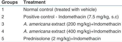
Gastro protective effect of
Agave
americana
Linn. leaf extract in
indomethacin-induced enterocolitis in rats
Basheerahmed Abdulaziz Mannasaheb
1*, Preeti Venkat Rao Kulkarni
2Ibrahim Shaikh
3, Mashood Ahmed Sangreskopp
2, Chetan Savant
21Department of Pharmacology, Al-Maarefa College of Pharmacy, Riyadh, K.S.A, 2Department of Pharmacology,
SET’s College of Pharmacy, Dharwad, India, 3Depatment of Pharmacology, College of Pharmacy, Najran
University, K.S.A.
Abstract
Objective: The present study was undertaken to evaluate the gastro protective effect of Agave americana Linn. leaf extract in indomethacin-induced enterocolitis in rats using prednisolone as standard drug. Methods: Male Wistar rats were pretreated with A. americana leaf extract in the dose of 200 and 400 mg/kg orally daily for 7 days. On 8th and 9th day indomethacin was administered (7.5 mg/kg, s.c.) to induce enterocolitis. The treatment was continued until 11th day. On 11th day animals were sacrificed and dissected open to remove ileum, cecum, and colon. Flushed gently with saline, cut open and inflammation was assessed based on the physical parameters, scoring of ulcer, myeloperoxidase (MPO), and lipidperoxidase (LPO) activity. Results: A. americana leaf extract (200 and 400 mg/kg) showed a significant decrease (P < 0.001) in ulcer scores for inflammation. Both prednisolone and A. americana have retained total body weight significantly (P < 0.01). Significant decrease (P < 0.001) in MPO and LPO activity was also observed. Histopathology of extract treated group showed reduced intensity of edema, inflammatory cellular infiltration, crypt damage and ulceration at mucosal and submucosal layers compared to induce control group. Conclusion: The above findings prove that, A. americana Linn. leaf extract has protective effect against indomethacin-induced enterocolitis.
Key words: Agave americana, indomethacin, lipidperoxides, myeloperoxidase, ulcer index
Address for correspondence:
Basheerahmed A.M, Department of Pharmacology, Al-Maarefa College of Pharmacy, Riyadh, Kingdom of Saudi Arabia - 11597. Phone: +00966-538199327. E-mail: abuhamza22@gmail.com
Received: 15-04-2014
Revised: 19-10-2015
Accepted:28-10-2015
INTRODUCTION
U
lcerative colitis (UC) and Crohn’s disease (CD) together constitute inflammatory bowel disease (IBD). CD (enterocolitis) may be localized in any part of the digestive tract and affects the entire intestinal wall, whereas UC is confined to the colon and rectum and the inflammation is restrained to the intestinal lining.[1] IBD is a growing worldwide health burden; specifically many developing countries have seen a dramatic rise in the incidence of IBD since 1990.[2] Although etiology of IBD is unknown, it appears that an abnormal response of the mucosal innate immune system to luminal bacteria may trigger inflammation which is perpetual by dysregulation of cellular immunity.[3-5] The most widely acceptedhypothesis in the pathogenesis of IBD is that the mucosal immune system shows an aberrant response toward luminal antigens such as dietary factors and/or bacteria in genetically
susceptible individuals. The chronic inflammatory process leads to disruption of the epithelial barrier, and the formation of epithelial ulceration. This permits easy access for the luminal microbiota and dietary antigens to cells resident in the lamina propria and stimulates further pathological immune cell responses. Cytokines are essential mediators of the interactions between activated immune cells and non-immune cells, including epithelial, and mesenchymal cells. The clinical efficacy of targeting tumor necrosis factor-alpha (TNF-α) clearly indicates that cytokines are the therapeutic targets in IBD patients.[6] CD and UC are caused
by excessive immune reactivity in the gut wall. Analysis
ORIGINAL
AR
of the type of immune responses ongoing in diseased gut has revealed important features.[7] CD is disease of young people with a peak incidence between the ages of 10 and 40 years. However, it affects people of any age and 15% of people are over the age of 60 at diagnosis. Up to 240,000 people are affected by IBD in the UK. Symptoms of CD includes diarrhea, right lower quadrant pain, fever, weight loss trans-mural inflammation-large ulcers, rectum spared, skip lesions, fistulae, and strictures.[8]
Immuno-modulators, such as azathioprine, 6-mercaptopurine, methotrexate or cyclosporin, are demonstrating increasing importance against steroid-resistant and steroid-dependent patients.[9] The drugs which are found to be effective
are sulfasalazine (azulfidine, salazopyrin) and its 5-aminosalicylic acid derivatives, glucocorticosteroids, immunomodulators/immunosuppressants, and other new potential drugs.[10]
There is widespread belief that the natural products are less toxic when compared to pure chemicals. Phyto-botanical and ethnobotanical research have focused for decades on the search for the single active principle in plants based on the assumption that plant has one or a few ingredients, which determine its therapeutic effects. However, the traditional system of medicine like Ayurveda, traditional Chinese medicine or the European Pharmacotherapy generally assumes that synergy of all ingredients of the plants will bring about the maximum therapeutic efficacy.[11] Recent data suggests that 80% drug molecules are natural products or natural compound inspired.[12]
The leaves of Agave americana Linn. (Family - Agavaceae) are used as hepatoprotective, antioxidant, antiseptic, and to relieve various liver diseases.[13] Agave leaves sap was used
internally in case of wounds and inflammation, and it also has anti-inflammatory activity.[14,15]
To best of our knowledge, no scientific data regarding the activity of A. americana on IBD is available in literature. Therefore, we have undertaken this work anti-IBD activity in indomethacin-induced enterocolitis in rats.
METHODOLOGY
Drugs and ReagentsPrednisolone, indomethacin, and trichloroacetic acid were purchased from Himedia, Mumbai. Other chemicals were purchased from Ranbaxy.
Plant Material
The fresh leaves of A. americana Linn. were collected from surrounding areas of Dharwad, Karnataka and authenticated
by Dr. Hebbar, Dharwad. The plant material was washed with running water and ground well using mortar and pestle. The obtained herbal juice was filtered and filtrate was subjected to rotary flash evaporator under reduced pressure to dryness. The dried extract was stored in desiccators until use.[16]
Preliminary Phytochemical Investigation
The A. americana leaf extract was subjected to preliminary phytochemical investigation as described in practical pharmacognosy by Kokate and Khandelwal.[17,18]
Animals
Male Wistar rats weighing 150-200 g were used for the present study. For acute toxicity study, female rats were used. The animals were purchased from Sri Venkateshwara Enterprises and maintained in the Animal House of SET’s College of Pharmacy, Dharwad for experimental purpose. The animals were maintained under controlled conditions of temperature (22 ± 2°C), humidity (50% ± 5%) and 12-h light-dark cycles. They were fed commercial stock diet and water, ad libitum. The animals were housed individually in sanitized polypropylene cages containing sterile paddy husk as bedding. Animals were habituated to laboratory conditions for 48 h prior to the experimental protocol to minimize if any of non-specific stress. All the studies conducted were approved by the Institutional Animal Ethical Committee of SET’s College of Pharmacy, Dharwad, Karnataka (Reg. No. 112/1999/CPCSEA) according to prescribed guidelines of Committee for the Purpose of Control and Supervision of Experiments on Animals, Government of India.
Determination of Acute Toxicity
The acute oral toxicity study was carried out as per the guidelines set by OECD 423. Animals (n = 3) were overnight fasted prior to dosing. The dose level to be used as the starting dose was selected from one of four fixed levels, 5, 50, 300, and 2000 mg/kg b.w. The test substance was administered in a single dose by gavage using intubation canula. Animals were observed individually after dosing at least once during the first 30 min, periodically during the first 24 h, with special attention given during the first 4 h, and daily thereafter, for a total of 14 days. Animals were observed for following profiles.
1. Behavioral profile - Alertness, restlessness, irritability and fearfulness
2. Neurological profile - Spontaneous activities, reactivity, touch and pain response
3. Autonomic profile - Defecation and urination.
Experimental Methods
Indomethacin-induced enterocolitis in rats
The animals were divided into five groups each containing six rats shown in Table 1.
Animals were pre-treated with A. americana extract for 7 days. On 8th and 9th day indomethacin (7.5 mg/kg) was administered to induce enterocolitis. Extract administration was continued until 11th day. On the 11th day the animals were sacrificed. Ileum, cecum, and colon were taken out to assess for inflammation based on physical parameters, macroscopic, and microscopic features. Quantification of inflammation was done using biochemical assay (myeloperoxidase [MPO] and lipidperoxidase [LPO]).[20,21]
Evaluation of the Disease
Change in body weight
As the weight loss is one of the clinical symptoms of enterocolitis all the rats from respective groups are weighed each day and the percentage of original weight is used for evaluation.[22]
Evaluation Based on Macroscopic Characters
Scoring of ulcer for ileum, cecum, and colon
The distal 10 cm portion of colon ileum and whole cecum was removed cut longitudinally and cleaned in physiological saline and were scored for macroscopic features using standard scoring pattern. Score for an individual rat was calculated as the combined score of ileum colon and caecum.[21]
Evaluation Based on Microscopic (histological) Characters
Caecum and ileum from each animal were removed and preserved in 10% formalin. The samples were submitted to Jeevan Regional Diagnostic Health Care and Research Centre Pvt. Ltd. (Belgaum, India) for histological examination.
Evaluation Based on Biochemical Parameters
Myeloperoxidase (MPO) assay for quantification of inflammation
The cecum was taken weighed and rinsed with ice-cold saline blotted dry weighed and excised. Minced tissue was homogenized in 10 volumes of ice-cold potassium phosphate buffer (pH 7.4) using Remi tissue homogenizer (RQ-127A). The homogenate was centrifuged at 3500 rpm for 30 min at 4°C (Remi centrifuge C23). The supernatant was discarded. 10 ml of icecold 50 mM potassium phosphates buffer (pH 6.0) containing 0.5% hexadecyltrimethyl ammonium bromide and 10 mM ethylenediaminetetraacetic acid was then added to the pellet. It was then subjected to one cycle of freezing and thawing and brief period (15 s) of sonication. After sonication solution was centrifuged at 15,000 rpm for 20 min (Remi centrifuge, R24). MPO activity was measured spectrophotometrically as follows: 0.1 ml of supernatant was combined with 2.9 ml of 50 mM phosphate buffer containing 0.167 mg/ml O-dianisidine hydrochloride and 0.0005% H2O2. The change in absorbance was measured spectrophotometrically (Shimadzu UV 1 60A UV-VIS spectrophotometer) at 460 nm. One unit of MPO activity is defined as the change in absorbance per minute by 1.0 at room temperature in the final reaction.[20]
Calculation of MPO activity:
MPO activity U g X
Weight of the piece of tissue taken /
(
)
=Where X 1 changes in absorbance per minute Volume of sup
= 0 ×
eernatant taken in the final reaction
Measurement of colonic LPO concentration
LPO an indicator of mucosal injury induced by reactive oxygen species was measured as thiobarbituric acid reactive substance (TBARS). LPO was measured by the TBA. Briefly 0.5 ml of tissue homogenate prepared were reacted with 2 ml of TBA reagent containing 0.375% TBA 15% trichloroacetic acid and 0.25 N HCl. Samples were boiled for 15 min cooled and centrifuged. Absorbance of the supernatants was spectrophotometrically measured at 532 nm. TBARS concentrations were calculated by the use of 1,3,3,3-tetra ethoxypropane as a standard. The results were expressed as μmol/g wet tissue weight.[21]
Statistical Analysis
All data were expressed as mean ± standard error of the mean of six rats per experimental group. Statistical analysis was performed using Graph pad prism 5.0 statistical software. Parametric one-way analysis of variance followed by Tukey’s post-test. The minimal level of significance was identified at P < 0.05.
Table 1: Grouping and treatment regimen for animals Groups Treatment
1 Normal control (treated with vehicle)
2 Positive control ‑ Indomethacin (7.5 mg/kg, s.c)
3 A. americana extract (200 mg/kg)+Indomethacin
4 A. americana extract (400 mg/kg)+Indomethacin
RESULTS
Phytochemical InvestigationThe phytochemical investigation of A. americana Linn. leaf extract showed the presence of steroids, triterpenoids, saponin glycosides, flavonoids, alkaloids and carbohydrates.
Acute Toxicity Studies
Acute toxicity study revealed that animals showed good tolerance (up to a dosage of 2000 mg/kg b.w) to single doses of A. americana. There were no signs and symptoms of restlessness, irritability, fearfulness, pain response, convulsions, defecation, urination, or coma. Therefore, two non-lethal doses (200 and 400 mg/kg b.w) of extract were selected.
Indomethacin-Induced Enterocolitis in Rats
Effect of A. americanaLinn. leaf extract on body weight
Weight loss is one of the clinical symptoms of enterocolitis all the rats from respective groups were weighed each day and the percentage of original weight is used for evaluation. Evaluation based on the body weight showed significant (P < 0.001) decrease in the body weight of the positive control group compared with normal group [Table 1]. The rats treated with the extract 200, 400 mg/kg and standard group showed significant (P < 0.01, P < 0.001 and P < 0.001, respectively) increase in body weight compared with positive control group. These values suggest that the body weight has been maintained with these animals compared to positive control group [Figure 1].
Ulcer scoring for ileum, cecum and colon
The standard drug and the extract have significantly reduced (P < 0.001) ulcer scores compared to positive control
[Table 2]. The values of the drug treated group (especially 400 mg/kg) were comparable with standard group. Score for an individual rat was calculated as the combined score of ileum, colon and caecum. Figures 2a-d shows the ulcer score of normal control, induced extract treated, and standard groups.
MPO and LPO activity
The MPO and LPO assay showed significant increase (P < 0.001) in MPO and LPO activity of positive control, whereas animals treated with extract and standard group have showed significant (P < 0.001) reduction compared to positive group. MPO and LPO activity of the drug-treated group was comparable with the standard group.
Figure 1: Effect of Agave americana Linn. leaf extract on body weight in indomethacin induced entero‑colitis (body weight in percentage of original weight)
Table 2: Effect of A. americana Linn. leaf extract on ulcer score MPO activity and LPO assay
in indomethacin induced entero‑colitis Groups Ulcer score MPO activity LPO assay
Normal 0 1.345±0.1 0.264±0.02
Positive
control 10.17±0.47*** 8.44±0.21*** 0.68±0.54*** 200 mg/kg
extract 3±0.2582
### 4.36±0.1### 0.509±0.33##
400 mg/kg
extract 2.33±0.21
### 3.10±0.07### 0.471±0.01##
Standard 2±0.25### 2.06±0.08### 0.404±0.01###
***Significant increase in score values P<0.001 compared to normal group. ###Significant decrease in score values P<0.001 compared
to positive control group. ##Significant decrease in score values P<0.01 compared to positive control group. A. americana: Agave americana, MPO: Myeloperoxidase, LPO: Lipidperoxidase
Figure 2: (a) Effect of Agave americana Linn. leaf extract on ulcer score in ileum, cecum and colon in normal control group, (b) effect of A. americana Linn. leaf extract on ulcer score in ileum, cecum, and colon in induced group, (c) effect of
A. americana Linn. leaf extract on ulcer score in ileum, cecum, and colon in extract treated group, (d) effect of A. americana
Linn. leaf extract on ulcer score in ileum, cecum, and colon in standard group
d c
Histopathological examination of ileum and cecum
Histopathology of ileum [Figure 3] in normal rat (a) showing normal mucosa with intact epithelial surface, whereas indomethacin-induced group (b) showing massive necrotic destruction of epithelium. Treatment with extract of A. americana Linn. leaf 200 mg/kg and 400 mg/kg (c and d, respectively) showing decreased epithelial damage, regeneration, and suppressed inflammatory reaction. Standard group showed suppressed inflammatory reaction (e).
Histological sections of normal caecum [Figure 4] of rat (a) shows intact epithelium, indomethacin-induced enterocolitis (b) shows inflamed caecum with severe infiltration and edema. Treatment with extract 200 and 400 mg/kg (c and d respectively) showing submucosal edema and few inflammatory cells and standard treated rat showing little submucosal edema (e).
DISCUSSION
IBD refers to relapsing chronic inflammatory diseases of which CD and UC constitute two major forms.[1] The
incidence of enterocolitis is approximately 10-20 per 105 per year in Western countries. The primary aims of medical therapy for patients with UC are directed at inducing and then maintaining remission of symptoms and mucosal inflammation to provide an improved quality of life with the least amount of steroid exposure.[23] According to traditional
claims A. americana Linn. leaf exhibits anti-inflammatory wound healing and antioxidant property.[14,15] Decrease in
body weight is one of the symptoms of enterocolitis. At the end of study body weight of induced rats was significantly decreased compared to normal group whereas extract and standard drug treated groups have retained significant body weight as shown in Table 1.
Mediators like TNF-α, interleukin 6 and LB4 plays an important role in the inflammation of the intestinal mucosa in animal models of IBD. Results have shown significant decrease in ulcer score of extract treated animals compared to positive control group proving its beneficial effect.
In IBD, the inflammatory process is probably derived from the chronic presence of numerous activated MPO containing phagocytes in the inflamed intestine. MPO is an indicator
Figure 3: (a‑e) Histopathology of ileum
d
c b
a
e
d
c b
a
of inflammation and mucosal damage. MPO assay has provided data supporting protective effect of extract showing a significant decrease in MPO activity compared to positive control.
Oxidative stress also plays an important role in the pathophysiology of IBD.[24] In patients with IBD the repeated
cycle of injury of intestinal mucosa has been shown to increase the risk of colon cancer.[14] LPO assay is an indicator
of oxidative stress and reactive metabolites. Results have shown there was significant increase in LPO activity in indomethacin group, whereas significant decrease in LPO activity has been observed in extract and standard drug treated groups.
Histopathological studies of ileum and cecum have shown sever necrosis and edema in induced group. Whereas extract treated groups have shown intact intestinal mucosa along with regeneration of intestinal epithelium cells providing protective mechanism. The major phytoconstituent present in A. americana are saponins. They are a vast group of glycosides, widely distributed in higher plants. Pharmacological properties of saponin include anti-inflammatory, antifungal, antibacterial, anti-parasitic, and antitumor activity.[25]
In all above studied, parameters extract treated groups have shown better results which are comparable with the standard prednisolone treated group. Possible mechanism of action may be by decreasing number of neutrophils and reduction in the synthesis of inflammatory mediators. A. americana is both anti-inflammatory and also has protective and regeneration property.
REFERENCES
1. Krzystek-Korpacka M, Neubauer K, Matusiewicz M. Platelet-derived growth factor-BB reflects clinical, inflammatory and angiogenic disease activity and oxidative stress in inflammatory bowel disease. Clin Biochem 2009;42(16-17):1602-9.
2. Luther J, Dave M, Higgins PD, Kao JY. Association between Helicobacter pylori infection and inflammatory bowel disease: A meta-analysis and systematic review of the literature. Inflamm Bowel Dis 2010;16:1077-84. 3. Ghosh P, Kandhare AD, Gauba D, Raygude KS,
Bodhankar SL. Determination of efficacy adverse drug reactions and cost effectiveness of three triple drug regimens for the treatment of H. pylori infected acid peptic disease patients. Asian Pacific J Trop Biomed 2012;Suppl 1:783-9.
4. Ghosh P, Kandhare AD, Raygude KS, Gauba D, Gosavi TP, Bodhankar SL. Cigarette smoking and H. pylori infection: A meta-analysis of literature. Der Pharm Lett 2012;4:128-34.
5. Raygude KS, Kandhare AD, Ghosh P, Gosavi TP,
Bodhankar SL. Consumption of alcohol and H. pylori infection: A cumulative meta-analysis of literature. Asian J Biochem Pharm Res 2011;3:338-45.
6. Andoh A, Yagi Y, Shioya M, Nishida A, Tsujikawa T, Fujiyama Y. Mucosal cytokine network in inflammatory bowel disease. World J Gastroenterol 2008;14:5154-61.
7. MacDonald TT, Monteleone G, Pender SL. Recent developments in the immunology of inflammatory bowel disease. Scand J Immunol 2000;51:2-9.
8. Carter MJ, Lobo AJ, Travis SP.; IBD Section, British Society of Gastroenterology. Guidelines for the management of inflammatory bowel disease in adults. Gut 2004;53 Suppl 5:V1-16.
9. Ogata H, Hibi T. Cytokine and anti-cytokine therapies for inflammatory bowel disease. Curr Pharm Des 2003;9:1107-13.
10. Kiron MD, Sherif AF. Current medical therapy of inflammatory bowel disease. World J Gastroenterol 2000;6:483-9.
11. Ulrich-Merzenich G, Panek D, Zeitler H, Vetter H, Wagner H. Drug development from natural products: Exploiting synergistic effects. Indian J Exp Biol 2010;48:208-19.
12. Bhutani KK, Gohil VM. Natural products drug discovery research in India: Status and appraisal. Indian J Exp Biol 2010;48:199-207.
13. Shanthasheela R, Chithra M, Vijayachitra R. Evaluation of hepatoprotective activity of combination of Anethumgraveolens and Agave americana on CCl4 intoxicated rats. Indian Drugs 2007;44:950-2.
14. Kurian JC. Plants that Heal. Pune: Oriental Watchman Publishing House; 1995. p. 27-8.
15. Peana AT, Moretti MD, Manconi V, Desole G, Pippia P. Anti-inflammatory activity of aqueous extracts and steroidal sapogenins of Agave americana. Planta Med 1997;63:199-202.
16. Vijayachitra R, Chitra M, Shanthasheela R. Efficacy of leaf extract of Agave americana against CCl4 induced hepatic damage in albino rats. J Sci Trans Environ Technov 2008;2:1-3.
17. Khandelwal KR. Practical Pharmacognosy Techniques and Experiments. 2nd ed. Pune: Nirali Prakashan; 2000. p. 26-9.
18. Kokate CK. Practical Pharmacognosy. 4th ed. New Delhi: Vallabh Prakashan; 1994: p 330-6.
19. OECD. Test No. 423: Acute Oral toxicity - Acute Toxic Class Method, OECD Guidelines for the Testing of Chemicals, Section 4: Health Effects. Paris: OECD Publishing; 2002.
20. Hagar HH, El-Medany A, El-Eter E, Arafa M. Ameliorative effect of pyrrolidinedithiocarbamate on acetic acid-induced colitis in rats. Eur J Pharmacol 2007;554:69-77.
22. Wirtz S, Neurath MF. Mouse models of inflammatory bowel disease. Adv Drug Deliv Rev 2007;59:1073-83. 23. Ke F, Yadav PK, Ju LZ. Herbal medicine in the treatment
of ulcerative colitis. Saudi J Gastroenterol 2012;18:3-10. 24. Mehrabani D, Bahrami F, Hosseini SV, Ashraf MJ,
Tanideh N, Rezaianzadeh A, et al. The healing effect of Teucrium polium in acetic acid-induced ulcerative colitis
in the dog as an animal model. Middle East J Dig Dis 2012;4:40-7.
25. Sparg SG, Light ME, van Staden J. Biological activities and distribution of plant saponins. J Ethnopharmacol 2004;94:219-43.