R E S E A R C H
Open Access
Contribution of spinal cord glial cells to
L.
amazonensis
experimental
infection-induced pain in BALB/c mice
Sergio M. Borghi
1,2, Victor Fattori
1, Felipe A. Pinho-Ribeiro
1, Talita P. Domiciano
1, Milena M. Miranda-Sapla
1,
Tiago H. Zaninelli
1, Rubia Casagrande
3, Phileno Pinge-Filho
1, Wander R. Pavanelli
1, Jose C. Alves-Filho
4,
Fernando Q. Cunha
4, Thiago M. Cunha
4and Waldiceu A. Verri Jr
1*Abstract
Background:The cellular and molecular pathophysiological mecha\nisms of pain processing in neglected parasitic infections such as leishmaniasis remain unknown. The present study evaluated the participation of
spinal cord glial cells in the pathophysiology of pain induced by Leishmania amazonensis infection in BALB/c
mice.
Methods: Mice received intra-plantar (i.pl.) injection of L. amazonensis (1 × 10⁵) and hyperalgesia, and paw edema were evaluated bilaterally for 40 days. The levels of TNF-α and IL-1β, MPO activity, and histopathology were assessed on the 40th day. ATF3 mRNA expression was assessed in DRG cells at the 30th day post-infection. Blood TNF-α and IL-1β levels and systemic parasite burden were evaluated 5–40 days after the infection. At the 30th day post-infection L. amazonensis, the effects of intrathecal (i.t.) treatments with neutralizing antibody anti-CX3CL1, etanercept (soluble TNFR2 receptor), and interleukin-1 receptor antagonist (IL-1ra) on infection-induced hyperalgesia and paw edema were assessed. In another set of experiments, we performed a time course analysis of spinal cord GFAP and Iba-1 (astrocytes and microglia markers,
respectively) and used confocal immunofluorescence and Western blot to confirm the expression at the protein level. Selective astrocyte (α-aminoadipate) and microglia (minocycline) inhibitors were injected i.t. to determine the contribution of these cells to hyperalgesia and paw edema. The effects of i.t. treatments with glial and NFκB (PDTC) inhibitors on spinal glial activation, TNF-α, IL-1β, CX3CR1 and CX3CL1 mRNA expression,
and NFκB activation were also evaluated. Finally, the contribution of TNF-α and IL-1β to CX3CL1 mRNA
expression was investigated.
(Continued on next page)
© The Author(s). 2019Open AccessThis article is distributed under the terms of the Creative Commons Attribution 4.0 International License (http://creativecommons.org/licenses/by/4.0/), which permits unrestricted use, distribution, and reproduction in any medium, provided you give appropriate credit to the original author(s) and the source, provide a link to the Creative Commons license, and indicate if changes were made. The Creative Commons Public Domain Dedication waiver (http://creativecommons.org/publicdomain/zero/1.0/) applies to the data made available in this article, unless otherwise stated. * Correspondence:[email protected];[email protected]
1Departament of Pathology, Biological Sciences Center, Londrina State
University, Rodovia Celso Garcia Cid, Pr 445, Km 380 Cx. Postal 10.011, Londrina, Paraná CEP 86057-970, Brazil
(Continued from previous page)
Results: L. amazonensis infection induced chronic mechanical and thermal hyperalgesia and paw edema in the infected paw. Mechanical hyperalgesia was also observed in the contralateral paw. TNF-α, IL-1β, MPO activity, and epidermal/dermal thickness increased in the infected paw, which confirmed the peripheral inflammation at the primary foci of this infection. ATF3 mRNA expression at the ipsilateral DRG of the infected paw was unaltered 30 days post-infection. TNF-αand IL-1βblood levels were not changed over the time course of disease, and parasitism increased in a time-dependent manner in the ipsilateral draining lymph node. Treatments targeting CX3CL1, TNF-α, and IL-1β inhibitedL. amazonensis-induced ongoing mechanical and thermal hyperalgesia, but not paw edema. A time course of
GFAP, Iba-1, and CX3CR1 mRNA expression indicated spinal activation of astrocytes and microglia, which was
confirmed at the GFAP and Iba-1 protein level at the peak of mRNA expression (30th day). Selective astrocyte and microglia inhibition diminished infection-induced ipsilateral mechanical hyperalgesia and thermal hyperalgesia, and contralateral mechanical hyperalgesia, but not ipsilateral paw edema. Targeting astrocytes, microglia and NFκB diminishedL. amazonensis-induced GFAP, Iba-1, TNF-α, IL-1β, CX3CR1 and CX3CL1 mRNA expression, and NFκB
activation in the spinal cord at the peak of spinal cord glial cells activation. CX3CL1 mRNA expression was also detected in the ipsilateral DRG of infected mice at the 30th day post-infection, and the i.t. injection of TNF-αor IL-1βin naïve animals induced CX3CL1 mRNA expression in the spinal cord and ipsilateral DRG.
Conclusions:L. amazonensisskin infection produces chronic pain by central mechanisms involving spinal cord astrocytes and microglia-related production of cytokines and chemokines, and NFκB activation contributes toL. amazonensisinfection-induced hyperalgesia and neuroinflammation.
Keywords:L. amazonensis, Hyperalgesia, Astrocytes, Microglia, NFκB
Background
Leishmaniasis represents a group of neglected diseases caused by the protozoan parasites fromLeishmaniagenus. The anthroponotic cutaneous leishmaniasis (CL) is the main form of the disease in humans [1] and is character-ized by the development of large cutaneous wounds and scars. This disease causes significant morbidity and is often associated with aesthetic-induced social dislocation and functional disorders [1, 2]. Despite the general as-sumption that skin wounds caused by leishmaniasis are painless, a growing body of evidence from pre-clinical [1– 4] and clinical studies [1,5–11] suggests that pain may be a neglected symptom in leishmaniasis. This evidence rises up the challenge of understanding the pain and painless mechanisms of leishmaniasis. In this sense, pre-clinical studies focusing on the pathophysiology ofLeishmania -in-duced pain are strongly encouraged.
Evidence shows that peripheral Leishmania (L.
major) infection causes pain in mice, whereas the higher L. major load the higher and chronic hyper-algesia [12]. L. major peripheral infection drives an immune response in the site of parasite inoculation culminating in an inflammatory response character-ized by the production of cytokines and growth fac-tors [3, 12, 13] with recognized pro-hyperalgesic function [14, 15]. These molecules can both activate and sensitize the primary nociceptor neurons, which make synapse with spinal cord neurons that transmit the peripheral nociceptive information to the brain [14, 15]. The spinal cord is an important structure
where the transmission of peripheral inputs to the cortex can be either suppressed or exacerbated by tis-sue resident cells [14, 15]. Recent data demonstrated that the pro-inflammatory and hyperalgesic cytokine tumor necrosis factor alpha (TNF-α) and the tran-scription factor nuclear factor kappa B (NFκB) syner-gize to maintain the L. amazonensis infection-driven hyperalgesic state in the spinal cord [2], which sup-ports the role of spinal cord neuroinflammation in leishmaniasis-induced pain.
Methods
Animals
The experiments were conducted only on health im-munocompetent male BALB/c mice, a prototype strain of susceptibility to Leishmania infection, weighing be-tween 20 and 25 g, 4–6 weeks old, obtained from Funda-ção Oswaldo Cruz (FIOCRUZ), Paraná State, Brazil, and from State University of Londrina (UEL), Paraná State, Brazil. The selective use of male mice considered the gender dimorphism in pain regulation in this species [20, 21]. The use of BALB/c in models of leishmaniasis is supported by literature showing host genetic back-ground influences the outcomes and the severity of the disease. BALB/c is a mouse strain that is highly suscep-tible toLeishmaniaspp.infection compared to other in-bred mice such as the relative resistant C57BL/6 and is often used to study pain mechanisms, immunopathol-ogy, and therapeutic approaches in experimental leish-maniasis [22–25]. Mice were carefully kept under pathogen-free conditions in cages with individual venti-lation in a rack system designed for mouse housing with regular shaving bedding, five animals per cage (32 × 20 × 21 cm, with 12.7 cm of internal height and 451 cm2 of floor area) (Alesco Indústria e Comércio LTDA, Monte Mor, São Paulo, Brazil), housed in standard clear plastic cages with free access to water and food (based on maize, Nuvilab CR-1 commercial food, Quimtia SA, Col-ombo, Paraná, Brazil), light/dark cycle of 12/12 h, exhausted air and controlled temperature (22 ± 2 °C), and were maintained in the vivarium of the Department of Pathology of State University of Londrina for at least 1 week before the experiments. Mice were used only once per experiment. For behavioral assessments, after the transport to the laboratory from the animal care fa-cility, animals were acclimatized to the testing room at least 1 h before the experiments, which were conducted during the light cycle. For euthanasia, at the end of ex-periments, mice were anesthetized with isoflurane 5% (Abbott Park, IL, USA) and terminally killed by cervical dislocation followed by decapitation always during the light cycle in the laboratory. The mice were continuously monitored regarding welfare-related assessment before, during, and after the experiments. Clinical signs such as body weight, erection of the back hairs (which occur when the animals are irritated or alarmed), diarrhea, lethargy, and paralysis were also recorded. Mice that present clinical signs of severe disease before the end of the experimental protocol were immediately euthanized by cervical dislocation, following the guidelines of the Ethics Committee on Animal Use (CEUA) of UEL.
Ethics statement
All animals were used according to the protocols ap-proved by the CEUA (the Animal Welfare Ethical
Review Board) of the State University of Londrina, registered under the number 1067.2015.64. Animals’ care and handling procedures were carried out follow-ing the Brazilian Council on Animal Experimentation (CONCEA), the Directive 2010/63/EU for animal ex-periments, and in accordance with the International Association for Study of Pain (IASP) guidelines. All efforts were made to minimize the number of animals used and their suffering.
L. amazonensispromastigotes culture and experimental infection
Promastigotes forms of L. (L.) amazonensis (MHOM/ BR/1989/166MJO; isolated and characterized from hu-man patient in the city of Maringá, Paraná, Brazil) in the stationary growth phase were obtained from homogen-ate of popliteal lymph nodes of infected BALB/c mice. The division of promastigote forms were cultured in 199 medium (Invitrogen-GIBCO) supplemented with 10% fetal bovine serum, 1 M Hepes, 0.1% L-glutamine, 1% penicillin-streptomycin solution, 10% sodium bicarbon-ate, and 1% human urine. Cultures were incubated in a BOD-type incubator at 25 °C in 25-cm2flasks. In our la-boratory, the use of 199 medium is well established as promastigote culture medium and follows the formula-tion and chemical composiformula-tion defined previously. Mice used in the present study were infected subcutaneously in the plantar region of the right hind paw withL.
ama-zonensis promastigote forms (1 × 10⁵/20μL) [2, 25]. All procedures related toL. amazonensismanipulations and infection protocols were conducted under Biosafety Level 2 protocols and guidelines.
Drugs and administration
The following materials were obtained from the sources indicated. Saline solution 0.9% was obtained from Gas-par Viana S/A (Fortaleza, CE, Brazil). Ketamin and xyla-zine where obtained from Syntec do Brazil (Santana de Parnaíba, SP, Brazil). Dimethyl sulfoxide (DMSO), ethyl-enediamine tetra acetic acid (EDTA), Tween 80, α-aminoadipate, minocycline, and pyrrolidine dithiocar-bamate (PDTC) were obtained from Sigma-Aldrich (St. Louis, MO, USA). Mouse neutralizing antibody anti-C-X3-C motif chemokine ligand 1 (CX3CL1, also
recombinant TNF-α (1 ng/5μL in saline) and IL-1β (1 ng/5μL in saline) were obtained from eBioscience (San Diego, CA, USA). α-Aminoadipate (selective astrocyte inhibitor, 30–100 nmol/5μL in saline), minocycline (microglia inhibitor, 50–150μg/5μL in saline), and pyr-rolidine dithiocarbamate (PDTC; NFκB inhibitor, 300μg/ 5μL in saline) were diluted immediately before use in 2% DMSO + 2% Tween 80 + 96% saline, 2% DMSO + 98% saline, and 20% DMSO + 80 % saline, respectively. Saline was used as vehicle (5μL) for treated groups de-scribed above with the exception of negative control group of anti-CX3CL1 antibody treatment that received
an isotype-matched antibody injection at the same con-centration of anti-CX3CL1 antibody (IgG, 5μL). Selected
doses used in the present study were defined considering its analgesic effects demonstrated earlier. Treatments were performed by intrathecal (i.t.) route to achieve a local effect on spinal sites. Drug administration was per-formed in unconscious animals (lumbar segment, L4–L6
zone) under anesthesia with isoflurane 5%, which was selected since it allows anesthesia during a brief period by inhalation (Abbott Park, IL, USA).
General experimental procedures
In the first set of experiments, mice (n= 12) were initially divided into control non-infected (n= 6) and in-fected groups (n= 6) to evaluate mechanical hyperalge-sia, thermal hyperalgehyperalge-sia, and paw edema (bilaterally in infected animals) during 40 days. At the end of this period (day 40), samples of control non-infected (right paw) and ipsilateral (right paw) and contralateral (left paw) paw tissues of infected animals were collected for the determination of tumor necrosis factor-α (TNF-α) and interleukin-1 beta (IL-1β) production and myeloper-oxidase (MPO) activity. Additionally, representative im-ages of control non-infected paw and ipsilateral and contralateral paw of infected mice together with the histological images and score of all experimental groups were provided at day 40 after the infection. Subse-quently, the mRNA expression of cyclic AMP-dependent transcription factor 3 (ATF3) in the dorsal root ganglia (DRG) cells was determined in control non-infected ani-mals and bilaterally in infected aniani-mals also at 30th day post-infection. Samples of ipsilateral DRG of chronic constriction injury (CCI) positive control group were in-cluded for comparison with the group infected with L.
amazonensis (n= 18; n= 6 for non-infected group,n= 6 for infected group, and n= 6 for CCI group). The tem-poral profile of blood TNF-α and IL-1β levels in com-parison with control group (5–40 days post-infection) as well as parasitism in ipsilateral draining lymph node, spleen, and contralateral lymph node (0–40 days post-infection) were evaluated (n= 36; n= 6 per group). Next, in another set of experiment (n= 30), i.t.
treatments targeting CX3CL1, TNF-α, and IL-1β with
neutralizing anti-CX3CL1 antibody (n= 6), etanercept (n
= 6), and IL-1ra (n= 6), respectively, were performed at the 30th day post-infection to evaluate their effects upon
L. amazonensis-induced mechanical hyperalgesia, ther-mal hyperalgesia, and paw edema in comparison with vehicle-treated control (n= 6) and non-infected group (n= 6) for up to 96 h after the treatments to determine the duration of pharmacological activity. In a second round of experiments, the temporal profile (5–40 days post-infection) of mRNA expression of glial fibrillary acidic protein (GFAP), ionized calcium-binding adapter molecule 1 (Iba-1), and C-X3-C motif chemokine
recep-tor 1 (CX3CR1) in the spinal cord of infected animals (n
= 30; n= 6 per day) were evaluated in comparison with control non-infected group (n= 6) to determine the peak of their expression during the course of the experimental leishmaniasis. After detecting the peak of mRNA expres-sion of GFAP and Iba-1 in control non-infected and in-fected mice, immunofluorescence of bilateral dorsal horn and in more detail only in ipsilateral side of the infected paw (n= 8;n= 4 for non-infected group and n= 4 for in-fected group) and Western blot (whole L4–L6 spinal
30th day post-infection, the effects of these inhibitors on GFAP, Iba-1, TNF-α, IL-1β, CX3CL1 and CX3CR1 mRNA
expression, and NFκB activation in spinal cord samples were evaluated (n= 60;n= 12 for non-infected group and
n= 12 for infected vehicle-treated,α-aminoadipate-treated, minocycline-treated, and PDTC-treated groups). Finally, CX3CL1 mRNA expression in bilateral DRG cells of
in-fected and control non-inin-fected animals was evaluated at the 30th day post-infection (n= 18; n= 6 for non-infected group, n= 6 for infected ipsilateral, and n= 6 for infected contralateral). One last round of experiment evaluated whether the i.t. injection of TNF-α and IL-1β induces CX3CL1 mRNA expression in DRG (3 h after) and spinal
cord (3–24 h after). Samples of TNF-α-stimulated (1 ng, i.t.,
n= 18) and IL-1β-stimulated (1 ng, i.t.,n= 18) were com-pared to naïve non-infected animals (naïve control [n= 6] and vehicle [n= 18]). Samples of spinal cord used in the present study were from the L4–L6 segment, responsible
for paw innervation. The experimental design, times of be-havioral analysis, paw edema measurement, sample collec-tion, and doses described above were based on previous studies [2,16]. Each mouse was considered a unit. Every ex-periment was performed twice to verify reproducibility, and the informednindicates the number of mice per group in each experiment. Experimenters were blinded to the treatments.
Electronic pressure meter test for mechanical hyperalgesia
The mechanical hyperalgesia test consisted of evoking in mice placed in acrylic cages (12 × 10 × 17 cm3) with wire grid floor a hind paw flexion reflex with a handheld force transducer adapted with 0.5 mm2 polypropylene tip (electronic anaesthesiometer; Insight, Ribeirão Preto, SP, Brazil) [26]. The endpoint is characterized by the re-moval of the paw followed by clear flinching movements. The intensity of the pressure was automatically recorded after the paw withdrawal, and the value for the response was obtained by averaging three measurements. Mice were tested before and after treatments. In the first set of experiments, mechanical hyperalgesia was evaluated before and during 40 days after experimental infection, and subsequently, in the next phase, it was evaluated only at day 30 before and after (1–7 h) i.t. treatment with vehicle, α-aminoadipate, or minocycline. Results are expressed asδ (Δ) withdrawal threshold (in grams) cal-culated by subtracting the mean measurements at indi-cated time points from the basal mean measurements [2]. Mean withdrawal threshold between control non-infected and infected groups analyzed was 9.1 ± 0.5 g (mean ± SEM; 10 groups;n= 60) before inoculation ofL. amazonensis. The experimenter was blinded to the treatments.
Evaluation of thermal hyperalgesia
Thermal hyperalgesia was evaluated in mice as described previously [27]. Thermal hyperalgesia evaluation proto-col was initially applied before and during 40 days after experimental infection, and subsequently, in the nest phase, it was evaluated only at day 30, before and after (1–7 hours) i.t. treatment with vehicle, α-aminoadipate, or minocycline. Briefly, mice were placed in a hot plate apparatus (EFF 361, Insight, Ribeirão Preto, SP, Brazil) maintained at 55 ± 1 °C. The reaction time was registered when the animal presents the behaviors of licking or flinching the infected hind paw. A maximum latency (cutoff ) was set at 15 s to avoid tissue damage.
Paw edema assessment
The paw edema was measured in mice as described pre-viously [27]. In the first set of experiments, paw edema was evaluated before and during 40 days after experi-mental infection, and subsequently, in the next phase, it was evaluated only at day 30, before and after (1–7 h) i.t treatment with vehicle, α-aminoadipate, or minocycline. The measurements were made using a caliper (Digmatic Caliper, Mitutoyo Corporation, Kanagawa, Japan). Paw thickness was expressed as the difference (Δreaction) in millimeters (mm) between the values obtained before (basal) and after the experimental infection.
Histopathology
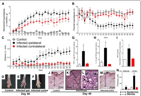
Paw tissue samples were removed at the 40th day post-infection, fixed in 4% formaldehyde and processed for paraffin embedding. Tissue longitudinal sections (5μm) were prepared in cryostat (CM1520, Leica Biosys-tem, Richmond, IL, USA) and slides stained with hematoxylin and eosin (H&E). The analysis of the slides (four slides per mice/four animal per group) was per-formed using light microscopy (Olympus Life Science, model CX31RTSF, Tokyo, Japan) with magnification of × 40 on the panels j, k, and m (scale bars 20μm) and × 100 on the panel l (scale bar 10μm) and presented in Fig. 1. Histopathological score of epidermis and dermis thickening was also performed for the experimental groups with magnification of × 200 and presented in panel n of Fig.1.
Cytokine measurement
cardiac puncture 5–40 days after the infection and added into sterile microtubes containing anticoagulant (EDTA, 5000 IU/mL) for subsequent centrifugation for separation of plasma (500g× 20 min × 4 °C). In brief, 96-well plates were coated overnight at 4 °C with immunoaffinity-puri-fied polyclonal sheep antibody especially for each cytokine evaluated. After blocking the plates, recombinant murine standards for each cytokine tested at various dilutions to-gether with the samples were added in duplicate and incubated overnight at 4 °C. Rabbit biotinylated immunoaffinity-purified antibodies anti-TNF-α and anti-IL-1β were added followed by incubation at room temperature for 1 h. Then, 50μL of avidin-HRP (1:5000 dilution) was added to each well; after 30 additional mi-nutes, the plates were washed and the color reagent o-phenylenediamine (200μL/well; Sigma-Aldrich, St. Louis, MO, USA) was added. After 5 min, the reactions were blocked with 1 M H2SO4 and measured
spectro-photometrically (MultiSkan GO Microplate Spectropho-tometer, ThermoScientific, Vantaa, Finland) at 450 nm. The results were expressed as picograms (pg) of cytokine per 100 mg of paw tissue and as picograms of cytokine per milliliter (mL) of plasma [28].
MPO activity assay
The leukocyte migration to paw tissue was determined 40 days after L. amazonensis infection using the MPO kinetic-colorimetric assay following previous description [28]. Samples of paw tissue were collected in 50 mM K2HPO4buffer (pH 6.0) containing 0.5% hexadecyl
tri-methylammonium bromide (HTAB) and kept at −86 °C until the next step. Samples were then homogenized and centrifuged (16,100g× 2 min × 4 °C), and 10μL of the resulting supernatant was mixed with 200μL of 50 Mm phosphate buffer (pH 6.0), containing 0.167 mg/mL of O-dianisidine dihydrochloride and 0.0005% of hydrogen peroxide (H2O2) and assayed spectrophotometrically for
MPO activity determination at 450 nm (MultiSkan GO Microplate Spectrophotometer, ThermoScientific, Van-taa, Finland). The MPO activity of paw tissue samples
was compared to a standard curve of neutrophils, and the results were presented as MPO activity (number of neutrophils × 104/mg of paw tissue).
Reverse transcription and quantitative polymerase chain reaction
Reverse transcription and quantitative polymerase chain reaction (RT-qPCR) was performed following the proto-col as described previously [2]. Spinal cord samples (L4–
L6 entire segments) were collected initially 5–40 days
and later only at day 30 after the infection withL.
ama-zonensis for the determination of temporal profile of GFAP and Iba-1 mRNA expression and evaluation of CX3CL1, GFAP, Iba-1, TNF-α, and IL-1βmRNA
expres-sion, respectively. The purity of total RNA was measured with a spectrophotometer (MultiSkan GO Microplate Spectrophotometer, ThermoScientific, Vantaa, Finland), and the wavelength absorption ratio (260/280 nm) was between 1.8 and 2.0 for all preparations. Reverse tran-scription of total RNA to cDNA and qPCR were carried out using Go Taq® 2-Step RT-qPCR system (Promega Corporation, Madison, WI, USA) following the manufac-turer’s instructions. The relative gene expression was measured using the comparative 2−(ΔΔCq) method. The primers used are presented in Table1. The expression of β-actin RNA was used as a reference gene to normalize data.
DNA extraction and parasite quantification by real-time qPCR
Real-time qPCR was conducted to evaluate the tissue parasite burden in ipsilateral draining lymph node, spleen, and contralateral lymph node starting from the day of the inoculation of the parasite until the 40th day post-infection. Organs were weighed, washed in PBS, and homogenized in lysis buffer (50 mM Tris-HCl [pH 7.6], 10 mM EDTA, 0.5% SDS, and 0.2 mg/mL proteinase K [Invitrogen, Carlsbad, CA]), followed by phenol-chloro-form extraction of DNA. Samples were then homogenized and incubated at 55 °C for 12 h and subsequently
Table 1Mouse mRNA primers used for RT-qPCR
Target gene Forward Reverse
β-actin 5′-AGCTGCGTTTTACACCCTTT-3′ 5′-AAGCCATGCCAATGTTGTCT-3′
GFAP 5′-GCGCTCAATGCTGGCTTCA-3′ 5′-TCTGCCTCCAGCCTCAGGTT-3′
Iba-1 5′-TGGAGTTTGATCTGAATGGAAAT-3′ 5′-CAGGGCAGCTCGGAGATAGCTTT-3′
CX3CL1 5′-ATTGGAAGACCTTGCTTTGG-3′ 5′-GCCTCGGAAGTTGAGAGAGA-3′
CX3CR1 5′-CACCATTAGTCTGGGCGTCT-3′ 5′-GATGCGGAAGTAGCAAAAGC-3′
TNF-α 5′-TCTCATCAGTTCTATGGCCC-3′ 5′-GGGAGTAGACAAGGTACAAC-3′
IL-1β 5′-GAAATGCCACCTTTTGACAGTG-3′ 5′-TGGATGCTCTCATCAGGACAG-3′
ATF3 5′-CGAAGACTGGAGCAAAATGATG-3′ 5′-CAGGTTAGCAAAATCCTCAAATAC-3′
extracted twice with phenol-chloroform-isoamyl alcohol (25:24:1). Two volumes of cold ethanol (Merck) were added to the aqueous phase, and samples were stored at −20 °C for 12 h. Samples were then centrifuged for 30 min at 10,000g, washed with 70% ethanol, dried at room temperature, and resuspended in 10 mM Tris HCl (pH 8.5). Real-time qPCR was performed by using Platinum SYBR Green qPCR SuperMix UDG with ROX reagent (Invitrogen Corporation, New York, NY) with 100 ng total genomic DNA (gDNA). The quantification of parasites was carried out through the use of specific Leishmania primers described in Table1. The samples were amplified with a Corbett Rotor-Gene thermal cycler under standard-ized steps. A standard curve constructed with DNA from culture samples of L. amazonensis promastigote forms was used for the determination of parasite load in evalu-ated tissues. Results were presented as parasites equiva-lents/100 nanograms (ng) of DNA per sample.
CCI model
For CCI protocol, mice were anesthetized with ketamine and xylazine (10μg/10 mL) followed by trichotomy in the surgery area. The incision was performed in the rear leg, and the sciatic nerve was exposed with a glass rod. A mod-erate constriction injury was performed around the sciatic nerve with a chrome suture according to the method de-scribed by Bennett and Xie [29] adapted to mice [16].
Western blot assay
At the 30th day after the infection withL. amazonensis, L4–L6entire segments of the spinal cord were dissected
and the whole sample homogenized in RIPA buffer con-taining protease and phosphatase inhibitors. The lysates were then homogenized and centrifuged (0.5gfor 10 min at 4 °C). The protein extracts were separated by SDS-PAGE and transferred onto a nitrocellulose mem-brane (GE Healthcare-Amersham, Pittsburgh, PA, USA). The membranes were then incubated in blocking buffer 95% non-fat milk in Tris-buffered saline with Tween 20 or 1% bovine serum albumin (BSA) for different times for each antibody at 4 °C in the presence of primary anti-body. β-Actin, GFAP, and IκBα were purchased from Cell Signaling Technology (Danvers, MA, USA), and Iba-1 and secondary antibody (anti-rabbit, HRP conju-gated) were purchased from Thermo Fisher Scientific (Waltham, MA, USA). Catalog numbers are indicated below. The antibodies and Western blot conditions were as follows: β-actin (#4970, 1:1000) on 12% gel and blocked with 5% non-fat milk; GFAP (#12389, 1:1000) on 12% gel and blocked with 5% non-fat milk; Iba-1 (#PA5-27436, 1:1000) on 15% gel and blocked with 5% non-fat milk; and total IκBα (#9242, 1:1000) on 10% gel and blocked with 5% BSA. The molecular masses of pro-tein were confirmed by Precision Plus Propro-tein Standards
(Bio-Rad, Hercules, CA, USA). After washing in Tris-buffered saline (TBS) with Tween 20, the mem-brane was incubated with secondary antibody (anti-rab-bit, #31460, 1:2000) for 2 h at room temperature. Protein was visualized by chemiluminescence with ECL detec-tion reagent (Luminata™ Forte, Millipore, USA). The membranes were reprobed with antibody againstβ-actin for use as loading control in addition to loading the same amount of protein.
Spinal cord immunofluorescence
On day 30 after the infection withL. amazonensis, mice were perfused through the ascending aorta with saline followed by 4% of paraformaldehyde. After the perfusion, L4–L6segments of the spinal cord were dissected out and
post-fixed and then replaced overnight with 30% sacarose. The spinal cord segments were embedded in optimum cutting temperature (O.C.T.) using Tissue-Tek® com-pound (Sakura® Finetek USA, Torrance, CA), and 7-μm sections were cut in a cryostat (CM1520, Leica Biosystem, Richmond, IL, USA) and processed for immunofluores-cence (four slides per mice/four animal per group). All of the sections were blocked with a buffer solution (500μL per slide containing PBS plus 0.1% Tween 20 plus 5% BSA) for 2 h at room temperature and subsequently incu-bated overnight at −4 °C with a solution containing pri-mary antibodies against GFAP (#180063; 1:500 dilution; Invitrogen, Life Technologies, Carlsbad, CA, USA) and Iba-1 (#PA5-27436; 1:500 dilution, Invitrogen, Life Tech-nologies, Carlsbad, CA, USA). Next, a new incubation with secondary antibody (Alexa Fluor 488, #A-110088 1:1000 dilutions, Thermo Fisher Scientific, Waltham, MA, USA) was performed for 1.5 h at room temperature. The slide assembly was carried out using ProLongTM Gold Antifade Mountant with DAPI melting media (#P36931, Thermo Fisher Scientific, Waltham, MA, USA). Immuno-fluorescence analyses were performed in the dorsal horn of the spinal cord. Dashed lines were used in some repre-sentative images for the demonstration of the gray and white matter areas in the dorsal horn of the spinal cord in which leishmaniasis-induced glial cell activation was ob-served and analyzed. For quantification of GFAP and Iba-1 immunostainings in Figs.3 and 4, equivalent areas of the spinal cord in control non-infected and infected mice were selected. For Figs.3and4, magnification of × 10 on the panels d and e (scale bars 250μm) and × 20 on the panels f–k (scale bars 100μm with gradual additional zoom on the panels h–k). The images and analysis were performed using a confocal microscope (SP8, Leica Microsystems, Mannheim, Germany) [16].
NFκB activation assay
described previously [2]. Spinal cord samples (L4–L6
en-tire segments) were collected at day 30 after the infec-tion with L. amazonensis and the whole sample homogenized in ice-cold lysis buffer (Cell Signaling Technology, Beverly, MA, USA). The homogenates were centrifuged (16,000g× 10 min × 4 °C) and the superna-tants use to assess the levels of total and phosphorylated NFκB p65 subunit by ELISA using PathScan kits (Cell Signaling Technology, Beverly, MA, USA) according to the manufacturer’s instructions. The results were expressed as total-p65/phosphor-p65 ratio measured spectrophotometrically (MultiSkan GO Microplate Spec-trophotometer, ThermoScientific, Vantaa, Finland) at 450 nm.
Statistical analysis
Results are presented as means ± SEM of measurements made on three to six mice in each group depending on the analysis, per experiment, and are representative of two separate experiments. Two-way analysis of variance (ANOVA) was used to compare the groups and doses at all times when responses were measured at different times after the parasite injection. Analyzed factors were treatments, time, and time versus treatment interaction, and when interaction was significant, one-way ANOVA followed by Tukey’s post hoc was performed for each time point. Differences between responses were evalu-ated by one-way ANOVA followed by Tukey’s post hoc for data of single time point. Statistical differences were considered significant whenP< 0.05.
Results
L. amazonensisi.pl. infection induces chronic mechanical and thermal hyperalgesia associated to the development of paw edema and increases TNF-α, IL-1β, and MPO activity levels in the paw tissue at the 40th day post-infection The measurements of mechanical hyperalgesia, thermal hyperalgesia, and paw edema were performed between 2 and 40 days after i.pl. infection with L. amazonensis (Fig.1a–f ). Samples of paw tissue were collected at day 40 after the infection to evaluate TNF-α and IL-1β produc-tion, MPO activity levels, and histopathological analysis (Fig.1g–m).L. amazonensisinfection induced chronic ip-silateral mechanical and thermal hyperalgesia compared to control non-infected animals starting at days 6 and 10, respectively, and persisting until day 40 post-infection (p < 0.05, Fig.1a, b). Interestingly, chronic mechanical hyper-algesia but not thermal hyperhyper-algesia (p< 0.05, Fig. 1a, b) was detected in the contralateral paw of the infected ani-mals compared to control non-infected aniani-mals from the 10th to 40th days post-infection. These results suggest that pain induced by leishmaniasis could involve the acti-vation of the ipsilateral spinal cord side to the infection and also a minor activation of the contralateral spinal cord
side to the infection. The paw edema became evident from day 18 onwards, remaining significantly different from the control non-infected animals until the 40th day post-infection (p< 0.05, Fig.1c). Paw edema was not ob-served in the contralateral paw of infected animals (Fig. 1c). Representative pictures from control non-in-fected animals, swollen ipsilateral paw, and contralateral paw of infected animals at day 40 post-infection are pre-sented respectively in Fig. 1d–f. L. amazonensis infection also increased the levels of TNF-α and IL-1β, and MPO activity in the ipsilateral, but not contralateral paw tissue of infected mice when compared to control non-infected animals at day 40 after the infection (p< 0.05, Fig.1g–i). Histopathological images of ipsilateral paw tissue of in-fected animals (Fig.1k, l) show the presence of amastigote forms of L. amazonensis inside the vacuoles of macro-phages (arrows). Paw tissue of control non-infected (Fig.1j) and contralateral paw of infected animal (Fig.1m) presented regular histology. Histopathological score pre-sented in Fig.1n shows increased thickening of the epider-mis and derepider-mis in ipsilateral paw compared to control non-infected and contralateral paw of infected animals. In addition to supporting the hyperalgesic and edematogenic effects of L. amazonensis infection [2], these data evi-denced increases in TNF-αand IL-1βproduction in paral-lel to increased neutrophil recruitment (as reflected by MPO assay) in infected paw tissue. Therefore, although leishmaniasis induces chronic ulcerative infection, these results confirm that there is consistent peripheral infection-induced inflammation even at early time points when skin ulceration is absent.
L. amazonensisi.pl. infection does not change the mRNA expression of ATF3 in the DRG
The extent that DRGs were affected by L. amazonensis i.pl. infection was evaluated 30 days of infection [2]. There was no significant induction of ATF3 mRNA expression in DRG samples of infected mice compared to non-infected mice (Additional file 1: Figure S1). CCI-induced ATF3 mRNA expression in the DRG was used as a positive control of ATF3 mRNA expression (Additional file 1: Figure S1). The absence of increased mRNA expression of ATF3 in ipsilateral DRG suggests that at least during this period of the disease course (30th day), there is no nerve damage, since this tran-scription factor is expressed by injured neurons [30].
of TNF-α and IL-1β over the course of the disease (Additional file 2: Figure S2). The i.pl. infection with L.
amazonensisdoes not modify the blood levels of TNF-α (Additional file2: Figure S2A) and IL-1β(Additional file 2: Figure S2B) during the experimental protocol in com-parison with non-infected animals, suggesting that the systemic levels of these hyperalgesic cytokines have no decisive role in the development of neuroinflammation in the spinal cord in the present model. Regarding para-sitism, we investigated parasite load at different time points (0, 5, 10, 20, 30, and 40 days) post-infection in im-mune organs distant from the site of inoculation of the parasite (ipsilateral draining lymph node, spleen, and contralateral lymph node) to analyze the occurrence of systemic infection (Additional file3: Figure S3). Mice re-ceived i.pl. injection of parasites on day 0. The presence
of parasites was not detected in samples collected at the day of infection (Additional file 3: Figure S3A–C). The presence of parasites occurred in all evaluated organs at the fifth day post-infection onwards. However, the para-sitism remained stable except by the ipsilateral draining lymph node that presented a progressive and time-dependent increase of parasitism (Additional file 3: Figure S3A–C). These results might be related to the time-dependent increase of infection in the paw [2].
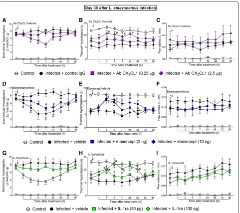
The i.t. treatments with anti-CX3CL1 antibody, etanercept, and IL-1ra inhibitedL. amazonensis-induced ongoing mechanical and thermal hyperalgesia without affecting paw edema
Mice were treated by i.t. route with vehicles (control IgG or saline) or anti-CX3CL1 antibody, etanercept, and
IL-1ra at day 30 after L. amazonensis infection, and mechanical hyperalgesia, thermal hyperalgesia, and paw edema were evaluated (Fig. 2). This time point was se-lected because it is when TNF-αexpression in the spinal cord reaches its peak [2]. The dose of 0.25μg of anti-CX3CL1 antibody had no effect on mechanical
hyperalgesia and thermal hyperalgesia, while the dose of 2.5μg significantly inhibited mechanical hyperalgesia and thermal hyperalgesia between 3–48 and 1–24 h after
the treatment, respectively. The anti-hyperalgesic effect of the 2.5μg of anti-CX3CL1 antibody was also
signifi-cant compared to the 0.25μg dose at the seventh and at the first hours after the treatment for mechanical hyper-algesia and thermal hyperhyper-algesia, respectively (p< 0.05, Fig.2a, b). Antibody (IgG) control treatment presents no effect. The dose of 3 ng of etanercept inhibited mechan-ical hyperalgesia between 5 and 24 h and thermal hyper-algesia between 3 and 5 h after the treatment. On the
Fig. 2Neutralizing antibody anti-CX3CL1, etanercept, and IL-1ra i.t. treatments inhibitL. amazonensis-induced hyperalgesia, but not paw edema.
Mechanical (a,d, andg) and thermal (b,e, andh) hyperalgesia and paw edema (c,f, andi) were measured in control non-infected and infected mice at day 30 after the infection, and subsequently, infected mice received i.t. injection of antibody anti- CX3CL1 (0.25–2.5μg), etanercept (3–10
ng), IL-1ra (30–100 pg), or vehicles (control IgG for Ab CX3CL1 and saline for the other drugs) followed by measurement of mechanical and
thermal hyperalgesia and paw edema. Results are presented as mean ± SEM of six mice per group per experiment and are representative of two separate experiments. *p< 0.05 compared to control non-infected mice;#p< 0.05 compared to vehicle-treated infected mice; **p< 0.05
compared to the lower doses of antibody anti- CX3CL1, etanercept, IL-1ra, and vehicle-treated infected mice (one-way ANOVA followed by Tukey
other hand, significant inhibition of mechanical hyper-algesia and thermal hyperhyper-algesia occurred for a longer duration with the dose of 10 ng of etanercept, which was observed between 1–48 and 3–24 h after the treatment, respectively, in comparison with the ve-hicle (saline)-treated group (p< 0.05, Fig. 2d, e). The effect of 10 ng of etanercept was also significant com-pared to the lower dose at the fifth hour after the treatment for mechanical hyperalgesia (p< 0.05, Fig. 2d). The dose of 30 pg of IL-1ra had no effect on mechanical hyperalgesia, but inhibited thermal hyper-algesia at the third hour after the treatment, whereas the dose of 100 pg inhibited mechanical hyperalgesia and thermal hyperalgesia between 1–48 and 3–48 h after the treatment, respectively, in comparison with the vehicle (saline)-treated group (p< 0.05, Fig. 2g, h). The dose of 100 ng of IL-1ra also reduced mechanical hyperalgesia compared to the dose of 30 ng between 1 and 24 h after the treatment (p< 0.05, Fig. 2g). None of the treatments affected L. amazonensis-induced paw edema compared to vehicle-treated groups (Fig. 2c, f, and i). These results suggest the participa-tion of spinal cord CX3CL1, TNF-α, and IL-1β in the
mechanism of L. amazonensis-induced pain.
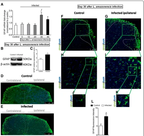
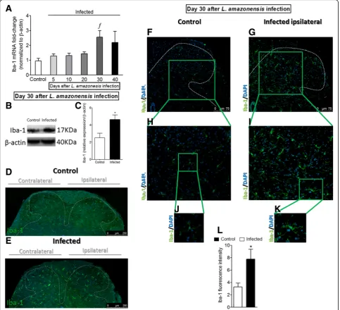
L. amazonensisi.pl. infection induces the activation of astrocytes in the spinal cord
Whether L. amazonensis i.pl. infection induces the acti-vation of spinal cord astrocytes is unknown. In this sense, the temporal profile of spinal cord GFAP mRNA expression over the course of disease (5–40 days post-infection) was investigated (Fig. 3a). RT-qPCR data showed no changes during the first 20 days when com-pared to control non-infected animals. However, signifi-cant increases in GFAP mRNA expression were detected at day 30 after the infection when compared to previous days (p< 0.05, 5–20) and control non-infected animals, followed by a slight reduction at day 40, without signifi-cant difference compared to other groups in this last day of evaluation (Fig. 3a). To confirm RT-qPCR data re-garding astrocytes activation (GFAP, Fig. 3a) at day 30 afterL. amazonensis infection, Western blot (Fig. 3b, c) and immunofluorescence (Fig. 3d–l) assays were per-formed in spinal cord samples of control non-infected and infected animals. Western blot analysis of the spinal cord tissues showed astrocyte activation in infected mals that was not detected in control non-infected ani-mals (p< 0.05, GFAP, Fig.3b, c). Immunofluorescence of the spinal cord evidenced that the activation of astro-cytes (GFAP) occurred bilaterally in infected animals, which was not observed in control non-infected ani-mals (Fig. 3d, e). Infected mice presented an increase of GFAP intensity of fluorescence in both gray and white matter areas of the ipsilateral side of the
infection in the spinal cord (Fig. 3g, i, k, and l) com-pared to non-infected mice (Fig. 3f, h, j, and l). To-gether with the upregulation of GFAP expression, astrocyte activation or astrogliosis is characterized by cellular hypertrophy observed as enlarged cell bodies and thickening of process [31], and this was also ob-served in spinal cord samples of infected mice (p< 0.05, Fig. 3i, k). L. amazonensis infection induces spinal cord activation of astrocytes (Fig. 3k) not ob-served in control non-infected animals (Fig. 3j).
L. amazonensisi.pl. infection induces the activation of microglia in the spinal cord
Following the same sequence of experiments as pre-sented in the previous subsection, mice received i.pl. in-oculation of L. amazonensis to evaluate whether the infection induces the activation of spinal cord microglia (Fig. 4 and Additional file 4: Figure S4). The temporal profile of Iba-1 mRNA expression during the course of disease was similar to that observed for GFAP. There was no statistical difference in the Iba-1 mRNA expres-sion between 5 and 20 days after the infection when compared to the control group, while significant in-crease was observed at day 30 post-infection (p< 0.05). In the following time point (40th day), Iba-1 mRNA ex-pression decreased slightly and presented no statistical difference compared to other groups (Fig. 4a). Spinal cord Western blot (Fig. 4b, c) and immunofluorescence (Fig. 4d–l) analysis at day 30 afterL. amazonensis infec-tion confirmed RT-qPCR data and demonstrated micro-glial activation in the infected group in comparison with control non-infected animals (p< 0.05, Iba-1, Fig.4b–l), but contrary to what was evidenced for astrocytes (GFAP, Fig. 3g), microglial activation was detected al-most exclusively in the gray matter area of the ipsilateral side of the infection in the spinal cord. Interestingly, as observed for astrocytes (GFAP, Fig.3d, e), immunofluor-escence of the spinal cord demonstrated bilateral micro-glial activation (Iba-1) in infected animals and not in control non-infected animals (Fig. 4d, e). Microglia acti-vation is often reported as Iba-1 upregulation associated to profound changes in cell shape, reflected by hyper-trophy of the cell body and increase of the branching processes [32], which can be observed in Fig. 4i and k (p< 0.05). Therefore, L. amazonensis infection induces spinal cord activation of microglia (Fig. 4j–l). CX3CR1
mRNA expression did not present statistical difference compared to the control group until the 10th day; how-ever, a significant increase was observed between 20 and 30 days. At the 40th day, no statistical difference was ob-served between the groups (Additional file1: Figure S4). CX3CR1 contributes to microglia activation [15]; thus,
the increase of CX3CR1 mRNA expression before Iba-1
Fig. 3Detection of spinal cord GFAP induced by i.pl.L. amazonensisinfection. GFAP mRNA expression was determined in control non-infected and infected mice after the infection (5–40 days) by RT-qPCR (a). On day 30 after the infection (peak of GFAP mRNA expression), Western blot analysis of the whole spinal cord was conducted to confirm GFAP expression at day 30 post-infection (three mice per group,bandc). Subsequently, spinal cord samples were stained with primary antibody for astrocytes (GFAP, green) and regular nucleus (DAPI, blue) detection (four mice per group/four slides per mice). Representative immunostainings of the spinal cord of control non-infected and infected mice are shown ind–k(dandemagnification × 10, scale bar 250μm; panelsf–kmagnification × 20, scale bar 100μm, with gradual zoom for panelsh
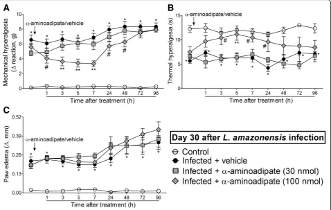
The i.t. treatment with the selective astrocyte inhibitor α-aminoadipate diminishesL. amazonensis-induced mechanical and thermal hyperalgesia without affecting paw edema
Mice were treated by i.t. route with vehicle or α-aminoadipate (30–100 nmol) 30 days after L.
amazonensis infection, and mechanical hyperalgesia, thermal hyperalgesia, and paw edema were evaluated (Fig. 5). The dose of 30 nmol ofα-aminoadipate did not affect mechanical hyperalgesia, thermal hyperalgesia, and paw edema (Fig.5a–c). On the other hand, i.t. treat-ment with 100 nmol of α-aminoadipate significantly Fig. 4Detection of spinal cord Iba-1 induced by i.pl.L. amazonensisinfection. Iba-1 mRNA expression was determined in control non-infected and infected mice after the infection (5–40 days) by RT-qPCR (a). On day 30 after the infection (peak of Iba-1 mRNA expression), Western blot analysis of the whole spinal cord was conducted to confirm Iba-1 expression at day 30 post-infection (four mice per group,bandc). Subsequently, spinal cord samples were stained with primary antibody for microglia (Iba-1, green) and regular nucleus (DAPI, blue) detection (four mice per group/four slides per mice). Representative immunostainings of the spinal cord of control non-infected and infected mice are shown ind–k(dande, magnification × 10, scale bar 250μm; panelsf–kmagnification × 20, scale bar 100μm, with gradual zoom for panelsh
inhibited L. amazonensis-induced mechanical hyperalge-sia for up to 48 h after the treatment and thermal hyper-algesia between 3 and 24 h after the treatment in comparison with the vehicle-treated group (p< 0.05, Fig. 5a, b). The anti-hyperalgesic effect of 100 nmol of α-aminoadipate in thermal hyperalgesia was also signifi-cant compared to the dose of 30 nmol at 5 h (p< 0.05, Fig. 5b). The treatment with α-aminoadipate did not affect L. amazonensis-induced paw edema (Fig. 5c). These results demonstrate that targeting spinal cord as-trocytes inhibitsL. amazonensis-induced hyperalgesia.
The i.t. treatment with the microglia inhibitor minocycline diminishesL. amazonensis-induced mechanical and thermal hyperalgesia without affecting paw edema Mice were treated by i.t. route with vehicle or minocycline (50–150μg) at the 30th day afterL. amazonensisinfection to determine the participation of spinal cord microglia in the mechanical hyperalgesia, thermal hyperalgesia, and paw edema in this infection (Fig.6). The dose of 50μg of minocycline did not alter leishmaniasis-induced mechan-ical and thermal hyperalgesia (Fig. 6a, b). On the other
hand, i.t. treatment with 150μg of minocycline inhibited
L. amazonensis-induced mechanical and thermal hyper-algesia between 1–24 h and 5–24 h, respectively (p< 0.05, Fig.6a, b). There was also significant inhibition of mech-anical hyperalgesia comparing the doses of 50 and 150μg of minocycline at the third and fifth hour after treatment (p< 0.05, Fig.6a). Minocycline treatment did not alter L.
amazonensis-induced paw edema (Fig. 6c). Therefore, inhibiting spinal cord microglial activity diminished L.
amazonensis-induced hyperalgesia.
The i.t. treatment with the selected effective analgesic doses ofα-aminoadipate and minocycline diminishesL.
amazonensis-induced contralateral mechanical hyperalgesia The two previous experiments evaluating the dose-re-sponse effects of α-aminoadipate and minocycline upon ipsilateral hyperalgesia and paw edema showed that the higher doses tested for both drugs presented a satisfac-tory analgesic effect on L. amazonensis-induced hyper-algesia. Thus, we tested the effects of these same doses (150 nmol and 150μg, respectively) upon the contralat-eral mechanical hypcontralat-eralgesia in infected mice 7 h after Fig. 5α-Aminoadipate i.t. treatment inhibitsL. amazonensis-induced hyperalgesia, but not paw edema. Mechanical (a) and thermal (b)
hyperalgesia and paw edema (c) were measured in control non-infected and infected mice on day 30 after the infection, and subsequently, infected mice received i.t. injection ofα-aminoadipate (selective astrocyte inhibitor, 30–100 nmol) or vehicle for measurement of mechanical and thermal hyperalgesia and paw edema. Results are presented as mean ± SEM of six mice per group per experiment and are representative of two separate experiments. *p< 0.05 compared to control non-infected mice;#
the treatments (Additional file 5: Figure S5). Both α-aminoadipate and minocycline inhibited contralateral mechanical hyperalgesia from 3 to 48 h after the treat-ment at the 30th day post-infection (Additional file 5: Figure S5A). As expected, treatments with these glial in-hibitors had no effect on contralateral thermal hyper-algesia and paw edema (Additional file5: Figure S5B and C), since these parameters were not altered previously in the infected animals in the contralateral side. These data corroborate that activated spinal cord astrocytes and microglia (Figs.3 and 4) mediate the mechanical hyper-algesia observed in the contralateral paw.
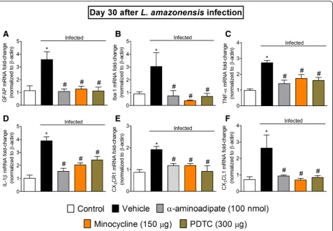
Targeting spinal cord astrocytes, microglia, and NFκB inhibitsL. amazonensis-induced spinal cord GFAP, Iba-1, TNF-α, IL-1β, CX3CR1, and CX3CL1 mRNA expression
Considering the results obtained in dose-response experi-ments forα-aminoadipate and minocycline presented earlier, the doses of 100 nmol and 150μg, respectively, were selected for the experiments in this section. The dose of the NFκB in-hibitor PDTC of 300μg per mouse by i.t. route was selected
in a dose-response curve from a previous study [2]. Mice re-ceived i.t. treatment with α-aminoadipate, minocycline, or PDTC at the 30th day after L. amazonensis infection, and samples of the spinal cord (L4–L6) were collected 7 h after
the treatments for RT-qPCR analysis of GFAP (Fig. 7a), Iba-1 (Fig. 7b), TNF-α (Fig. 7c), IL-1β (Fig. 7d), CX3CR1
(Fig. 7e), and CX3CL1 (Fig. 7f) mRNA expression.
Leish-maniasis induced significant increase of spinal cord GFAP, Iba-1, TNF-α, IL-1β, CX3CR1, and CX3CL1 mRNA
expres-sion, which were inhibited byα-aminoadipate, minocycline, and PDTC treatments (p< 0.05, Fig.7a–e). These results in-dicate that spinal cord astrocytes, microglia, and NFκB acti-vation underlies the mRNA expression of pro-hyperalgesic cytokines and chemokines in leishmaniasis.
Leishmaniasis induces CX3CL1 mRNA expression in the ipsilateral DRG and i.t. injection of TNF-αand IL-1β stimulates CX3CL1 mRNA expression in the ipsilateral DRG and in the spinal cord
To determine the mechanism of CX3CL1 upregulation
in the spinal cord of L. amazonensis-infected mice, we Fig. 6Minocycline i.t. treatment inhibitsL. amazonensis-induced hyperalgesia, but not paw edema. Mechanical (a) and thermal (b) hyperalgesia and paw edema (c) were measured in control non-infected and infected mice on day 30 after the infection, and subsequently, infected mice received i.t. injection of minocycline (microglia inhibitor, 50–150μg) or vehicle for measurement of mechanical and thermal hyperalgesia and paw edema. Results are presented as mean ± SEM of six mice per group per experiment and are representative of two separate experiments. *p< 0.05 compared to control non-infected mice;#p< 0.05 compared to vehicle-treated infected mice; **p< 0.05 compared to 50μg minocycline
investigated firstly the CX3CL1 mRNA expression in
ip-silateral and contralateral DRG of infected mice to deter-mine in which side there would be upregulation. An increase of CX3CL1 mRNA expression was observed in
the ipsilateral DRG only (Fig. 8a). Considering that in-hibition of glial cells reduced TNF-α and IL-1β mRNA expression (Fig. 7b, d), we tested whether these cyto-kines would represent a possible mechanism of induc-tion of CX3CL1 mRNA expression in the spinal cord.
TNF-α and IL-1β were injected by i.t. route (1 ng for both) in naïve non-infected mice, and CX3CL1 mRNA
expression was evaluated by RT-qPCR. CX3CL1 mRNA
expression was observed in the DRG as early as 3 h after cytokine injection and after 24 h in the spinal cord (Figs. 8b, c). Together, these data suggest a mechanism involving spinal TNF-α and IL-1β in CX3CL1
produc-tion in the spinal cord after L. amazonensis peripheral infection. This effect may represent a positive feedback loop in which the production of TNF-α and IL-1β by glia stimulates the release of CX3CL1 by neurons and
astrocytes [17,33] that perpetuate the cycle by the acti-vation of microglia via CX3CR1 and, consequently, more
TNF-αand IL-1βrelease.
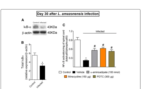
Targeting spinal cord astrocytes and microglia inhibitsL. amazonensis-induced spinal cord NFκB activation
Spinal cord NFκB has a key role in L. amazonensis -in-duced hyperalgesia [2]. Figure7 shows that the inhibition of glial cells diminishes the mRNA expression of hyper-algesic molecules similarly to NFκB inhibition. Therefore, we investigated the activation of spinal cord NFκB in con-trol non-infected and infected animals as well as whether targeting spinal cord glial cells would also result in NFκB inhibition using Western blot and ELISA assays (Fig. 9). Western blot analysis demonstrated that in control non-infected mice, total IκBα expression is higher when compared to spinal cord samples of infected mice (p< 0.05, Fig. 9a). Since IκBα has the function of inhibiting NFκB in the cytoplasm, these data suggest that infection induces NFκB activation. Next, mice were treated by i.t. Fig. 7α-Aminoadipate, minocycline, and PDTC i.t. treatments inhibitL. amazonensis-induced spinal cord GFAP, Iba-1, TNF-α, IL-1β, CX3CR1, and
CX3CL1 increased mRNA expression. On day 30 after the infection, spinal cord GFAP (a), Iba-1 (b), TNF-α(c), IL-1β(d), CX3CR1 (e), and CX3CL1 (f) mRNA
Fig. 8L. amazonensisi.pl. infection induces CX3CL1 mRNA expression in the ipsilateral DRG, and i.t. injection of TNF-αand IL-1βinduces CX3CL1
mRNA expression in the ipsilateral DRG and spinal cord. CX3CL1 mRNA expression was determined in control non-infected and bilaterally in
infected mouse on the 30th day after the infection by RT-qPCR (a).b,cThe CX3CL1 mRNA expression in DRG (3 h) and spinal cord (3–24 h) after
i.t. injections of TNF-αand IL-1βin naïve non-infected animals. Results are presented as mean ± SEM of six mice per group per experiment and are representative of two separated experiments fora,b, andc. *p< 0.05 compared to control non-infected mice (one-way ANOVA followed by Tukey post hoc)
Fig. 9L. amazonensisi.pl. infection decreases spinal cord total IκBαexpression, andα-aminoadipate, minocycline, and PDTC i.t. treatments inhibit
L. amazonensis-induced spinal cord NFκB activation. On day 30 after the infection, spinal cord total IκBαprotein expression was evaluated by Western blot (four mice per group,aandb), and NFκB activation was evaluated in control non-infected and infected mice 7 h after treatments with vehicle,α-aminoadipate, minocycline, or PDTC by ELISA (c). Results are presented as mean ± SEM of four mice per group per experiment and are representative of two separate experiments foraandb. Forc, results are presented as mean ± SEM of six mice per group and are representative of two separated experiments. *p< 0.05 compared to control non-infected mice;#p< 0.05 compared to vehicle treated-infected
route with α-aminoadipate and minocycline at the 30th day ofL. amazonensis infection, and spinal cord samples were collected 7 h after treatment and processed for ELISA (Fig.9b). The inhibition of astrocytes and microglia diminished the NFκB activation as observed by an in-crease in total NFκB p65 subunit/phosphorylated NFκB p65 subunit ratio (p< 0.05, Fig.9b). As we have previously observed [2], the control i.t. treatment with PDTC dimin-ished leishmaniasis-induced spinal cord activation of NFκB (p< 0.05, Fig.8b). These results indicate that the ac-tivity of glial cells involve the activation of NFκB in the spinal cord during leishmaniasis, which likely explains why targeting glial cells reduces mechanical hyperalgesia, thermal hyperalgesia, and the mRNA expression of hyper-algesic molecules.
Discussion
Pathophysiological features of pain processing in neglected parasitic infectious diseases such as leishman-iasis remain poorly investigated. The idea that cutaneous leishmaniasis induces painless ulcers likely dampened studying pain in leishmaniasis. However, it is striking that animal models show clear nociceptive behavior [3, 12, 13]. Further, growing body of clinical evidence sup-ports that pain is a symptom of leishmaniasis independ-ently of the region of the body [1, 5–11]. This compelling evidence of pain highlights the importance of understanding the nociceptive mechanisms of leish-maniasis to improve the treatment of pain in human leishmaniasis.
The relation between viral or bacterial infections and glial activation during pain processing was previously in-vestigated using disease models and in vitro analysis [34–36]. CL models show similarities to the human dis-ease; however, immunity, disease outcome, parasite strains, and mice strains limit the homogeneity of data. To the best of our knowledge, this is the first study dem-onstrating that infection with L. amazonensis induces spinal cord neuroinflammation dependent on glia activa-tion that accounts to hyperalgesia. The choice of using only male mice was based on previous demonstration that distinct cell populations in the spinal cord are re-sponsible in mediating mechanical hypersensitivity de-pending on the gender [20, 21] and also because glial inhibitor minocycline has no effect in female mice re-garding nociception [20].
L. amazonensisi.pl. infection induces prolonged mech-anical and thermal hyperalgesia in mice in the ipsilateral side of the infection, which corroborates our previous results [2] and others using L. major infection [3, 12, 13]. Interestingly, we observed that L. amazonensis also induced prolonged mechanical hyperalgesia in the contralateral side of the infection, although less intense in comparison with the ipsilateral side. The induction of
thermal hyperalgesia was observed in the ipsilateral paw, but not in the contralateral paw toLeishmaniainfection. It is also possible that the neuroinflammatory plastic changes in the spinal cord are responsible for this select-ive sparing of the contralateral side to sensitization to thermal stimulus by yet undetermined mechanisms [15, 35]. For instance, in a model of CFA-induced masseter muscle inflammation in rats, unilateral injection of CFA caused bilateral allodynia and increased TRPV1 expres-sion in the ipsilateral trigeminal ganglia, but not in the contralateral trigeminal ganglia [37]. The intraplantar in-jection of CFA also causes bilateral mechanical hyper-algesia and thermal hyperhyper-algesia in the ipsilateral side only [38]. Thus, stimulus was not capable of inducing the same alterations in the contralateral side as in the ip-silateral side to the stimulus. No ulcerative lesions were observed during the experiments, which are important to allow studying the nociceptive phase of disease [2, 9, 13]. Furthermore, L. amazonensisinfection did not alter ipsilateral ATF3 mRNA expression in DRG cells at the 30th day post-infection indicating no neuronal lesion in contrast with neuropathic pain conditions such as CCI of the sciatic nerve.
L. amazonensisinduced an increase of MPO activity in the ipsilateral side of the infection indicating neutrophil/ monocyte recruitment. TNF-α and IL-1β protein levels also increased in the ipsilateral paw skin tissue at the 40th day of infection, which were not observed in the contralat-eral non-infected paw skin. Blood TNF-αand IL-1βlevels did not change during the course of infection, which sug-gests that systemic inflammation does not explain the neuroinflammatory events observed in the spinal cord. On the other hand, resident and recruited innate immune cells produce TNF-αand IL-1βat the site of infection [14, 39], which contribute to host protection and killing mech-anisms inLeishmaniainfection [40,41] as well as may ac-count to nociception [42–45]. In fact, it is rational to suppose that TNF-αand IL-1βproduced at the infection primary foci explain the hyperalgesia observed before spinal cord glia activation. TNF-α and IL-1β could sensitize nociceptor neurons at an early stage leading to later spinal cord processing of nociceptive information and activation of glial cells that would boost nociception by releasing additional hyperalgesic molecules in the spinal cord [15, 18, 19], therefore contributing to leishmaniasis-induced pain. At the 30th day post-infec-tion, single i.t. treatments with neutralizing anti-CX3CL1,
etanercept, or IL-1ra inhibited hyperalgesia but not paw edema, which is in line with the notion that peripheral in-puts can induce spinal cord neuroinflammation that will enhance nociception [15,18,19].
The immunofluorescence data demonstrate L.
pain models in which the injury is unilateral such as spinal nerve ligation (SNL), bilateral detection of glial activity is not observed for up to 28 days after SNL [46]. Although the injection of parasite was performed in only one paw, the parasite may spread and infect other tis-sues. Time-dependent increase of parasitism was de-tected only in the ipsilateral draining lymph node. Parasites were also detected in the contralateral lymph node and spleen; however, the parasite load remained stable until the 40th day post-infection. In agreement with our data, evidence supports that the main organs affected up to 90 days post-infection are paw tissue (site of the inoculation of the parasite) and ipsilateral draining lymph node in leishmaniasis [23]. These results suggest that parasite load parallels, at least in part, the observed pain responses [2]. Corroborating this rationale, parasite load increases over time in the draining lymph node of the ipsilateral paw with detectable parasitism starting at the 5th day and increasing up to the 40th day. The im-mune cellular/molecular events in periphery contribute to produce neuroplasticity in the spinal cord and persistent pain, which might account for long-lasting bilateral pain detected here [15]. Depending on the intensity of local peripheral activation of nociceptor neurons, spinal cord activation in the ipsilateral side can spread to the contra-lateral side [15]. In the present experimental condition, this is likely to be occurring considering there was minor mechanical hyperalgesia and no thermal hyperalgesia in the contralateral paw, which should be comparable to the ipsilateral side in case of infection spread.
Astrocytes are distributed in the CNS with low levels of GFAP expression and in non-overlapping and orga-nized manner with only the individual most distal tips interdigitation with one another in grey matter. Diseases of the CNS, nerve lesion, activated microglia, cell dam-age, ischemia, and neuronal hyperactivity can induce astrogliosis, which encompasses molecular and func-tional changes resulting in progressive modifications ranging from hypertrophy to proliferation and scar for-mation, and overlapping individual fields. In moderate astrogliosis, there is hypertrophy, increase of GFAP ex-pression, but the individual astrocyte domains are pre-served and there is no pronounced overlapping of processes. In severe astrogliosis, there is hypertrophy with high increase of GFAP expression together with proliferation and overlapping of processes [47].L.
ama-zonensisinduced a moderate astrogliosis at the 30th day of infection as observed by increase of GFAP expression, hypertrophy, and no pronounced overlapping of astro-cyte processes. Upon a pathological event, microglia mi-grate and proliferate with increasing Iba-1 expression, transform morphologically into more branched and ramified cells than in the resting state, and expand their surveillance area invading the territory of other
microglial cells that normally occupy defined territories [48]. The expression of CX3CR1 by microglia is also a
critical step to the activation of these cells [15]. These phenotypic changes characteristic of microgliosis also occurred inL. amazonensisinfection.
Spinal cord astrocytes and microglia are described as key cells in the mechanism of pain processing in inflam-matory, neuropathic, and cancer models [15,16,18,19]. α-Aminoadipate and minocycline are effective inhibitors used in models of glial cell-dependent chronic pain such as SNL [49, 50]. In the present study, α-aminoadipate and minocycline reduced leishmaniasis-induced ipsilat-eral mechanical hypipsilat-eralgesia and thermal hypipsilat-eralgesia and also contralateral mechanical hyperalgesia. Thus, spinal astrocytes and microglia contribute to L.
In neuropathic pain, neuronal-glial interactions lead to central sensitization by mechanisms involving the release of primary sensory neuron-derived CX3CL1, which binds
to its receptor CX3CR1 expressed by microglia and
acti-vates these cells. In turn, microglia produce hyperalgesic mediators such as TNF-α and IL-1β that together with other pro-nociceptive molecules activate and sensitize spinal cord nociceptor neurons. Astrocytes are also in-volved in the mechanism of central sensitization and under TNF-α-dependent activation represent important sources of glutamate that further contributes to activate second-order neurons [17,18]. CX3CL1 is described as a
key chemokine for enhanced nociceptive response in the spinal cord with increased protein and mRNA expres-sion [17, 54–56]. Herein, the inhibition of leishmaniasis-induced activation of astrocytes, microglia, and NFκB reduced spinal cord CX3CL1 mRNA
expres-sion as well as Leishmania infection induces CX3CL1
mRNA expression in the ipsilateral DRG of infected mice. Accordingly, using naïve non-infected mice, we demonstrated that the i.t. injection of hyperalgesic doses of TNF-α and IL-1β [16] stimulate CX3CL1 mRNA
ex-pression in the spinal cord, which corroborates previous in vitro data using human astrocytes [57]. Although
CX3CL1 may be induced in spinal cord astrocytes, it is
mainly constitutively expressed by DRG neurons and their projections into the spinal cord. Its receptor, CX3CR1, may be found in peri-neuronal glia in DRG,
but it is primarily expressed by spinal cord dorsal horn microglia with a prominent upregulation in these glial cells in neuropathic pain models, suggesting a neuronal-microglial signaling in spinal cord upon patho-logical conditions [17, 33, 58]. Spinal cord CX3CR1
mRNA expression was detected between 20 and 30 days post-L. amazonensisinfection, which is rational since its expression is necessary for microglial activation that was showed to start at day 30 [15]. Evidence in other models of pain observed CX3CL1 expression by astrocytes and
neurons in the spinal cord although the neuronal ex-pression is more widely demonstrated [15, 17, 33, 59]. TNF-α and IL-1β also induced CX3CL1 mRNA
expres-sion, and glial inhibition reduced TNF-αand IL-1β pro-duction, thus suggesting that glial cells regulate CX3CL1
mRNA expression via, at least in part, TNF-α and IL-1β duringL. amazonensisinfection. Another possible mech-anism contributing to hyperalgesia is the retrograde axonal transport of TNF-αand its receptors TNFR1 and TNFR2 from the periphery to the spinal cord [60]. At
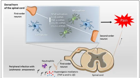
Fig. 10Schematic proposition of the mechanisms involved inL. amazonensisinfection-induced spinal cord events leading to hyperalgesia in BALB/c mice. The peripheral infection withL. amazonensisleads to an immune reaction in infected paw tissue characterized by neutrophil recruitment (increased MPO activity) and elevated production of pro-inflammatory cytokines TNF-αand IL-1βat the primary infection focus, which are well-known sensitizers of first-order neurons. Nociceptive signaling reaches the dorsal horn of spinal cord resulting in increased mRNA expression of CX3CL1, CX3CR1, microglia, and astrocytes activation and consequent NFκB-dependent TNF-αand IL-1βincreased mRNA expression.