R E S E A R C H
Open Access
Targeting late-stage non-small cell lung
cancer with a combination of DNT cellular
therapy and PD-1 checkpoint blockade
Linan Fang
1,2, Dalam Ly
2,3, Si-si Wang
4, Jong Bok Lee
2,3, Hyeonjeong Kang
2, Hao Xu
4, Junlin Yao
2,7,
Ming-sound Tsao
5,6, Wei Liu
1*and Li Zhang
2,3,6,8*Abstract
Background:Though immune checkpoint blockade (ICB) against PD-1 has shown success in the treatment of lung cancer, not all patients respond. We have previously shown that adoptive transfer of double negative T (DNT) cells expanded from healthy donors can target leukemia but their role in treating established lung cancer is not clear. Here we explore the role of human DNT cells in targeting late-stage established lung cancer either alone or in combination with Nivolumab (anti-PD-1 antibody) and describe underlying mechanisms.
Methods:DNT cells from resected lung cancer tissue of patients were analyzed by flow cytometry to determine their infiltration and PD-1 expression. Expansion capacity and anti-tumor function of lung cancer patient and healthy donor DNT cells were compared. Late-stage lung cancer xenograft models were developed to determine the anti-tumor effect of DNT cells alone or in combination with anti-PD-1 antibody, and the level of tumor-infiltrating DNT cells was quantified by histology and characterized by flow cytometry.
Results:Patient-derived tumor infiltrating lymphocytes contained a lower frequency of DNT cells with a higher expression of PD-1 relative to normal lung tissue. Ex vivo expanded patient- and healthy donor-derived DNT cells showed similar levels of cytotoxicity against lung cancer cells in vitro. Healthy donor-derived DNT cells significantly inhibited the growth of late-stage lung cancer xenografts, which was further augmented by anti-PD-1 through increased DNT cell tumor infiltration.
Conclusion:This study supports the use of DNT cells for adoptive cellular therapy against lung cancer either alone or in combination with anti-PD-1.
Keywords:Double negative T cells, Adoptive cell therapy, Lung cancer, NSCLC, Checkpoint blockade, Anti-PD-1, Nivolumab, Combination therapy
Background
Lung cancer is the leading cause of cancer mortality worldwide, with 85% of patients diagnosed with non-small-cell lung cancer (NSCLC), many presenting at an advanced stage of the disease [1, 2]. For decades, platinum-based chemotherapy was the only available systemic therapy for advanced NSCLC. However, the median survival of chemotherapy-treated patients was a
modest 8–10 months [3]. Immunotherapies, which har-ness the host immune response to treat cancers have re-cently generated great excitement in lung cancer treatment, but currently approved therapies, such as im-mune checkpoint blockades (ICB), are most effective in select patient populations which express high PD-L1 or harbor high tumor mutation burdens and infiltrating im-mune cells [4,5].
Adoptive cellular therapy (ACT) involves isolation and ex vivo expansion of cytotoxic immune cells, with or without genetic modification, for infusion into patients with cancer and may provide a new therapeutic option for patients who are not responsive to standard
© The Author(s). 2019Open AccessThis article is distributed under the terms of the Creative Commons Attribution 4.0 International License (http://creativecommons.org/licenses/by/4.0/), which permits unrestricted use, distribution, and reproduction in any medium, provided you give appropriate credit to the original author(s) and the source, provide a link to the Creative Commons license, and indicate if changes were made. The Creative Commons Public Domain Dedication waiver (http://creativecommons.org/publicdomain/zero/1.0/) applies to the data made available in this article, unless otherwise stated.
* Correspondence:drweiliu@outlook.com;lzhang@uhnresearch.ca
1
Department of Thoracic Surgery, The First Hospital of Jilin University, Changchun, China
2Toronto General Hospital Research Institute, University Health Network,
Toronto, ON, Canada
treatments [6]. Currently, there are several ACTs pro-posed for cancer therapy such as tumor-infiltrating lym-phocytes (TILs), chimeric antigen receptor (CAR)- or T cell receptor (TCR)-modified T cells and cytotoxic in-nate lymphocytes such as cytokine-induced killer (CIK) cells,γδ-T or natural killer T cells [6–10]. Despite break-through success in targeting B cell leukemia and lymph-oma [11], clinical success for ACT in solid tumors is limited due to challenges in obtaining sufficient numbers of tumor-reactive T cells and the immunosuppressive tumor microenvironment [12, 13]. The cell surface expressed programmed cell death 1 (PD-1) receptor has been implicated in tumor immune evasion of many can-cer types through mediating inhibitory signals upon en-gagement of its ligand, PD-L1, expressed on tumors [14–17]. Indeed, the success of ICB antibodies targeting this pathway has led to clinical reduction of tumor size and improvements in overall patient survival, but as men-tioned above, the response rates remain low [18–21].
Previously we have demonstrated that ex vivo expanded human peripheral blood T cells expressing CD3, without CD4, CD8, and NK T cell marker expression, termed double negative T (DNT) cells, have potent activity against lung cancer and leukemia cells in patient-derived xenograft (PDX) models [22–25]. We have developed protocols allowing for large-scale ex vivo expansion of clinical grade DNT cells and demonstrated that allogeneic DNT cells ex-panded from healthy donors are able to target a broad range of cancer cells in a donor non-restricted manner in vitro and in PDX models. Moreover, infusion of allogeneic DNT cells did not induce a host-versus-graft reaction nor cause graft versus host disease [24,26]. These unique fea-tures of DNT cells make them different from conventional T cells and support their potential use as a new “off-the--shelf” ACT for cancers [26]. Based on these findings, a first-in-human clinical trial using ex vivo expanded DNT cells from healthy donors to treat high risk acute myeloid leukemia has been initiated (NCT03027102).
Whereas the anti-cancer activity of DNT cells has been demonstrated, little is known about the presence of DNT cells in patient lung tumors and how immune checkpoint blockade may regulate them. Here, we show that DNT cells are found amongst TILs of lung cancer patients and express PD-1. We further demonstrate that DNT cell therapy can inhibit the growth of late-stage established lung cancers in xenograft models and that addition of anti-PD-1 therapy further augments DNT cell-mediated anti-tumor function and increases their in-filtration into tumor xenografts. Together, these data support the use of DNT cells as adoptive cellular therapy for NSCLC either alone or in combination with anti-PD-1 and show, for the first time, that anti-PD-1 antibody can increase tumor infiltration of adoptively transferred DNT cells.
Methods
Patient lung tissue sample collection and analysis
Resected human lung cancer tissue, cancer adjacent normal tissue (3–5 cm away from tumor) and grossly normal appearing lung tissue (> 10 cm away from tumor) from treatment naïve NSCLC patients were collected upon in-formed patient consent approved (NO.2016–408) by Ethics Committee, The First Hospital of Jilin University. Patient demographics are shown in Additional file 1: Table S1. Upon receiving samples, tissues were micro-dissected on ice and digested in a HBSS solution supplied with collage-nase I and DNase I (Collagecollage-nase I: 100u/ml, DNase I: 1μg/ ml) at 37 °C for 30 min. Dissected tissue suspensions were washed in cold PBS and filtered through cell strainers for single cell suspension. Red blood cell lysis was performed if necessary. T cell analysis of lung cancer tissue were per-formed using antibodies against human CD45 (clone HI30), CD3 (UCHT1), CD4 (SK3), CD8 (RPA-T8), PD-1 (EH12.1), CD45RA (HI100), CD27 (O323) and analyzed on BD LSR Fortessa and Canto Plus flow cytometer. Further details of all the antibodies used in this study are shown in Additional file1: Table S2).
T cell expansion
DNT cells were expanded as previously reported [22,24]. Briefly, heparinized lung cancer patient or healthy donor derived peripheral blood was collected upon informed consent approved (#05–0221) by Research Ethics Board, University Health Network. CD4 and CD8 cells were de-pleted by using CD4- and CD8-antibody depletion cock-tails (Stem cell Technologies) and DNT cell enriched population were cultured on anti-CD3 (OKT3, 5μg/ml) coated plates for 3 days in RPMI-1640 supplemented with 10% FBS and 250 unit /ml IL-2 (Proleukin, Novartis Phar-maceuticals). Cells were maintained in fresh media con-taining IL-2 (250 unit/ml), OKT3 (100 ng/ml) on day 7, 10, 12 and 14. Purity and PD-1 expression were analyzed by flow cytometry (Thermo Fisher Attune NxT, or BD Accuri C6) at indicated time points using anti-human antibodies CD3 (HIT3a), CD4 (RPA-T4), CD8 (SK1), PD-1 (EH12.2H7).
Co-culture and cytotoxicity assay
Lung cancer cell lines NCI-H460, A549, and NCI-H520, were obtained from ATCC. Lung adenocarcinoma patient-derived xenograft cell line XDC137 was derived as previously reported [27, 28]. All the NSCLC cells were maintained in DMEM/F12(Gibco) supplemented with 10% FBS and screened for PD-L1 expression using anti-PD-L1 (29E.2A3) antibody by flow cytometry. A549 cell line were transduced with lentiviral PD-L1 (EX-OL03086-LX304) or GFP (EX-EGFP-LX304) ex-pression vectors, respectively (both from GeneCopoeia). For DNT cell PD-1 induction assays, 1 × 10⁵ DNT cell
were cultured alone or with 1 × 10⁵ NSCLC cells in 6-well plates for 1–5 days at 37 °C with 5% of CO2 in RPMI with 10% FBS and analyzed for PD-1 expression by flow cytometry. For intracellular cytokine staining in the absence of additional stimulation, DNT cells were in-cubated with Protein Transport Inhibitor Cocktail (eBiosciences) for 4 h prior to staining. For cytotoxicity assays, NCI-H460, XDC137, A549-control, A549-PD-L1 cell lines were labelled with 5μM DiO (Life Technolo-gies) dye and co-cultured with effector cells at different effector: target (E: T) ratios for 12-16 h. In some assays, anti-PD-1 (Nivolumab, 10μg/ml) or isotype control (hu-man IgG4, 10μg/ml) were incubated for 30 min at room temperature with DNT cells prior to co-culture. Co-cultured cells were collected and lung cancer cyto-toxicity was detected by TO-PRO-3 (life Technologies) dye incorporation and detected by flow cytometry by gating on DiO+ labelled cells. % specific killing by DNT cells was calculated by the formula: % Specific killing = %DiOþTO−PRO−3þ
with DNT−%DiOþTO−PRO−3þwithout DNT
100−%DiOþTO−PRO−3þwithout DNT 100 %.
In vivo xenograft mice experiments
NOD.Cg-Prkdcscid Il2rgtm1Wjl/SzJ (NSG) mice were maintained at UHN animal facility. 6–8-week-old males were irradiated (250 cGy) 1 day prior to tumor inocula-tion. To establish late-stage xenograft models, 1 × 10⁶ NCI-H460 or XDC137 cells were injected subcutane-ously in both flanks of NSG mice in a solution of 50% Matrigel. After the tumor volume reached ~100mm3, tumor bearing mice were treated 3 times with 2 × 10⁷ DNT, or CD8 T cells, or with PBS as controls through subcutaneous (s.c.) peritumoral injection or intravenous (i.v.) tail-vein injection, with or without 10 mg/kg anti-PD-1 (Nivolumab, Bristol-Myers), intraperitoneal (i.p.) injections, starting one day before DNT cell trans-fer and repeated every 5 days to the end of the experi-ment. To facilitate DNT cell survival, all treatment groups were supplemented with human recombinant IL-2 (104U / dose, twice per week to the end of the ex-periment). Tumors were measured using digital calipers and tumor volumes were calculated using the formula length × width [2] × 0.5. When humane endpoint was reached according to institutional guidelines (tumor diam-eter of 1 .5cm), mice were sacrificed for humane reasons and tumor bearing mice survival curves were plotted.
Histology and immunohistochemistry analysis
Tumor xenografts were fixed in 10% Formalin and par-affin embedded tissues were sent to the Applied Molecu-lar Profiling Laboratory at Princess Margaret Cancer Centre for H&E or anti-human CD3 antibody staining. Sections were digitally scanned and analyzed using Aperio Image-scope (Leica Biosystems). Necrotic areas
observed in the H&E stained tissues were quantified by determining percent of necrotic area per whole tumor area. Density of CD3+ infiltrating DNT cells were evalu-ated by positive CD3 staining pixel density per whole tumor area.
Xenograft tumor infiltrating lymphocyte analysis
To analyze tumor infiltrating DNT cells, xenograft tu-mors were resected and digested as described above. Tumor tissues were filtered through 40-um nylon mesh cell strainers and red blood cells were lysed if needed. Necrotic debris were removed using a Dead Cell removal Kit (Milte-nyi). Cells were stained with anti-human CD45 (HI30), NKG2D (1D11), DNAM-1 (11A8), PD-1 (EH12.2H7). For intracellular staining, single cell suspensions were stimu-lated with PMA/ionomycin cocktail followed by protein transport inhibition (eBiosciences) for 4 h and stained with IFN-γ(B27), TNF-α(Mab11), GranzymeB (GB11) and Per-forin (B-D48). For CD107a analysis, anti-CD107a (H4A3) was added to intracellular stimulation cocktail. Frequency of infiltrating cells determined by total CD45+ cells in tumor x percentage of marker positive frequency.
Statistical analysis
All graphs and statistical analysis were generated using GraphPad Prism 5. Unpaired student t-test was used when comparing two groups and 1-way ANOVA were used in comparing three groups. *, p< 0.05; **, p< 0.01; ***,p< 0.001 indicate significance between groups. Error bars represent mean ± SEM.
Results
DNT cells infiltrate patient lung adenocarcinoma and have cytotoxic function
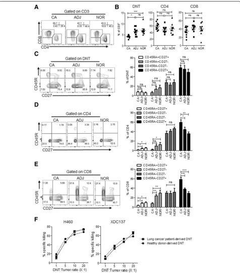
Fig. 1DNT cells infiltrate lung cancer and are cytotoxic to lung cancer cells. Flow cytometric analysis of T cells from different lung tissue compartments, cancer (CA), adjacent (ADJ), or grossly normal lung tissue (NOR), resected from lung cancer patients (n= 10).aRepresentative contour plots of lung tissue-derived T cell subsets by gating on CD3+cells.bFrequency of lung tissue derived T cell subsets, each point represents data from an individual patient. *p< 0.05, **p< 0.01, ***p< 0.001 by one-way ANOVA. Representative flow cytometry plot and cumulative frequency of DNT (c), CD4 (d) and CD8 (e) T cell subsets expressing CD45RA and CD27, presented as mean ± SEM of 9 evaluable patients. *p< 0.05, **p< 0.01, ***p< 0.001 by two-tailed unpaired t-test relative to cancer (CA) tissue.fLung cancer patient-derived or healthy donor-derived DNT cells were co-cultured with indicated lung cancer cell lines at various DNT cell to tumor ratios. % specific killing of target cells is shown. The results represent two independent experiments each with triplicate cultures
did observe a significantly higher frequency of central memory phenotype amongst CD4 and CD8 T cells within cancer tissue relative to adjacent and normal lung tissue (Fig.1d and e).
Given the presence of DNT cells in tumor tissue, we determined whether DNT cells derived from lung cancer patients have anti-tumor function. Using our well-estab-lished DNT cell expansion protocol, by which we have previously expanded DNT cells from peripheral blood of both leukemia patients [22] and healthy donors [24], we attempted to selectively expand DNT cells from tumor samples, but failed, possibly due to the low frequency of DNT cells obtained from tumors and/or exhaustion of the obtained DNT cells. However, DNT cells expanded from peripheral blood of lung cancer patients resulted in high purity (> 90%) but with a lower yield than those de-rived from healthy donors (Additional file 2: Figure S1). Importantly, DNT cells expanded from both lung cancer patients and healthy donors displayed potent and compar-able cytotoxicity against established lung cancer cell line, NCI-H460, and patient xenograft-derived cell line XDC137 (Fig. 1f ). Next, we compared the potency of anti-tumor activity mediated by CD4, CD8 and DN T cells expanded from the same donor in in vitro killing assays against the two cell lines and found that while all ex-panded T cell subsets showed cytotoxicity towards lung cancer cell lines, DNT cells induced the highest degree of cytotoxicity (Additional file2: Figure S2A).
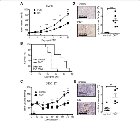
Ex vivo expanded DNT cells from healthy donors can target advanced late-stage lung cancer xenografts To determine whether DNT cells can target late-stage lung cancer in vivo, we generated two late-stage xeno-graft models. An NSCLC established cell line NCI-H460 and a patient-derived adenocarcinoma xenograft cell line XDC137 were inoculated subcutaneously (s.c.) into the flanks of sublethally irradiated NSG mice and let grown to ~100mm3. Tumor-bearing mice were then treated subcutaneously with 3 peritumoral injections of ex vivo expanded DNT cells or CD8 T cells in 3–4 days inter-vals. For the more aggressive NCI-H460 model, the PBS treated control tumor reached end-point by 20 days post treatment (Fig. 2a). However, DNT cell treatment re-sulted in a significant reduction of tumor growth as early as 6 days post 1st DNT cell injection. At 20 days post DNT cell treatment NCI-H460 tumor volume was re-duced by 43.3 ± 15.9%, from 834.2 ± 234.8 mm3 in the control group to 473.2 ± 132.9 mm3 in the DNT cell treated group (Fig.2a). In contrast, injection of an equal number of CD8 T cells was not able to reduce tumor growth during this observation period (Additional file2: Figure S2B). Additionally, DNT cell-mediated inhibition of tumor growth led to a significant increase in the survival of NCI-H460 tumor-bearing mice, with a humane endpoint
extending from median 24 days to 38 days (Fig. 2b). Though patient-derived xenograft model XDC137 grew much slower than NCI-H460, with humane endpoint not being reached by 71 days of observation, DNT cell treat-ment significantly reduced XDC137 xenograft volume from 160.8 ± 39.5mm3 in the PBS control group to 86.2 ± 34.8mm3in the DNT cell treated group (Fig.2c), resulting in a 46.4 ± 21.6% reduction in tumor volume. These results show that adoptive transfer of healthy donor-derived DNT cells can significantly inhibit the growth of both aggressive and slow growing lung cancer xenografts. As DNT cells were found in lung cancer patient TILs, we next deter-mined whether DNT cells would be detectable within tumor xenografts at experimental endpoints. Using immu-nohistochemical staining for human CD3+ cells, we de-tected DNT cells infiltrating both aggressive xenograft, NCI-H460 (Fig. 2d) and slower growing xenograft, XDC137 (Fig.2e), at days 21 and day 71, respectively.
Tumor infiltrating and ex vivo expanded DNT cells express PD-1
With the observation that significantly fewer DNT cells were found in the patient TILs than in adjacent or nor-mal tissue (Fig. 1b), we hypothesized that the immuno-suppressive tumor microenvironment may prevent DNT cell infiltration. Consistent with this hypothesis, PD-1 was expressed on DNT cells within resected lung tissue, similar to that seen for CD4+and CD8+T cells (Fig.3a). Further, a significantly higher proportion of DNT cells expressed PD-1 within tumors compared to adjacent or normal tissue (CA: 55.5 ± 11.7% vs ADJ: 36.1 ± 14.5% and NOR: 35.5 ± 9.1%). Though tumor-infiltrating DNT cells expressed PD-1, they were the least frequent PD-1+ T cell subset and showed the most variability in PD-1 ex-pression compared to CD4+and CD8+T cells (CD4: 65.8 ± 7.1%, CD8: 67.2 ± 7.2%, DNT: 55.5 ± 11.7%, Fig.3b).
tissues (Fig.3a), and lung cancer cell lines express differ-ent levels of PD-L1 (Additional file2: Figure S3A), we de-termined if co-culture of DNT cells with lung cancer cells was sufficient to induce PD-1 expression. Consistent with the observations in patients, in vitro coculture with 4 dif-ferent PD-L1+ lung cancer cell lines (A549, H460, H520 and XDC137, Additional file2: Figure S3A), all resulted in a significant increase in PD-1+DNT cells when compared with DNT cells cultured alone (Fig.3e and Additional file2:
Figure S3B). PD-1 induction was not dependent on the level of PD-L1 expression on lung cancer cells as H520 expressed the lowest level of PD-L1 (Additional file 2: Figure S3A) but induced a similar level of PD1+DNT cells as H460 which showed a very high level of PD-L1 expres-sion (Fig.3e and Additional file2: Figure S3A). Prolonged co-cultures with lung cancer cells did not further increase PD-1+DNT cells for any given cell line (Additional file 2: Figure S3B). Co-culture with lung cancer cell lines also Fig. 2Ex vivo expanded DNT cells inhibit late stage tumor growth in xenograft models. NSG mice were inoculated subcutaneously with NCI-H460 (a, b, d) or XDC137 (cande) in 50% Matrigel solution and grown to ~100mm3. After tumors were established, tumor bearing mice were randomized into groups and treated with peritumoral injection of IL-2 with or without DNT cells on day 0, 3 and 6. (aandc) Tumor volume was measured at indicated time points. Arrows indicate DNT cell treatments. Results represent one of three independent experiments, each consisting of 5 mice per treatment group (a), or one experiment consisting of 3 mice per treatment group (c).bSurvival of mice receiving IL-2 (control) or IL-2 + DNT cells (DNT)dande.Immunohistochemistry staining with anti-human CD3 antibody on resected tumor xenografts. Representative sections of CD3+DNT cells within tumor xenograft from both groups are shown at 21 days for NCI-H460 xenografts (d) and at 71 days for XDC137 xenografts (e) Quantified CD3+staining density of whole xenograft sections, as determined by digital analysis of positive stain per area analyzed. Each dot represents one mouse and horizontal bars represent the mean ± SEM. Data shown are representative of 2 separate experiments. *p< 0.05, **p< 0.01, ***p< 0.001, by two-tailed unpaired t-test (a, c, dande) or log-rank (b)
increased intracellular expression of IFNγ and TNFα in DNT cells (Additional file2: Figure S4), suggesting the acti-vation of these T cells by lung cancer cells.
Anti-PD-1 treatment enhances DNT cell-mediated anti-tumor activity
With the propensity of DNT to upregulate PD-1 and cy-tokines expression in the presence of lung cancer, we sought to determine if addition of anti-PD-1 may aug-ment DNT cell-mediated anti-tumor activity in vivo. To observe whether anti-PD-1 can benefit adoptive DNT therapy in vivo, PD-L1 expressing NCI-H460 lung can-cer cell line was subcutaneously implanted and estab-lished to ~ 100 mm3 and DNT cells, with or without anti-PD-1, were administered using two methods, either locally by s.c. peritumoral injection or systemically by intravenous (i.v.) tail vein injection as shown schematic-ally in Fig. 4a and Additional file 2: Figure S5A,
in an additional 32.6 ± 20.0% reduction in tumor volume (from 572.5 ± 186.5 mm3 in the DNT cell alone treated group to 385.9 ± 114.3 mm3when in combination) by day 20 (Additional file2: Figure S5B). Importantly, combination therapy prolonged the survival of both s.c. peritumoral inoc-ulated DNT cell treated mice from median 38 days to 48.5 days (Fig.4c) and i.v. inoculated DNT cell treated mice from median 33 days to 38 days (Additional file 2: Figure S5C). Analysis of hematoxylin and eosin (H&E) stained tumor tis-sue shortly after DNT treatment revealed that though tumor size remained similar (181.0 ± 53.7 mm3 for DNT cell treated vs 152.2 ± 54.7 mm3 for DNT cell and anti-PD-1
treated), anti-PD-1 significantly increased the propor-tion of necrotic area detected within tumors from mice receiving combination treatment (64.9 ± 11.7% vs 41.3 ± 14.5%; Fig. 4d), with a similar result observed for i.v. inoculated DNT cells (42.1 ± 10.4% vs 22.4 ± 7.2%; Additional file 2: Figure S5D). These results suggest that DNT cells inhibit tumor growth by ac-tively targeting tumor cells and causing tumor necro-sis, and that this activity was enhanced by anti-PD1 therapy. Overall, these results show that addition of anti-PD-1 augments the ability of DNT cells to re-duce tumor growth and increase survival of mice. Fig. 4Anti-PD-1 antibody enhances the efficacy of DNT cell-mediated inhibition of late-stage tumor growth. NSG mice were inoculated subcutaneously with NCI-H460 in 50% Matrigel solution and grown to ~100mm3. After tumors were established, tumor bearing mice were randomized and received peritumoral injection of DNT cells and IL-2 on day 0, 3 and 6, without or with anti-PD-1 antibody (10 mg/kg repeated every 5 days i.p., starting one day prior to 1st DNT cell infusion).aSchematic diagram of the treatment protocol of NCI-H460 xenograft model.b
Tumor volume was measured at indicated time points (n= 8 for each group).cSurvival of the mice treated with PBS control or DNT cells with or without anti-PD-1 (n = 8 for each group).d. Representativeh&estaining of xenografts from indicated treatment groups 9-days post DNT cell infusion (n= 6 for each group; 3 tumors each with 2 random sections).ePercent necrotic area in tumors from indicated treatment groups calculated by histological analysis (n = 6 for each group; 3 tumors each with 2 random sections). Representative results shown as mean and SEM of 2 separate experiments. *p < 0.05, **p < 0.01, ***p < 0.001, by two-tailed unpairedttest (b), log-rank test (c),or one-way ANOVA (e)
Anti-PD-1 treatment increases DNT cell infiltration into tumor xenografts
To understand how anti-PD-1 augmented DNT cell-medi-ated tumor growth inhibition, we first determined whether the presence of anti-PD-1 altered in vitro cytotoxicity of DNT cells to lung cancer cell lines expressing different levels of PD-L1 (Additional file 2: Figure S7A). We found that addition of anti-PD-1 to the cocultures did not alter DNT cell cytotoxicity towards lung cancer cell lines H460, XDC137 and A549 natively expressing PD-L1, but signifi-cantly increased killing of PD-L1 overexpressing cell line A549-PD-L1 (Additional file2: Figure S7B). To analyze how anti-PD-1 enhanced DNT cell treatment towards lung cancer xenografts in vivo we analyzed tumor infiltrating DNT cells post treatment. Consistent with PD-1 induction on DNT cells by lung cancer in vitro (Fig.3e), flow cytometric analysis of xenograft infiltrating DNT cells showed a 2-fold increase in PD-1 expression compared to DNT cells prior to infusion (Fig. 5a). Further, anti-PD-1 treatment abrogated PD-1 ex-pression on xenograft infiltrating DNT cells as shown by the lack of staining using anti-PD1 clone EH12.2H7 that recog-nizes a Nivolumab shared epitope of PD-1 [33,34] (Fig.5a), suggesting that the Nivolumab treatment effectively blocked the PD-1 epitope on tumor infiltrating DNT cells.
To determine whether anti-PD-1 treatment affects tumor infiltration of DNT cells, we quantified DNT cell infiltration of tumor xenografts by histological analysis. Mice receiving combination treatment of DNT cells and anti-PD-1 antibody had a 5.9 ± 1.2-fold increase in the number of tumor infiltrating DNT cells relative to mice that received DNT cells alone (Fig.5b). Similarly, i.v. fusion of DNT cells also resulted in a 1.7 ± 0.3-fold in-crease in DNT cells accumulating in tumor xenografts (Additional file 2: Figure S5E). These data indicate that anti-PD-1 treatment can increase the accumulation of DNT cells in tumor tissue. We next analyzed whether anti-PD-1 treatment could alter the phenotype of tumor infiltrating DNT cells. To this end, tumor infiltrating DNT cells were isolated from mice receiving different treatments and expression of cytolytic molecules known to be involved in DNT cell anti-tumor responses were analyzed by flow cytometry [24, 25, 35]. We found that DNT cells expressing NKG2D and DNAM1 were present in both control and anti-PD-1 treated mice but were more abundant in mice receiving combination therapy than those receiving DNT cells alone, though differences did not reach statistical significance (Fig.5c). Similarly, mice that received anti-PD-1 showed a greater number of TNFα+ and IFNγ+
DNT cells in the tumor (Fig. 5d). Importantly, consistent with the cytotoxic na-ture of DNT cells, anti-PD-1 treatment significantly in-creased the frequency of CD107a+, perforin+, and granzyme B+ DNT cells within tumors (Fig. 5e). These data suggest that anti-PD-1 treatment increases the
accumulation of DNT cells within tumors expressing molecules involved in anti-tumor responses.
Discussion
Adoptive cellular therapy based on DNT cells emerges as a promising therapeutic option for hematological and lung malignancies [22–26]. Here we show that adoptive transfer of DNT cells significantly inhibited growth of late-stage lung tumor xenografts and enhanced the sur-vival of recipient mice. Moreover, we show that anti-PD-1 increased the accumulation of cytotoxic DNT cells within tumor xenografts. These results collectively demonstrate the potential of DNT cells to benefit NSCLC patients, particularly those receiving ICB treat-ment with limited response due to lack of TILs.
Tumor infiltrating CD8+ and CD4+ T cells remain an important predictor of patient outcomes and responsive-ness to anti-PD-1 therapy, with recent discoveries highlighting a role for TCF7+CD8+T cells in predicting re-sponsiveness [36–39]. However, the role of non-conventional T cells, such as DNT cells, in solid tumor remains largely un-explored. By examining lung cancer patients’resected lung tissues we found that DNT cells were present within early stage lung adenocarcinoma (Fig.1 a and b) and exhibited a predominate central/effector memory phenotype (Fig. 1d). Further, whereas we observed no significant difference be-tween conventional CD4 and CD8 T cells in their infiltration, a lower frequency of DNT cells was found infiltrating tumors relative to adjacent and normal tissue, suggesting that the tumor microenvironment may be more hostile to DNT cells (Fig.1b). Of note, whereas significantly higher frequencies of central memory CD4+and CD8+T cells were found in can-cer tissue relative to adjacent and normal lung tissue (Fig.1d and e), this was not the case for DNT cells (Fig.1c). While our attempts to directly measure tumor infiltrating DNT cell cytotoxicity against lung cancer failed due to the limited number of DNT cells available for expansion, indirect evi-dence from DNT cells expanded amongst total TILs from pancreatic and glioma patient tumors showed intracellular IFNγand TNFαexpression upon stimulation by autologous tumor [40,41], suggesting that tumor infiltrating DNT cells are likely to be cytotoxic.
Fig. 5Anti-PD-1 antibody enhances infiltration of cytotoxic DNT cells into tumor xenografts. Tumor-bearing NSG mice received peritumoral injection of DNT cells with or without anti-PD1 treatment.A.Representative flow cytometric analysis of DNT cells pre-infusion and tumor infiltrating DNT cells 21 days post infusion. The data shown represent results from 2 independent experiments.bImmunohistochemistry analysis of DNT cells. Nine days post DNT cell infusion, tumor xenografts were harvested and stained with anti-human CD3 antibody and quantified by Aperio Image-scope. Representative staining and analysis of tumor infiltrating DNT cells in indicated treatment groups are shown. Each dot represents one mouse and horizontal bars represent the mean ± SEM. Data shown are representative of 2 separate experiments.c-eFlow cytometry analysis of tumor infiltrating DNT cells. Frequency of NKG2D+or DNAM-1+DNT cells (c).IFNγ+and TNFα+DNT cells (d), perforin+, granzyme B+and CD107a+DNT cells (e). Representative results shown as mean ± SEM from 3 tumors of 2 separate experiments are shown. (*p< 0.05 by two-tailed unpairedt-test)
cells exerted a greater cytotoxicity against lung cancer in vitro compared to CD4 and CD8 T cells from the same donor (Additional file 2: Figure S2A). Together, these findings suggest that the use of healthy donor DNT cells is more practical and will make DNT-cell therapy more readily available.
Importantly, DNT cells, but not CD8 T cells, signifi-cantly inhibited late-stage H460 lung tumor growth in vivo (Fig.2a and Additional file2: Figure S2B) and pro-longed survival of tumor bearing mice (Fig.2b and d). In the case of slow growing patient-derived xenograft cell line, XDC137, DNT cell treatment limited the growth of the tumor for over 70 days of the observation period and DNT cells were found infiltrating the tumor at this time point (Fig.2e), suggesting that adoptive transfer of DNT cells could lead to a long-lasting anti-tumor immunity. Interestingly, though adoptive cellular therapy shows promise in clinical trials, rarely do preclinical studies show complete tumor regression in xenograft models [43–46]. Similarly, DNT cell therapy significantly inhib-ited tumor growth but did not eradicate late-stage lung cancer xenografts. This may be due to the lack of other components of the immune system in immunodeficient mice which may not support memory T cell formation or may be due to the immunosuppressive tumor micro-environment [47].
Given the role of the tumor microenvironment in regu-lating T cells [12, 13], we found that tumor-infiltrating DNT cells had a higher expression of PD-1 relative to ad-jacent and normal tissue (Fig.3a). Consistent with this ob-servation, DNT cells co-cultured with lung cancer cells increased PD-1 expression (Fig. 3e and Additional file2: Figure S3B). Additionally, xenograft infiltrating DNT cells also showed higher PD-1 expression compared to pre-infusion cells (Fig. 5a). Collectively, our findings are consistent with the observation that tumor recognition and activation of T cells lead to upregulation of PD-1 [17, 48] and suggest that expression of PD-1 on DNT cells is regulated in a similar manner. Interestingly, patient-derived tumor infiltrating DNT cells expressed a lower level of PD-1 than conventional CD4+ and CD8+ T cells (Fig. 3b). In line with this, we found that stimulation of CD4 T cells in vitro resulted in sustained PD-1 expression, which differs from what was observed for DNT and CD8 T cells (Fig.3d). These findings show differences in PD-1 regulation between T cell subsets and suggest the possibil-ity that DNT cells may be more resistant to tumor micro-environmental changes in vivo than conventional T cells.
Observations from patients responsive to ICB suggest that blocking PD-1 greatly increased the number and function of CD8+T cells infiltrating the tumor bed [49]. Interestingly, we found that the addition of anti-PD-1 to DNT and lung cancer cell co-cultures only increased killing of PD-L1 over expressing cells but not the lung
cell lines natively expressing PD-L1 (Additional file 2: Figure S7). Though initially surprising, this observation was consistent with results published by others using gamma/delta T cells and CIK cells [44, 46] and suggests that tumor natively expressed PD-L1 may not have enough density to alter innate T cell function in vitro. Similar to observations in patients receiving ICB, we found that anti-PD-1 blockade also led to greater numbers of DNT cells within tumors (Fig.5b and Additional file2: Figure S5E), suggesting that DNT cells were regulated by the PD-1/PD-L1 pathway. Whether anti-PD-1 blockade increased the ability of DNT cells to migrate to xenografts or survive within xenografts was not directly explored, but given the role of PD-1 engagement in regulating T cell ac-tivation [14] and apoptosis [15, 16], and that DNT cells could infiltrate tumors in the absence of ICB (Fig.2d and e), anti-PD-1 blockade may allow for continued DNT cell survival within tumors.
Tumor recognition by DNT cells was shown to be dependent on ligation of NKG2D and DNAM1 receptors by innate ligands preferentially expressed on malignant cells [24, 25]. In addition to increasing the number of DNT cells within tumor xenografts, we found that anti-PD-1 treatment resulted in increased NKG2D+ and DNAM1+DNT cells, capable of cytolytic granule secretion (Fig. 5). This increase in tumor-recognizing DNT cells within xenografts coincided with an increase in tumor ne-crosis (Fig.4d and Additional file2: Figure S5D), supporting direct engagement and lysis of lung cancer xenografts by DNT cells. Indeed, addition of anti-PD-1 to adoptively trans-ferred DNT cells significantly enhanced DNT cell-mediated tumor inhibition and prolonged the survival of tumor bear-ing mice (Fig.4and Additional file2: Figure S5). Taken to-gether, these data support the notion that combination therapy of anti-PD-1 and DNT cells is beneficial to DNT cell therapy of solid tumors such as lung cancer.
already receiving immune checkpoint blockade and may be ideal for patients characterized as having “immune deserts”.
Conclusions
We demonstrated for the first time that patient-derived tumor infiltrating lymphocytes contained a lower fre-quency of DNT cells with a higher expression of PD-1 relative to normal lung tissue. Our data show that DNT cells are cytotoxic to lung cancer cells in vitro and can inhibit the progression of late-stage lung cancer in vivo. DNT cell treatment in combination with anti-PD-1 re-sulted in increased DNT cell-mediated anti-tumor activ-ity in vivo by increasing the frequency of effector DNT cells in tumors. These results highlight the effect of DNT cells and combinatorial potential of DNT cellular therapy with anti-PD-1 checkpoint blockade for the treatment of lung cancer.
Additional files
Additional file 1:Table S1.Patients’clinical characteristics. Ten newly diagnosed lung cancer patients who were treated with surgery alone were enrolled in this study. The patient age, gender, pathological classification and stage are shown.Table S2.List of flow cytometric antibodies used in the study. (DOCX 15 kb)
Additional file 2:Figure S1.Purity and yield of lung cancer patient-and healthy donorderived DNT cells. Lung cancer patient-derived DNT cells (n= 4) or healthy donor-derived DNT cells (n= 5) were expanded as described in the Materials and Methods and analyzed for their phenotype (A) and yield (B) 14 days after expansion.Figure S2.A. Healthy donor de-rived DNT, CD4 and CD8 T cells were co-cultured with indicated lung cancer cell lines at various T cell to tumor cell ratios for 12-14 h. % spe-cific killing of target cells is shown. The results represent 3 independent experiments from 3 different donors, each with triplicate cultures. Results shown as mean ± SEM. *p< 0.05, **p< 0.01, ***p< 0.001 indicate the dif-ferences in the cytotoxicity of DNT versus CD8 T cells at indicated E:T ra-tios by two-tailed unpaired t-test. B. NSG mice were inoculated subcutaneously with NCI-H460 in 50% Matrigel solution and grown to ~ 100 mm3. After tumors were established, tumor bearing mice were ran-domized into groups and treated with peritumoral injection of IL-2 with or without DNT or CD8 T cells from the same donor on day 0, 3 and 6. Tumor volume was measured at indicated time points. Results represent one of two independent experiments, each had 5 mice per treatment group. Results shown as mean ± SEM. *p < 0.05, **p< 0.01 show the dif-ference in tumor volume at indicated timepoints between DNT and CD8 treated groups by two-tailed unpaired student t-test.Figure S3.A. The expression of PD-L1 on NSCLC cell lines. NSCLC cell lines were stained with either anti-human PD-L1 (blue histograms) or control (red histo-grams), numbers represent MFI. B. Time course of PD-1 expression on ex-panded DNT cells, cultured alone or with various NSCLC cell lines for varying time points. Results represent the data obtained from 2 different donors.Figure S4.Intracellular cytokine analysis of DNT cells, either cul-tured alone or with various NSCLC cell lines. Expanded DNT cells were culture alone or co-cultured with indicated NSCLC cell lines for 48 h and prior to analysis of indicated intracellular cytokines. % INF-γand TNF-α positive DNT cells are shown.Figure S5.Anti-PD-1 antibody enhances ef-ficacy of DNT cell mediated antitumor response and DNT cell tumor infil-tration in late stage tumor xenograft model. NSG mice were inoculated subcutaneously with NCI-H460 in 50% Matrigel solution and tumors were allowed to grow to ~ 100 mm3. After tumors were established, tumor bearing mice were randomized and received intravenous injection of IL-2 (control treatment) or DNT cell plus IL-2 on days 0, 3 and 6. Additionally,
some mice received PBS or anti-PD-1 antibody (10 mg/kg repeated every 5 days i.p., starting one day prior to 1st DNT cell infusion to the end of the experiment). A. Schematic diagram of the treatment protocol of NCI-H460 xenograft model. B. Tumor volume were measured at indicated time points (n= 10 for each group). C. Humane end point survival of treated mice (n = 10 for each group). D. Representative H&E staining of xenografts from indicated treatment groups 9 days post DNT cell infusion and percent necrotic area in tumors from indicated treatment groups cal-culated by histological analysis. E. Immunohistochemical analysis of CD3+ human T cells in tumor xenografts 9 days post DNT cell infusion. Repre-sentative staining and analysis of tumor infiltrating DNT cells in indicated treatment groups. Results shown as mean ± SEM, from untreated, DNT (n= 6) or DNT plus anti-PD1 treatment groups (n = 6). Results shown are representative of 2 separate experiments. **p < 0.01, ***p < 0.001, by two-tailed unpaired t-test (B), by log-rank test (C) or by one-way ANOVA (D and E).Figure S6.Treatment with anti-PD-1 alone has no effect on NCI-H460 xenograft growth or mouse survival. A. NSG mice were inoculated subcutaneously with NCI-H460 in 50% Matrigel solution and grown to ~ 100 mm3, and treated with 10 mg/kg anti-PD-1 or PBS i.p. every 5 days to the end of the experiment. Tumor volume and recipient survival were monitored (n= 5 for each group).Figure S7.A. The expression of PDL1 on NSCLC cell line H460, XDC137, A549 control vector and A549 PD-L1 overexpressing cell line. NSCLC cell lines were stained with either anti hu-man PD-L1(blue histograms) or control (red histograms), values represent MFI. B. DNT cells were co-cultured with indicated lung cancer cell lines at a 5 to 1 DNT cell to tumor cell ratio. % specific killing of target cells is shown. Results shown as mean ± SEM. The results represent 3 independ-ent experimindepend-ents each with triplicate cultures. **p < 0.01, by two-tailed un-paired t-test. (PDF 827 kb)
Abbreviations
ACT:Adoptive cellular therapy; ADJ: Adjacent tumor tissue; CA: Cancer tissue; CAR: Chimeric antigen receptor; CIK: Cytokine-induced killer; DNAM 1: DNAX accessory molecule 1; DNT: Double negative T cell; E:T: Effector to target; FBS: Fetal bovine serum; HBSS: Hanks’balanced salt solution; i.v.: Intravenous; ICB: Immune checkpoint blockade; IFNγ: Interferon gamma; NKG2D: Natural-killer group 2; NOR: Grossly normal appearing tissue; NSCLC: Non-small cell lung cancer; PBS: Phosphate-buffered saline; PD-1: Programmed cell death protein 1; PD-L1: Programmed cell death-ligand 1; PDX: Patient-derived xenograft; s.c.: Subcutaneously; TCR: T cell receptor; TILs: Tumor-infiltrating lymphocytes; TNFα: Tumor necrosis factor alpha
Acknowledgements
We would like to thank Dr. Cheryl D’Souza for comments and critical reading of the manuscript. Dr. Kewei Zhang, Dr. Yang Li for collecting clinical samples, Dr. Sebastiao Martins for examination of histology, and all study participants.
Funding
This work was supported by the by the Canadian Cancer Society (grant # 704121 to LZ), Canadian Institutes of Health Research grant (to LZ), the Ontario Research Fund Research Excellence grant (RE-03-020 to MST), Scientific Research Foundation in the Science and Technology Development Plan of Jilin Province (#20190701043GH to WL and LZ) and the National Natural Science Foundation of China (#81502202 to Si-si Wang). Li Zhang is the Inaugural Maria H. Bacardi Chair in Transplantation. Ming-Sound Tsao is the M. Qasim Choksi Chair in Lung Cancer Translational Research.
Availability of data and materials
All data generated or analyzed during this study are included in this published article (and its additional files).
Authors’contributions
LF, DL, WL and LZ conceived and designed the study. LF, DL, SW, HK, HX, JL and JY performed the experiments. LF and DL analyzed and interpreted the data. WL provided NSCLC patient samples for the study. MT generate lung cancer patient derived cell line for the study. LF and DL wrote the manuscript. JL, MT and LZ provided feedback and edited the manuscript. LZ, MT and SW provided funding support. All authors read and approved the final manuscript.
Ethics approval and consent to participate
Resected human lung cancer tissues from treatment naïve NSCLC patients were collected upon informed patient consent approved (NO.2016–408) by Ethics Committee, The First Hospital of Jilin University. Heparinized lung cancer patient or healthy donor derived peripheral blood was collected upon informed consent approved (#05–0221) by Research Ethics Board, University Health Network.
Consent for publication
All authors consent for publication.
Competing interests
Li Zhang is one of the inventors on 7 DNT cell technology-related patents and serves as a scientific consultant for WYZE BIOTECH Co. Ltd. LF, DL, HK, JB and LZ are inventors of patent applications using DNT cells to treat cancers. Other authors declare no conflict of interest.
Publisher’s Note
Springer Nature remains neutral with regard to jurisdictional claims in published maps and institutional affiliations.
Author details
1Department of Thoracic Surgery, The First Hospital of Jilin University,
Changchun, China.2Toronto General Hospital Research Institute, University Health Network, Toronto, ON, Canada.3Department of Immunology,
University of Toronto, Toronto, ON, Canada.4Department of Translational Medicine, The First Hospital of Jilin University, Changchun, China.5Princess
Margaret Cancer Centre, University Health Network, Toronto, Ontario, Canada.6Department of Laboratory Medicine and Pathobiology, University of
Toronto, Toronto, ON, Canada.7Present address: Sir Run Run Shaw Hospital, College of Medicine, Zhejiang University, Hangzhou, China.8Toronto General
Research Institute, Princess Margaret Cancer Research Tower, 101 College St. Rm 2-807, Toronto, Ontario M5G 1L7, Canada.
Received: 26 November 2018 Accepted: 1 March 2019
References
1. Herbst RS, Morgensztern D, Boshoff C. The biology and management of non-small cell lung cancer. Nature. 2018;553:446–54.
2. Molina JR, Yang P, Cassivi SD, Schild SE, Adjei AA. Non-small cell lung Cancer: epidemiology, risk factors, treatment, and survivorship. Mayo Clin Proc. 2008;83:584–94.
3. Schiller JH, et al. Comparison of four chemotherapy regimens for advanced non-small-cell lung cancer. N Engl J Med. 2002;346:92–8.
4. Cristescu R, et al. Pan-tumor genomic biomarkers for PD-1 checkpoint blockade-based immunotherapy. Science. 2018;362.
5. Pitt JM, et al. Resistance mechanisms to immune-checkpoint blockade in Cancer: tumor-intrinsic and -extrinsic factors. Immunity. 2016;44:1255–69. 6. June CH, Riddell SR, Schumacher TN. Adoptive cellular therapy: a race to
the finish line. Sci Transl Med. 2015;7:–280ps7.
7. Ben-Avi R, et al. Establishment of adoptive cell therapy with tumor infiltrating lymphocytes for non-small cell lung cancer patients. Cancer Immunol Immunother. 2018;67:1221–30.
8. Chen D, et al. Cytokine-induced killer cells as a feasible adoptive immunotherapy for the treatment of lung cancer. Cell Death Dis. 2018; 9(366).
9. Hoeres T, Smetak M, Pretscher D, Wilhelm M. Improving the efficiency of Vγ9Vδ2 T-cell immunotherapy in Cancer. Front Immunol. 2018;9(800). 10. Takami M, Ihara F, Motohashi S. Clinical application of iNKT cell-mediated
anti-tumor activity against lung Cancer and head and neck Cancer. Front Immunol. 2018;9(2021).
11. Kochenderfer JN, Rosenberg SA. Treating B-cell cancer with T cells expressing anti-CD19 chimeric antigen receptors. Nat Rev Clin Oncol. 2013; 10:267–76.
12. Joyce JA, Fearon DT. T cell exclusion, immune privilege, and the tumor microenvironment. Science. 2015;348:74–80.
13. Binnewies M, et al. Understanding the tumor immune microenvironment (TIME) for effective therapy. Nat Med. 2018;24:541–50.
14. Zou W, Wolchok JD, Chen L. PD-L1 (B7-H1) and PD-1 pathway blockade for cancer therapy: mechanisms, response biomarkers, and combinations. Sci Transl Med. 2016;8:328rv4.
15. Dong H, et al. Tumor-associated B7-H1 promotes T-cell apoptosis: a potential mechanism of immune evasion. Nat Med. 2002;8:793–800. 16. Chen L. Co-inhibitory molecules of the B7-CD28 family in the control of
T-cell immunity. Nat Rev Immunol. 2004;4:336–47.
17. Keir ME, Butte MJ, Freeman GJ, Sharpe AH. PD-1 and its ligands in tolerance and immunity. Annu Rev Immunol. 2008;26:677–704.
18. Topalian SL, Drake CG, Pardoll DM. Immune checkpoint blockade: a common denominator approach to cancer therapy. Cancer Cell. 2015;27: 450–61.
19. Pfirschke C, et al. Immunogenic chemotherapy sensitizes tumors to checkpoint blockade therapy. Immunity. 2016;44:343–54. 20. Koyama S, et al. Adaptive resistance to therapeutic PD-1 blockade is
associated with upregulation of alternative immune checkpoints. Nat Commun. 2016;7:10501.
21. Lanitis E, Dangaj D, Irving M, Coukos G. Mechanisms regulating T-cell infiltration and activity in solid tumors. Ann Oncol Off J Eur Soc Med Oncol. 2017;28:xii18–32.
22. Merims S, et al. Anti leukemia effect of ex vivo expanded DNT cells from AML patients: a potential novel autologous T cell adoptive immunotherapy. Leukemia. 2011;25:1415–22.
23. Chen B, Lee JB, Kang H, Minden MD, Zhang L. Targeting chemotherapy-resistant leukemia by combining DNT cellular therapy with conventional chemotherapy. J Exp Clin Cancer Res CR. 2018;37(88).
24. Lee J, et al. Allogeneic human double negative T cells as a novel immunotherapy for acute myeloid leukemia and its underlying
mechanisms. Clin. Cancer Res. Off. J. Am. Assoc. Cancer Res. 2018;24:370–82. 25. Yao J, et al. Human double negative T cells target lung cancer via
ligand-dependent mechanisms that can be enhanced by IL-15. J Immunother Cancer. 2019;7(17).
26. Lee JB, et al. Developing allogeneic double-negative T cells as a novel off-the-shelf adoptive cellular therapy for Cancer. Clin. Cancer Res. Off. J. Am. Assoc. Cancer Res. 2019.https://doi.org/10.1158/1078-0432.CCR-18-2291. 27. John T, et al. The ability to form primary tumor xenografts is predictive of
increased risk of disease recurrence in early-stage non–small cell lung Cancer. Clin Cancer Res. 2011;17:134–41.
28. Stewart EL, et al. Clinical utility of patient-derived xenografts to determine biomarkers of prognosis and map resistance pathways in EGFR-mutant lung adenocarcinoma. J Clin Oncol Off J Am Soc Clin Oncol. 2015;33:2472–80. 29. Daley D, et al.γδT cells support pancreatic oncogenesis by restrainingαβT
cell activation. Cell. 2016;166:1485–1499.e15.
30. Appay V, van Lier RAW, Sallusto F, Roederer M. Phenotype and function of human T lymphocyte subsets: consensus and issues. Cytom Part J Int Soc Anal Cytol. 2008;73:975–83.
31. Sallusto F, Geginat J, Lanzavecchia A. Central memory and effector memory T cell subsets: function, generation, and maintenance. Annu Rev Immunol. 2004;22:745–63.
32. Lee J-B, et al. Developing allogeneic double negative T cells as a novel off-the-shelf adoptive cellular therapy for cancer. Clin Cancer Res Submitted after revision. 2018.
33. Zelba H, et al. Accurate quantification of T-cells expressing PD-1 in patients on anti-PD-1 immunotherapy. Cancer Immunol Immunother CII. 2018.
https://doi.org/10.1007/s00262-018-2244-7.
34. Kamphorst AO, et al. Proliferation of PD-1+ CD8 T cells in peripheral blood after PD-1-targeted therapy in lung cancer patients. Proc Natl Acad Sci U S A. 2017;114:4993–8.
35. Martínez-Lostao L, Anel A, Pardo J. How do cytotoxic lymphocytes kill Cancer cells? Clin Cancer Res Off J Am Assoc Cancer Res. 2015;21:5047–56. 36. Galon J, et al. Type, density, and location of immune cells within human
colorectal tumors predict clinical outcome. Science. 2006;313:1960–4. 37. Ohtani H. Focus on TILs: prognostic significance of tumor infiltrating
lymphocytes in human colorectal cancer. Cancer Immun. 2007;7(4). 38. Dieu-Nosjean M-C, et al. Long-term survival for patients with non-small-cell
lung cancer with intratumoral lymphoid structures. J Clin Oncol Off J Am Soc Clin Oncol. 2008;26:4410–7.
39. Sade-Feldman M, et al. Defining T cell states associated with response to checkpoint immunotherapy in melanoma. Cell. 2018;175:998–1013.e20. 40. Meng Q, et al. Expansion of tumor-reactive T cells from patients with
41. Liu Z, et al. Tumor-infiltrating lymphocytes (TILs) from patients with glioma. Oncoimmunology. 2017;6:e1252894.
42. Liang H, Chu X, Zhao J, Xing G, Si Y. Elevated peripheral blood B lymphocytes and CD3+CD4-CD8- T lymphocytes in patients with non-small cell lung cancer: a preliminary study on peripheral immune profile. Oncol Lett. 2018;15:8387–95.
43. Moon EK, et al. Blockade of programmed death 1 augments the ability of human T cells engineered to target NY-ESO-1 to control tumor growth after adoptive transfer. Clin. Cancer Res. Off. J. Am. Assoc. Cancer Res. 2016;22: 436–47.
44. Dai C, et al. Implication of combined PD-L1/PD-1 blockade with cytokine-induced killer cells as a synergistic immunotherapy for gastrointestinal cancer. Oncotarget. 2016;7:10332–44.
45. Guo Y, et al. PD1 blockade enhances cytotoxicity of in vitro expanded natural killer cells towards myeloma cells. Oncotarget. 2016;7:48360–74. 46. Iwasaki M, et al. Expression and function of PD-1 in humanγδT cells that
recognize phosphoantigens. Eur J Immunol. 2011;41:345–55. 47. Spitzer MH, et al. Systemic immunity is required for effective Cancer
immunotherapy. Cell. 2017;168:487–502.e15.
48. Gros A, et al. PD-1 identifies the patient-specific CD8+tumor-reactive repertoire infiltrating human tumors. J Clin Invest. 2014;124:2246–59. 49. Tumeh PC, et al. PD-1 blockade induces responses by inhibiting adaptive
immune resistance. Nature. 2014;515:568–71.
50. Zaretsky JM, et al. Mutations associated with acquired resistance to PD-1 blockade in melanoma. N Engl J Med. 2016;375:819–29.