Open Access
Review
Cancer, inflammation and the AT1 and AT2 receptors
Gary Robert Smith*
1and Sotiris Missailidis
2Address: 1Research Department, Perses Biosystems Limited, University of Warwick Science Park, Coventry, CV4 7EZ, UK and 2Chemistry
Department, The Open University, Walton Hall, Milton Keynes MK7 6AA, UK
Email: Gary Robert Smith* - gary.smith@persescomms.com; Sotiris Missailidis - S.Missailidis@open.ac.uk * Corresponding author
Abstract
The critical role of inappropriate inflammation is becoming accepted in many diseases that affect man, including cardiovascular diseases, inflammatory and autoimmune disorders, neurodegenerative conditions, infection and cancer.
This review proposes that cancer up-regulates the angiotensin II type 1 (AT1) receptor through systemic oxidative stress and hypoxia mechanisms, thereby triggering chronic inflammatory processes to remodel surrounding tissue and subdue the immune system. Based on current literature and clinical studies on angiotensin receptor inhibitors, the paper concludes that blockade of the AT1 receptor in synergy with cancer vaccines and anti-inflammatory agents should offer a therapy to regress most, if not all, solid tumours.
With regard to cancer being a systemic disease, an examination of supporting evidence for a systemic role of AT1 in relationship to inflammation in disease and injury is presented as a logical progression. The evidence suggests that regulation of the mutually antagonistic angiotensin II receptors (AT1 and AT2) is an essential process in the management of inflammation and wound recovery, and that it is an imbalance in the expression of these receptors that leads to disease.
In consideration of cancer induced immune suppression, it is further postulated that the inflammation associated with bacterial and viral infections, is also an evolved means of immune suppression by these pathogens and that the damage caused, although incidental, leads to the symptoms of disease and, in some cases, death.
It is anticipated that manipulation of the angiotensin system with existing anti-hypertensive drugs could provide a new approach to the treatment of many of the diseases that afflict mankind.
Review
Tumour and Inflammation
Tumour has been linked with inflammation since 1863, when Rudolf Virchow discovered leucocytes in neoplastic tissues and made the first connection between inflamma-tion and cancer [1]. Since then, chronic inflammainflamma-tion has been identified as a risk factor for cancer and even as a
means to prognose/diagnose cancer at the onset of the dis-ease. Examples of such association include the Human papiloma virus (HPV) and cancer [2], including cervical [3], cancers of the oesophagus [4] and larynx [5], Helico-bacter pylori Helico-bacterial infection and gastric adenocarci-noma [6], the hepatitis B virus, cirrhosis and hepato-cellular carcinoma [7], Schistosoma haematobium and
Published: 30 September 2004
Journal of Inflammation 2004, 1:3 doi:10.1186/1476-9255-1-3
Received: 05 July 2004 Accepted: 30 September 2004
This article is available from: http://www.journal-inflammation.com/content/1/1/3
© 2004 Smith and Missailidis; licensee BioMed Central Ltd.
cancer of the bladder [8], asbestos induced inflammation and bronchogenic carcinoma or mesothelioma in humans [9].
Several reports implicate inflammation as a significant risk factor in cancer development: asbestos, cigarette smoke and inflammation of the bowel and pancreas are but a few well-known examples given [1,10]. These papers demonstrate that the inflammation environment is one that would support tumour development and is consist-ent with that observed in tumour sites. The relationship of cancer with inflammation is, however, not limited to the onset of the disease due to chronic inflammation. Schwartsburd [11] goes a step further and proposes that chronic inflammation occurs due to tumour environment stress and that this would generate a protective shield from the immune system. It has been recently demon-strated that the tumour microenvironment highly resem-bles an inflammation site, with significant advantages for the progression of tumour, including the use of cytokines, chemokines, leucocytes, lymphocytes and macrophages to contribute to both vassal dilation and neovascularisa-tion for increased blood flow, the immunosuppression associated with the malignant disease, and tumour metas-tasis [1,11]. Furthermore, this inflammation-site tumour-generated microenvironment, apart from its significant role in cancer progression and protection from the immune system, has a considerable adverse effect to the success of the various current cancer treatments. It has recently been demonstrated that the inflammatory response in cancer can greatly affect the disposition and compromise the pharmacodynamics of chemotherapeu-tic agents [12].
It is evident that cancer is using natural inflammatory processes to spread and, unlikely as it seems at first, it is proposed that this is through the use of the angiotensin II type 1 (AT1) receptor.
AT receptors and inflammation
Angiotensin II (Ang II) is a peptide hormone within the renin-angiotensin system (RAS), generated from the pre-cursor protein angiotensinogen, by the actions of renin, angiotensin converting enzyme, chymases and various carboxy- and amino-peptidases [13]. Ang II is the main effector of the RAS system, which has been shown to play an important role in the regulation of vascular homeosta-sis, with various implications for both cardiovascular dis-eases and tumour angiogenesis. It exerts its various actions to the cardiovascular and renal systems via interactions with its two receptor molecules, angiotensin II type 1 receptor (AT1) and angiotensin II type 2 receptor (AT2) [13]. AT1 and AT2 receptors have been identified as seven transmembrane-spanning G protein-coupled receptors [13], comprising an extracellular, glycosylated region
con-nected to the seven transmembrane α-helices linked by
three intracellular and three extracellular loops. The car-boxy-terminal domain of the protein is cytoplasmic and it is a regulatory site. AT1 is 359 amino acids, while AT2 is 363 amino acids being ~30% homologous to AT1 and are both N-linked glycosylated post-translationally. Various studies have looked at the pharmacological properties of the two receptors and the expression of those receptors on various cell lines. Their affinity for the angiotensin II pep-tide and their ability to perform their physiological func-tions has been characterised using radioligand binding analyses and Scatchard plots. The results have indicated that both receptors have high binding affinities for the AngII peptide. The AT1 receptor has demonstrated a Kd of 0.36 nM for the AngII peptide [14], whereas the AT2 receptor has demonstrated a Kd of 0.17 nM respectively, under similar studies [15].
AT1 receptors are expressed in various parts of the body and are associated with their respective functions, such as blood vessels, adrenal cortex, liver, kidney and brain, while AT2 receptors are highest in fetal mesenchymal tis-sue, adrenal medulla, uterus and ovarian follicles [13]. The opposing roles of the AT1 and AT2 receptors in main-taining blood pressure, water and electrolyte homeostasis are well established. It is, however, becoming recognised that the renin-angiotensin system is a key mediator of inflammation [16], with the AT receptors governing the transcription of pro-inflammatory mediators both in resi-dent tissue and in infiltrating cells such as macrophages.
In addition to the mediators reviewed by Suzuki et al
(2003) [16], a number of vital molecules in inflammatory processes are induced by the AT1 receptor. These include interleukin-1 beta (IL-1b) in activated monocytes [17],
Tumour Necrosis Factor-alpha (TNF-α) [18],
Plasmino-gen Activator Inhibitor Type 1 (PAI-1) [19] and adrenom-edullin [20] all of which have been shown to have active participation in various aspects of cancer development.
Activation of AT1 also causes the expression of TGF-β
[21,22] and a review of literature indicates this may be a unique capability for this receptor. TGF-β is a multifunc-tional cytokine that is produced by numerous types of tumours and amongst its many functions is the ability to promote angiogenesis, tissue invasion, metastasis and immune suppression [23]. It has been postulated that the low response rates achieved in cancer patients undergoing immunotherapy is in part caused by tumour expression of TGF-β and this is supported by inhibition of the antigen-presenting functions and anti-tumour activity of dentritic cell vaccines [24].
utilise for drug delivery [25]. This would imply something unusual about the presentation of angiotensin receptors; however it is predominantly over expression of the vaso-constrictor AT1 that is reported in association with human cancers of the breast [26], pancreas [27], kidney [28], squamous cell carcinoma [29], keratoacanthoma [29], lar-ynx [30], adrenal gland [30], and lung [31]. AT2 has been identified as expressed in preference to AT1 in only one case, in an earlier paper on colorectal cancer [32].
Is it evolution that causes over expression of AT1?
In the 'Hallmarks of Cancer', the authoritative work by Douglas Hanahan and Robert A. Weinberg, the evolution-ary acquired capabilities necessevolution-ary for cancer cells to become life-threatening tumours are described. Further-more, it is suggested that cancer researchers should look not just at the cancer cells, but also at the environment in which they interact, with cancers eliciting the aid of fibroblasts, endothelial cells and immune cells [33].
Sustained angiogenesis, tissue invasion and metastasis are the latter of six necessary steps in tumour progression, as described in the 'Hallmarks of Cancer' [33]. These envis-aged evolutionary steps allow cancers to progress from growths of <2 mm to full tumors. A single evolutionary step, however, upregulation of AT1 would provide a con-siderable advantage to cancer cells that have learnt to evade the apoptosis and growth regulatory effects of
TGF-β. Supporting this hypothesis is the observed genetic
change from non-invasive cancer esophageal cell line T.Tn to invasive cancer cell line T.Tn-AT1. This genetic change concerns 9 genes, all of which are known to influence inflammation signalling (8 down and 1 up regulated) [34].
Is it environment?
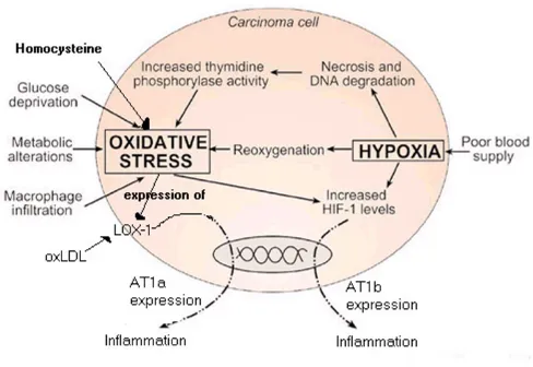
The alternative basis under which induction of AT1 in tumours may occur is by looking at the environment under which the cancer is developing. Stresses and cell damage on the growing tumour boundary could poten-tially be causing the expression of AT1. Evidence that appears to support this view can be found in a study of AT1 expression in breast cancers [35]. In this case, in situ carcinoma has over-expressed AT1 receptors in addition to expressing proteins for yet more AT1. In the invasive carcinoma, high proportions of AT1 receptors are found on the tumour boundary, but in this case protein genera-tion for AT1 is very noticeably absent. How could this behaviour be explained? Perhaps the answer lies in oxida-tive stress and hypoxia.
The formation of oxidised LDL by monocytes and macro-phages at the sites of tissue damage has been established in a recent report by Jawahar L. Mehta and Dayuan Li [36]. In this study, the ox-LDL LOX-1 receptor is noted to be
induced by fluid shear stress (4 hrs), TNF-α (8 hrs) and self-induced by ox-LDL (12 hrs). Of particular interest is that activation of LOX-1 by ox-LDL induces the expression of the AT1 receptor [36]. This key role of ox-LDL regarding AT1 is demonstrated by HMG Co-A reductase inhibitor causing the down-regulation of the AT1 receptor with con-sequential reduction in inflammatory response [37]. Also of interest is that another marker of many diseases, homo-cysteine, enhances endothelial LOX-1 expression [38].
Hypoxia has been demonstrated to induce the expression of both AT1b (AT1a and AT1b are subsets of AT1) and AT2 receptors in the rat carotid body and pancreas [39,40]. The expression of AT1 and AT2 receptors has been studied during the development and regression of hypoxic pul-monary hypertension [41]. Hypoxia has been shown to strongly induce the expression of AT1b but not AT1a. The expression of AT2 is believed to protect the cell from apoptosis and this effect has been demonstrated in the
brain when AT1 is antagonized [42]. Since HIF-1α
gov-erns many hypoxia driven transcriptions [43], its control of AT1b and AT2 expression can be hypothesized. AT1 activation has also been shown to increase the activity of HIF-1α [43], and is consistent with other cases of AT1 pro-viding a positive feedback mechanism. Since hypoxia counts for the expression of AT1b, the speculation that the AT1a subtype is induced by oxidative stress is tempting, although a review of literature appears absent in this regard and further investigation is required to confirm this hypothesis.
A review of hypoxia and oxidative stress in breast cancer cites the chaotic flow of blood in the tumor environment with resultant periods of hypoxia and reperfusion [44]. Reperfusion after myocardial infarction or cerebral ischemia is known to cause the generation of ROS. Hence, summarised in figure 1, the tumor environment thus offers both hypoxia and oxidative stress mechanisms for induction of AT1. It would however seem likely that genetic factors speed up the progression of the more aggressive forms of cancer.
A combination therapy for cancer
The evidence relating to over-expression of AT1 with can-cer progression is compelling. To this effect, AT1 blockade has been hypothesised as the mechanism to overcome cancer associated complications in organ graft recipients [45]. Additionally, a study undertaken in 1998 suggested that hypertensive patients taking ACE inhibitors were sig-nificantly less at risk of developing cancer than those tak-ing other hypertensive treatments [46].
angiogenesis. Reduction of MCP-1 was noted [48], as was the expression of many pro-inflammatory cytokines. The activity of tumour-associated macrophages was also noticed to be severely impaired [48]. The importance in reducing the action of tumour-associated macrophages in extracellular matrix decomposition is not to be underesti-mated, since, in this action, they further progress remod-elling by releasing stored TGF-β [49]. The similarity of action of tumour associated macrophages with those in the tissue healing and repair environment has been noted [49]. The tumour suppressant action of tranilast, an AT1 antagonist, [50] has been more widely explored [51-54]. In one study on the inhibition of uterine leiomyoma cells, Tranilast also induced p21 and p53 [55]. Similarly, the AT1 blocker losartan has been shown to antagonise plate-lets, which are thought to modulate cell plasticity and angiogenesis via the vascular endothelial growth factor
(VEGF) [56]. It has been postulated that losartan and other AT1 blockers can act as novel angiogenic, anti-invasive and anti-growth agents against neoplastic tissue [56]. Furthermore, it has been shown that angiotensin II induces the phosphorylations of mitogen-activated pro-tein kinase (MAPK) and signal transducer and activator of transcription 3 (STAT3) in prostate cancer cells. In con-trast, AT1 inhibitors have been shown to inhibit the pro-liferation of prostate cancer cells stimulated with EGF or angiotensin II, through the suppression of MAPK or STAT3 phosphorylation [57]. Angiotensin II also induces (VEGF), which plays a pivotal role in tumour angiogen-esis and has been the target of various therapeutics, including antibodies and aptamers [58]. Although the role of angiotensin II in VEGF-mediated tumour develop-ment has not yet been elucidated, an ACE inhibitor signif-icantly attenuated VEGF-mediated tumour development,
AT1 expression in cancer
Figure 1
accompanying the suppression of neovascularisation in the tumour and VEGF-induced endothelial cell migration [59]. Perindopril, another ACE inhibitor has also been shown to be a potent inhibitor of tumour development and angiogenesis through suppression of the VEGF and the endothelial cell tubule formation [60].
The powerful direct and indirect suppression effects of TNF-α [61], IL-1β [62] and TGF-β [63] on APC presenting cells, NK, T and B cell have been reviewed [64]. The expression of these mediators makes an effective immune response most unlikely.
Despite this, it has long been established that the body does have the capability to recognise cancer cells and develop antigens. Dentritic cell vaccines for instance have been developed and have demonstrated limited effect in treating established tumours. The effectiveness of one such approach was greatly enhanced leading to complete
regression of tumours in 40% of cases when TGF-β was
neutralised using TGF-β monoclonal antibodies in
syn-ergy with a dentritic cell vaccine [24].
Strong evidence suggests that tumour cells over-express AT1 receptors and compelling evidence has been pre-sented on the implications of AT1 in cancer progression. Although still at a theoretical stage, this evidence leads to the formulation of the hypothesis that effective blockade of AT1 with a tight binding receptor antagonist, in combi-nation with NSAIDs to further control the inflammation, and immunotherapy, such as cancer vaccines, would pro-vide an effective treatment. Most, if not all, solid tumours utilise inflammation processes, which, through the over-expression and activation of AT1 and the subsequent expression of a number of inflammatory cytokines and chemokines, allow for tumour protection from the immune system through immunosuppression, as well as tumour progression and metastasis. Blocking these path-ways through inhibition of AT1 using one of the commer-cially available AT1 inhibitors, whilst lifting the induced protective effect of immunosuppression and further reducing inflammation with the use of NSAIDs will both inhibit tumour progression and allow currently devel-oped immunotherapies, such as cancer vaccines, to pro-mote their therapeutic effect uninhibited. The role of AT1 post-metastasis, given the observation that AT1 protein expression ceases, as demonstrated in the breast cancer study, requires further investigation [35]. However, the premise for the necessity of immunosuppression by can-cer is none the less fundamental and this is encouraging for the prospects of regression of cancers that have pro-gressed to metastasis by combinational AT1 blockade/ immune therapy.
Learning from Cancer: wound management
Cancer is a systemic disease, one that can affect every part and organ in the body and, as presented in this review so far with regards to the role of AT1 in cancer, AT1 upregu-lation is of the utmost importance in the activation of inflammation. Systemically, therefore, what purpose does this upregulation of AT1 serve? The release of ACE and extended expression of AT1 and AT2 during the healing process following vascular injury helps to answer this question [65]. AngII is demonstrated to promote migra-tion and proliferamigra-tion of smooth muscle cells, as well as production of extracellular matrix through AT1 activation. In this work [65], the AT1 and AT2 receptors are recog-nized as having a substantial role in the tissue repair and healing processes of injured arteries. Although further lit-erature in regard to the role of AT1 and AT2 in the healing process appears absent and additional studies are required, it appears rational that a systemic agent for the management of inflammation and healing would be one associated with the vascular system.
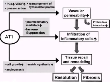
The activation of AT1 (shown in figure 2) has a powerful pro-inflammatory effect [16], promoting the expression of many pro-inflammatory mediators, such as cytokines, chemokines and adhesion molecules through the activa-tion of signalling pathways. The influx, proliferaactiva-tion and behaviour of immune cells are steered away from an effec-tive immune response to pathogens (thereby achieving immunosuppression) but instead towards activities con-sistent with a wound environment. Through the activa-tion of these pathways [16], AT1 effectively elicits this response with local effects intended to initiate wound recovery through destruction of damaged cells, remodel-ling, the laying down of fibrous material and angiogen-esis. AT1 acts in three ways, as indicated in figure 3. Firstly, via the up-regulation of growth factors that leads to increased vascular permeability. Secondly, through the increase of pro-inflammatory mediators that leads to uti-lisation of immune cells such as macrophages in their response to wound mode. Thirdly, through the generation of other factors which promote cell growth, angiogenesis and matrix synthesis. The observation that cancer resem-bles a wound that never heals is therefore substantiated.
Confirming the systemic role of the AT1 receptor in inflammation and disease
With the role of AT1 in cancer established, when the liter-ature of other diseases is reviewed, it is reasonable to anticipate that the role of this receptor is system-wide with regard to inflammation. Interest in the wider implications of the AT1 receptor within disease is gradually increasing and these studies further substantiate a systemic role for AT1 as a key inductor of inflammation and disease. In these studies, a wide variety of pro-disease mediators,
molecules and PAI-I are shown to be induced by AT1 (Table 1).
It is clear that a number of diseases, including heart and kidney disease, diseases associated with the liver and pan-creas, as well as diseases of the skin, bone, the brain and most of the autoimmune and inflammatory disorders, are all affected by the AT1 blockade. It is worth noting at this stage that many of these diseases are often considered to be associated with ageing and with fibrosis. An investiga-tion of the acinvestiga-tion of IGF-1 in the regulainvestiga-tion of expression of AT2 leads to an explanation of this association.
Role of IGF-1 in regulating AT receptors
The majority of studies on AT1 are related to cardiovascu-lar disease, for which AT1 receptor antagonists were
gen-erated as treatment. Regarding AT2, although there has been increased research and interest in its role, this area appears little explored. That which has been learnt so far about the interplay and regulation of these receptors lends itself to a potentially useful model for the management of inflammation:
The expression of AT1 and AT2 receptors on fibroblasts present in cardiac fibrosis is investigated [79]. These types of fibroblast are noted for their expression of AT1 and AT2 receptors. The presence of IL-1b, TNF-α and lipopolysac-charides, through induction of NO and cGMP, all serve to down-regulate AT2 with no effect on AT1 leading to a quicker progression of fibrosis. Interestingly, the continu-ance in the presence of pro-inflammatory signals serves to delay expression of AT2. This is confirmed in a separate
AT1 signalling
Figure 2
local effects of AT1 activation
Figure 3
local effects of AT1 activation. Activation of AT1 leads to growth factors causing increased vascular permeability, pro-inflammatory mediators that lead to utilisation of immune cells such as macrophages in their response to wound mode and other factors that promote cell growth, angiogenesis and matrix synthesis during fibrosis and resolution.
Table 1: AT1 as a key inductor of inflammation and disease. A wide range of pro-inflammatory mediators, cytokines, chemokines and surface adhesion moleculesinvolved in a number of diseases are induced by AT1 and thus inhibited by its blockade.
Disease Mediators inhibited by AT1 blockade Reference
Cardiovascular disease NFκB, 'markers of oxidation inflammation and fibrinolysis' 66
Cardiovascular disease TGF-β 67
Cardiovascular disease TNF-α, IL-6, ICAM-1, VCAM-1 18
Cardiovascular disease PAI-1 19
Cardiovascular disease Surface adhesion molecules 68,69
Cardiovascular disease MCP in Hypercholesterolemia associated endothelial dysfunction 70
Kidney disease None noted in this study. 71
Pancreatitis (Key markers of the disease) 72
Liver fibrosis and cirrhosis 'TGF-β and pro-inflammatory cytokines' 21
Skin disease None noted in this study. 73
Osteoporosis 'Markers of inflammation' 74
study of AT2 expression in proliferating cells. TGF-β1 and bFGF are shown as powerful inhibitors of AT2 expression, whilst IGF-1 is shown to induce the expression of AT2 [80].
IGF-1 is principally produced by the liver from GH (Growth Hormone) and circulates in the blood (decreas-ing with age) and is important in the regulation of immu-nity and inflammation [81]: IGF-1 is also capable of being produced by fibroblasts and macrophages on induction
by pro-inflammatory cytokines, including TNF-α and
IL-1b. In addition to the induction of AT2, IGF-1 can be seen as responsible for mediating the actions of many active cells in the immune/inflammation response [81]. Of note is that TNF-α and IL-1b also affect the circulating expres-sion of IGF-1 by feedback on the release of GH from the anterior pituitary.
The controlling role of AT receptors in inflammation and healing
Significant evidence has been shown that AT1 receptors are upregulated during disease and that AT2 receptor expression follows behind AT1 expression during injury and healing. Given the opposing roles of AT1 and AT2 it can thus be postulated that the interplay of these receptors plays a significant part in judging the current local status of appropriate versus inappropriate inflammation and in providing feedback to the rest of the body. Indeed it is anticipated that prolonged expression of AT1 combined with a lack of AT2 expression results in sustained chronic inflammation and fibrosis.
Overall, the role of the AT receptors in managing and monitoring the healing process is complex, with many positive and negative feedback mechanisms both within the site of inflammation/healing and with the rest of the systems in the body. An attempt to summarise these systemic signalling inter-relationships is given in figure 4. Note the glucocorticoid inhibition of AT1 pro-inflamma-tory activities via NFκB. This model, although hypotheti-cal, provides an explanation of the mechanisms whereby ox-LDL and homocysteine exert their pro-inflammatory effects. Further supporting this model is evidence that a lack of IGF-1 presence contributes to degenerative arthritis [81], septic shock [81], cardiovascular diseases [82] and inflammation of the bowel [83]. The introduction of IGF-1 is also proposed for protection against Huntington's [84], Alzheimer's [85] and Parkinson disorders [86]. Upregulation of IGF-1 has been noted in patients with chronic heart failure who undertake a programme of stretching exercise, thus providing benefits against cardiac cachexia [87].
Conclusions
The invasiveness and immunosuppression of many can-cers appears dependent on inflammation and the upregu-lation of AT1. Two mechanisms for upreguupregu-lation of AT1 are discussed: 1) evolutionary changes to take advantage of this pro-inflammatory control mechanism, 2) AT1 expression induced by an alternating environment of hypoxia and oxidative stress. Immunosuppression as a common protection mechanism of solid tumours against immune responses has been verified from current litera-ture and experimental procedures, as has the implication of cytokines and chemokines in tumour growth and metastasis. Given the involvement of AT1 in the immuno-suppression and inflammatory processes, as well as in the expression of the pro-inflammatory cytokines and chem-okines, it becomes evident that the AT1 receptor is essen-tial for tumour protection and progression. A combination therapy consisting of AT1 receptor antago-nists, NSAID for further control of the inflammation and immune therapy in the form of tumour vaccines should provide a novel and successful treatment for solid tumours.
In the renin-angiotensin system, the angiotensin II recep-tors AT1 and AT2 seem to have opposing functions. The actions of AT1 being principally pro-inflammatory whilst AT2 provides protection against hypoxia, draws inflammatory action to a close and promotes healing. The various direct and indirect mechanisms for feedback between the receptors, their induced products and the external hormonal system in the control of inflammation and healing are summarised in a highly simplified model which none the less can be used to explain how many key promoters and inhibitors of disease exert their effects.
From a review of the current disease literature, it has been demonstrated that the role of AT1 and AT2 in inflammation is not limited to cancer-associated inflam-mation, but is generally consistent and system wide. Potential therapy by manipulation of these receptors, although at an early stage, has been demonstrated for some of these diseases and it is proposed that this approach will provide an effective basis for the treatment of autoimmune, inflammatory and neurodegenerative disorders using existing drugs. AT1 receptor blockade should, in addition, provide a treatment to alleviate the damage caused by bacterial and viral infections, where their destructive action is through chronic inflammation. Given the importance of the immune suppressant effect of inflammation in cancer, it is anticipated that AT1 block-ade should also serve to elicit a more effective immune response to other invaders that seek to corrupt the wound recovery process.
List of abbreviations used
TGF-β Transforming Growth Factor Beta
AT1 Angiotensin II Type 1 receptor
AT2 Angiotensin II Type 2 receptor
IGF-1 Insulin-like Growth Factor 1
LOX-1 Lectin-like Oxidized Low-Density Lipoprotein Receptor 1
HIF-1a Hypoxia Induced Factor 1 Alpha
HMG CoA 3-Hydroxy-3-Methyl-Glutaryl Coenzyme A
bFGF basic Fibroblast Growth Factor
ROS Reactive Oxygen Species (most notably O2-.)
Competing interests
Gary R Smith is a founding director of Perses Biosystems Ltd. The goals of the company are to drive laboratory and clinical research into the role of angiotensin receptors in
Systems view of the AT receptor role
Figure 4
disease management. Although we envisage these activi-ties to be humanitarian (non-profit making) in nature, our long-term ambition is to identify additional drug tar-gets and agents that could work in combination with ACE inhibitors and AT1 blockers to treat most diseases.
Sotiris Missailidis is a Lecturer at the Chemistry Depart-ment of The Open University, with research focus on can-cer and had been the academic supervisor of Gary R Smith. There are no conflicting interests or financial implications related to the publication of this review article.
Authors' contributions
Gary R Smith performed the literature review and pro-posed the hypothesis that cancer utilises the Angiotensin system to trigger chronic inflammation as a means of spreading and avoiding the immune system. In addition to providing significant editorial contributions and litera-ture related comments, Sotiris Missailidis prompted Gary R Smith to undertake additional research with led to clar-ification of the role of hypoxia and oxidative stress in gov-erning AT receptor expression. This understanding led Gary R Smith to propose the hypothesis that inflamma-tion through the AT receptors is the cause of many of the diseases that affect mankind, including infectious dis-eases, which utilise inflammation to disrupt the immune system.
Acknowledgements
Many thanks to Jim Iley of the Open University not only for S807 Molecules in Medicine but also for suggesting the title of this paper.
References
1. Balkwill F, Mantovani A: Inflammation and cancer: back to Virchow?Lancet 2001, 357:539-545.
2. Munger K: The role of human papillomaviruses in human cancers.Frontiers in Bioscience 2002, 7:D641-D649.
3. Castle PE, Hillier S, Rabe L, Hildesheim A, Herrero R, Bratti M, Sher-man M, Burk R, Rodriguez A, Alfaro M, Hutchinson M, Morales J, Schiffman M: An Association of Cervical Inflammation with High-Grade Cervical Neoplasia in Women Infected with Oncogenic Human Papillomavirus (HPV). Cancer Epidemiol Biomarkers Prev 2001, 10:1021-1027.
4. Syrjanen KJ: HPV infections and oesophageal cancer.Journal of Clinical Pathology 2002, 55:721-728.
5. Aaltonen LM, Rihkanen H, Vaheri A: Human papillomavirus in larynx.Laryngoscope 2002, 112(4):700-707.
6. Naumann M, Crabtree J: Helicobacter pylori-induced epithelial cell signalling in gastric carcinogenesis.Trends in Microbiology
2002, 12:29-36.
7. Hilleman M: Critical overview and outlook: pathogenesis, pre-vention, and treatment of hepatitis and hepatocarcinoma caused by hepatitis B virus.Vaccine 2003, 21:4626-4649. 8. Rosin MP, Anwar WA, Ward AJ: Inflammation, chromosomal
instability and cancer: the schistosomiasis model.Cancer Res
1994, 54(7 Suppl):1929S-1933S.
9. Manninga C, Vallyathanb V, Mossman B: Diseases caused by asbes-tos: mechanisms of injury and disease development. Interna-tional Immunopharmacology 2002, 2:191-200.
10. Farrow B, Evers BM: Inflammation and the development of pancreatic cancer.Surgical Oncology 2002, 10:153-169.
11. Schwartsburd PM: Chronic inflammation as inductor of pro-cancer microenvironment: Pathogenesis of dysregulated feedback control.Cancer and Metastasis reviews 2003, 22:95-102. 12. Slaviero KA, Clarke SJ, Rivory LP: Inflammatory response: an
unrecognised source of variability in the pharmacokinetics and pharmacodynamics of cancer chemotherapy.Lancet Oncol
2003, 4:224-32.
13. Thomas WG, Mendelsohn FAO: Molecules in Focus Angiotensin receptors: form and distribution.IJBCB 2003, 35:774-779. 14. Martin MM, Victor X, Zhao X, McDougall JK, Elton TS:
Identifica-tion and characterizaIdentifica-tion of funcIdentifica-tional angiotensin II type 1 receptors on immortalized human fetal aortic vascular smooth muscle cells. Molecular and Cellular Endocrinology 2001,
183:81-91.
15. Moore SA, Patel AS, Huang N, Lavin BC, Grammatopoulos TN, Andres RD, Wayhenmeyer JA: Effects of mutations in the highly concerved DRY motif on binding affinity, expression, and G-protein recruitment of the human angiotensin II type-2 receptor.Molecular Brain Res. 2002, 109:161-167.
16. Suzuki Y, Ruiz-Ortega M, Lorenzo O, Ruperez M, Esteban V, Egido J:
Inflammation and angiotensin II.IJBCB 2003, 35:881-900. 17. Dorffel Y, Latsch C, Stuhlmuller B, Schreiber S, Scholze S, Burmester
GR, Scholze J: Preactivated Peripheral Monocytes in Patients with Essential Hypertension.Hypertension 1999, 34:113-117. 18. Tsutamoto T, Wada A, Maeda K, Mabuchi N, Hayashi M, Tsutsui T,
Ohnishi M, Sawaki M, Fujii M, Matsumoto T, Kinoshita M: Angi-otensin II type 1 receptor antagonist decreases plasma levels of tumor necrosis factor alpha, interleukin-6 and soluble adhesion molecules in patients with chronic heart failure. Journal of the American College of Cardiology 2000, 35:715-721. 19. Chen HC, Bouchie J, Perez A, Clermont A, Izumo S, Hampe J, Feener
E: Role of the Angiotensin AT1 Receptor in Rat Aortic and Cardiac PAI-1 Gene Expression.Arteriosclerosis, Thrombosis, and Vascular Biology 2000, 20:2297.
20. Mishima K, Kato J, Kuwasako K, Imamura T, Kitamura K, Eto T: Angi-otensin II modulates gene expression of adrenomedullin receptor components in rat cardiomyocytes.Life Sciences 2003,
73:1629-35.
21. Leung PS, Suen PM, Ip SP, Yip CK, Chen G, Paul BS, Lai PBS: Expres-sion and localization of AT1 receptors in hepatic Kupffer cells: its potential role in regulating a fibrogenic response. Regulatory Peptides 2003, 116:61-69.
22. Rosenkranz S: TGF-beta1 and angiotensin networking in car-diac remodeling.Cardiovasc Res 2004, 63:423-432.
23. Teicher B: Malignant cells, directors of the malignant process: Role of transforming growth factor-beta.Cancer and Metastasis reviews 2001, 20:133-143.
24. Kobie JJ, Wu RS, Kurt RA, Lou S, Adelman MK, Whitesell LJ, Ram-anathapuram LV, Arteaga CL, Akporiaye ET: Transforming Growth Factor β Inhibits the Antigen-Presenting Functions and Antitumor Activity of Dendritic Cell Vaccines. Cancer Research 2003, 63:1860-1864.
25. Maeda H, Fang J, Inutsuka T, Kitamoto Y: Vascular permeability enhancement in solid tumors: various factors, mechanisms involved and its implications.International Immunopharmacology
2003, 3:319-328.
26. Tahmasebi M, Puddefoot JR, Inwang ER, Goode AW, Carpenter R, Vinson GP: Transcription of the prorenin gene in Normal and Diseased Breast.Eur J Cancer 1998, 34:1777-1782.
27. Fujimoto Y, Sasaki T, Tsuchida A, Chayama K: Angiotensin II type 1 receptor expression in human pancreatic cancer and growth inhibition by Angiotensin type 1 receptor antagonist. FEBS Letters 2001, 495:197-200.
28. Goldfarb A, Diz I, Tubbs R, Ferrario M, Novick C: Angiotensin II receptor subtypes in the human renal cortex and renal cell carcinoma.J Urol 1994, 151(1):208-13.
29. Takeda H, Kondo S: Differences between Squamous Cell Car-cinoma and Keratoacanthoma in Angiotensin Type-1 Recep-tor Expression.American Journal of Pathology 2001, 158:1633-1637. 30. Marsigliante S, Resta L, Muscella A, Vinson GP, Marzullo A, Storelli C:
AT1 antagonist II receptor subtype in the human larynx and squamous laryngeal carcinoma.Cancer Letters 1996, 110:19-27. 31. Batra V, Gropalakrish V, McNeill J, Hickie R: Angiotensin II
33. Hanahan D, Weinberg RA: The Hallmarks of Cancer.Cell 2000,
100:57-70.
34. Kawamata H, Furihata T, Omotehara F, Sakai T, Horiuchi H, Shina-gawa Y, Imura J, Ohkura Y, Tachibana M, Kubota K, Terano A, Fuji-mori T: Identification of genes differentially expressed in a newly isolated human metastasizing esophageal cancer cell line, T.Tn-AT1, by cDNA microarray. Cancer Sci 2003,
94:699-706.
35. De Paepe, Verstraeten VLRM, De Potter CR, Vakaet LAML, Bullock GR: Growth stimulatory angiotensin II type-1 receptor is upregulated in breast hyperplasia and in situ carcinoma but not in invasive carcinoma.Histochem Cell Biol 2001, 116:247-254. 36. Mehta JL, Li D: Identification, Regulation and Function of a Novel Lectin-Like Oxidized Low-Density Lipoprotein Receptor.J Am Coll Cardiol 2002, 39:1429-1435.
37. Dechend R, Fiebler A, Lindschau C, Bischoff H, Muller D, Park JK, Dietz R, Haller H, Luft FC: Modulating Angiotensin II-Induced Inflammation by HMG Co-A Reductase Inhibition. Am J Hypertens 2001, 14:55S-61S.
38. Nagase M, Ando K, Nagase T, Kaname S, Sawamura T, Fuj T: Redox-Sensitive Regulation of LOX-1 Gene Expression in Vascular Endothelium.Biochemical and Biophysical Research Communications
2001, 281:720-725.
39. Chan WP, Fung ML, Nobiling R, Leung PS: Activation of local renin-angiotensin system by chronic hypoxia in rat pancreas. Molecular and Cellular Endocrinology 2000, 160:107-114.
40. Leung PS, Fung ML, Tam MS: Renin-angiotensin system in the carotid body.Int J Biochem Cell Biol 2003, 35:847-854.
41. Chassagne C, Eddahibi S, Adamy C, Rideau D, Marcotte F, Dubois-Rande JL, Adnot S, Samuel JL, Teiger E: Modulation of Angiotensin II Receptor Expression during Development and Regression of Hypoxic Pulmonary Hypertension.Am J Respir Cell Mol Biol
2000, 22:323-332.
42. Grammatopoulos T, Morris K, Ferguson P, Weyhenmeyer J: Angi-otensin protects cortical neurons from hypoxia-induced apoptosis via the angiotensin type 2 receptor.Molecular Brain Research 2002, 99:144-124.
43. Page EL, Robitaille GA, Pouyssegur , Richard DE: Induction of Hypoxia-Inducible Factor-1α by Transcriptional and Trans-lation Mechanisms.JBC 2002, 277:48403-48409.
44. Brown NS, Bicknell R: Hypoxia and oxidative stress in breast cancer. Oxidative stress: its effects on the growth, metastatic potential and response to therapy of breast cancer.Breast Cancer Res 2001, 3:323-327.
45. Maluccio M, Sharma V, Lagman M, Konijn G, Suthanthiran M: Angi-otensin II Receptor Blockade: A novel Strategy to Prevent Immunosuppressant-Associated cancer Progression. Trans-plantation Proceedings 2001, 33:1820-1821.
46. Lever AF, Hole DJ, Gillis CR, McCallum IR, McInnes GT, MacKinnon PL, Meredith PA, Murray LS, Reid JL, Robertson JWK: Do inhibitors of angiotensin-1-converting enzyme protect against risk of cancer?Lancet 1998, 352:179-184.
47. Miyajima A, Kosaka T, Asano T, Asano T, Seta K, Kawai T, Hayakawa M: Angiotensin II Type I Antagonist Prevents Pulmonary Metastasis of Murine Renal Cancer by Inhibiting Tumor Angiogenesis.Cancer research 2002, 62:4176-4179.
48. Egami K, Murohara T, Shimada T, Sasaki KI, Shintani S, Sugaya T, Ishii M, Akagi T, Ikeda H, Matsuishi T, Imaizumi T: Role of host angi-otensin II type I receptor in tumor angiogenesis and growth. J Clin Invest 2003, 112:67-75.
49. Yu Q, Stamenkovic I: Cell surface-localised matrix metallopro-teinase-9 proteolytically activates TGF-β and promotes tumor invasion and angiogenesis.Genes & Development 2000,
14:163-176.
50. Jin D, Takai S, Shiota N, Miyazaki M: Tranilast, an anti-allergic drug, possesses antagonistic potency to angiotensin II. Euro-pean Journal of Pharmacology 1998, 361:199-205.
51. Yashiro M, Chung Y, Sowa M: Tranilast [N-(3,4 dimethoxycin-namoyl) anthranilic acid] down regulates the growth of scir-rhous gastric cancer.Anticancer Res 1997, 17:895-900.
52. Noguchi N, Kawashiri S, Tanaka A, Kato K, Nakaya H: Effects of fibroblast growth inhibitor on proliferation and metastasis of oral squamous cell carcinoma.Oral Oncology 2003, 39:240-247. 53. Platten M, Wild-Bode C, Wick W, Leitlein J, Dichgans J, Weller M:
[N-(3,4-dimethoxycinnamoyl) anthranilic acid] (Tranilast) inhibits transforming growth factor-beta release and
reduces migration and invasiveness of human malignant gli-oma cells.Int J Cancer 2001, 93:53-61.
54. Hiroi M, Onda M, Uchida E, Aimoto T: Anti-tumor Effect of [N-(3,4-dimethoxycinnamoyl) anthranilic acid] (tranilast) on Experimental Pancreatic Cancer. J Nippon Med Sch 2002,
69:224-234.
55. Shime H, Kariya M, Orii A, Momma C, Kanamori T, Fukuhara K, Musakari T, Tsuruta Y, Takakura K, Nikaido T, Fujii S: Tranilast inhibits the proliferation of uterine leiomyoma cells vitro through G1 arrest associated with the induction of p21 (waf1) and p53.J Clin Endocrinol Metab 2002, 87:5610-5617. 56. Abali H, Gullu IH, Engin H, Haznedaroglu IC, Erman M, Tekuzman G:
Old antihypertensives as novel antineoplastics: angiotensin-I-converting enzyme inhibitors and angiotensin II type 1 receptor antagonists.Medical Hypotheses 2002, 59:344-348. 57. Uemura H, Ishiguro H, Nakaigawa N, Nagashima Y, Miyoshi Y,
Fuji-nami K, Sakaguchi A, Kubota Y: Angiotensin II receptor blocker shows antiproliferative activity in prostate cancer cells: A possibility of tyrosine kinase inhibitor of growth factor. Molec-ular Cancer Therapeutics 2003, 2(11):1139-1147.
58. Tucker CE, Chen LS, Judkins MB, Farmer JA, Gill SC, Drolet DW:
Detection and plasma pharmacokinetics of an anti-vascular endothelial growth factor oligonucleotide-aptamer (NX1838) in rhesus monkeys.Journal of Chromatography B 1999,
732(1):203-212.
59. Yoshiji H, Yoshii J, Ikenaka Y, Noguchi R, Yanase K, Tsujinoue H, Imazu H, Fukui H: Suppression of the renin-angiotensin system attenuates vascular endothelial growth factor-mediated tumor development and angiogenesis in murine hepatocel-lular carcinoma cells.Int J Oncol 2002, 20:1227-1231.
60. Yoshiji H, Kuriyama S, Kawata M, Yoshii J, Ikenaka Y, Noguchi R, Nakatani T, Tsujinoue H, Fukui H: The angiotensin-I-converting enzyme inhibitor perindopril suppresses tumor growth and angiogenesis: Possible role of the vascular endothelial growth factor.Clinical Cancer Research 2001, 7:1073-1078. 61. Szlosarek PW, Balkwill FR: Tumour necrosis factor α: a
poten-tial target for the therapy of solid tumours.The Lancet oncol
2003, 4:565-573.
62. Apte RN, Voronov E: Interleukin-1, a major pleiotropic cytokine in tumor-host interactions.Seminars in Cancer Biology
2002, 12:277-290.
63. Kobie JJ, Akporiaye ET: Immunosuppressive role of transform-ing growth factor beta in breast cancer.Clinical and Applied Immunology Reviews 2003, 3:277-287.
64. Adam JK, Odhav B, Bhoola KD: Immune responses in cancer. Pharmacology & Therapeutics 2003, 99:113-132.
65. Eto H, Biro S, Miyata M, Kaieda H, Obata H, Kihara K, Tei C: Angi-otensin II type 1 receptor participates in extracellular matrix production in the late stage of remodelling after vascular injury.Cardiovascular research 2003, 59:200-211.
66. Kwang KK, Jeong YA, Sueng HH, Dae SK, Dong KJ, Hyung SK, Mi-Seung S, Ta HA, In SC, Eak KS: Pleiotropic Effects of Angiotensin II receptor blockade in Hypertensive Patients.J Am Coll of Cardiol 2003, 42:905-910.
67. Saiura A, Sata M, Hirata Y, Nagai R, Makuuchi M: Tranilast inhibits transplant-associated coronary arteriosclerosis in a murine model of cardiac transplantation. European Journal of Pharmacology 2001, 433:163-168.
68. Costanzo A, Moretti F, Burgio VL, Bravi C, Guido F, Levrero M, Puri PL: Endothelial activation by angiotensin II through Nfkap-paB and P38 pathways: Involvement of NfkapNfkap-paB-inducible kinase (NIK), free oxygen radicals, and selective inhibition by aspirin.J Cell Physiol 2003, 195:402-410.
69. Takemori K, Ito H, Suzuki T: Effects of the AT1 Receptor Antag-onist on Adhesion Molecule Expression in Leukocytes and Brain Microvessels of Stoke-Prone Spontaneously Hyperten-sive Rats.Am J Hypertens 2000, 13:1233-1241.
70. Wassmann S, Hilgers S, Laufs U, Bohm M, Nickenig G: Angiotensin II type 1 receptor antagonism improves hypercholestero-lemia-associated endothelial dysfunction.Arteriosclerosis, Throm-bosis and Vascular Biology 2002, 22:1208.
Publish with BioMed Central and every scientist can read your work free of charge "BioMed Central will be the most significant development for disseminating the results of biomedical researc h in our lifetime."
Sir Paul Nurse, Cancer Research UK
Your research papers will be:
available free of charge to the entire biomedical community
peer reviewed and published immediately upon acceptance
cited in PubMed and archived on PubMed Central
yours — you keep the copyright
Submit your manuscript here:
http://www.biomedcentral.com/info/publishing_adv.asp
BioMedcentral 72. Tsang SW, Ip SP, Leung PS: Prophylactic and therapeutic
treat-ments with AT1 and AT2 receptor antagonists and their effects on changes in the severity of pancreatitis.IJBCB 2004,
36:330-339.
73. Stander H, Stadelmann , Luger T, Traupe H: Granulomatous blephartis successfully treated with Tranilast,.Br J Dermatol
2003, 149:222-224.
74. Perez-Castrillon JL, Silvia J, Justo I, Sanz A, Martin-Luquero M, Igea R, Escudero P, Pueyo C, Diaz C, Hernandez G, Duenas A: Effect of Quinapril, Quinapril-Hydroclorothiazide, and Enalapril on the Bone Mass of Hypertensive Subjects: Relationship with Angiotensin Converting Enzyme Polymorphisms. Am J Hypertens 2003, 16:453-459.
75. Burton T, Liang B, Dibrov A, Amara F: Transforming growth fac-tor-β-induced transcription of the Alzheimer β-amyloid pre-cursor proteingene involves interaction between the CTCF-complex and Smads. Biochemical and Biophysical Research Communications 2002, 295:713-723.
76. Ge J, Barnes NM: Alterations in angiotensin AT1 and AT2 receptor subtype levels in brain regions from patients with neurodegenerative disorders.European Journal of Pharmacology
1996, 297:299-306.
77. Savaskan E, Hock C, Olivieri G, Bruttel S, Rosenberg C, Hulette C, Muller-Spahn F: Cortical alterations of angiotensin converting enzyme, angiotensin II and AT1 receptor in Alzheimer's disease.Neurobiology of Aging 2001, 22:541-546.
78. Platten M, Eitel K, Wischhusen J, Dichgans J, Weller M: Involvement of protein kinase Cδ and extracellular signal-regulated kinase-2 in the suppression of microglial inducible nitric oxide synthase expression by [N-(3,4-dimethoxycinnamoyl) anthranilic acid] (Tranilast). Biochemical Pharmacology 2003,
66:1263-1270.
79. Tamura M, Chen YJ, Howard EF, Tanner M, Landon EJ, Myers PR:
Lipopolysaccharides and cytokines downregulate the angi-otensin II type 2 receptor in rat cardiac fibroblasts.European Journal of Pharmacology 1999, 386:289-295.
80. Li JY, Avallet O, Berthelon MC, Langlois D, Saez JM: Effects of growth factors on cell proliferation and angiotensin II type 2 receptor number and mRNA in PC12W and R3T3 cells. Molecular and Cellular Endocrinology 1998, 139:61-69.
81. Heemskerk VH, Daemen MARC, Buurman WA: Insulin-like growth factor (IGF-1) and growth hormone (GH) in immunity and inflammation.Cytokine & Growth Factor Reviews
1999, 10:5-14.
82. Khan AS, Sane DC, Wannenburg T, Sonntag WE: Growth hor-mone, insulin-like growth factor-1 and the aging cardiovas-cular system.Cardiovascular Research 2002, 54:25-35.
83. Katsanos KH, Tsatsoulis A, Christodoulou D, Challa A, Katsaraki A, Tsianos EV: Reduced serum insulin-like growth factor-1 (IGF-1) and IGF-binding protein-3 levels in adults with inflamma-tory bowel disease. Growth Hormone & IGF Research 2001,
11:364-367.
84. Humbert S, Saudou F: Huntingtin phosphorylation and signaling pathways that regulate toxicity in Huntington's disease. Clini-cal Neuroscience Research 2003, 3:149-155.
85. Gasparini L, Xu H: Potential roles of insulin and IGF-1 in Alzhe-imer's disease.Trends in Neurosciences 2003, 28:404-406. 86. Offen D, Shtaif B, Hadad D, Weizman A, Melamed E, Gil-Ad I:
Pro-tective effect of insulin-like-growth-factor-1 against dopamine-induced neurotoxicity in human and rodent neu-ronal cultures: possible implications for Parkinson's disease. Neuroscience Letters 2001, 316:129-132.