R E V I E W
Open Access
Human iPSC banking: barriers and
opportunities
Ching-Ying Huang
1, Chun-Lin Liu
1, Chien-Yu Ting
1, Yueh-Ting Chiu
1, Yu-Che Cheng
1, Martin W. Nicholson
1and
Patrick C. H. Hsieh
1,2,3*Abstract
The introduction of induced pluripotent stem cells (iPSCs) has opened up the potential for personalized cell therapies and ushered in new opportunities for regenerative medicine, disease modeling, iPSC-based drug discovery and toxicity assessment. Over the past 10 years, several initiatives have been established that aim to collect and generate a large amount of human iPSCs for scientific research purposes. In this review, we compare the construction and operation strategy of some iPSC banks as well as their ongoing development. We also introduce the technical challenges and offer future perspectives pertaining to the establishment and management of iPSC banks.
Keywords:Induced pluripotent stem cell (iPSC), Cell bank, Personalized medicine
Introduction
Since the generation of induced pluripotent stem cells (iPSCs) by Shinya Yamanaka and his colleagues in 2006 [1, 2], there has been an ever-growing interest in exploiting the full potential of these extraordinary cells. In culture, iPSCs are able to self-renew and dif-ferentiate into any cell type from all three germ layers (ectoderm, mesoderm, and endoderm), and import-antly, use of iPSCs avoids the ethical issues associated with embryonic stem cells. Furthermore, the develop-ment of iPSC technology allows for an almost unlim-ited amount of either healthy or disease-specific human pluripotent stem cells. Obtaining such cells is a major hurdle when employing primary, patient-derived
disease-affected cell types, which represent the ‘gold
standard’ for disease modeling [3]. Due to these char-acteristics, iPSCs hold great promise for use in bio-medical research and development.
Unfortunately, however, the high cost of generating and validating iPSCs hinders their use by many re-searchers. Therefore, there is a need for cell banks which
provide high-quality iPSCs to researchers who would otherwise be unable to generate and characterize these cells in their own labs. This review provides a compre-hensive comparison of the current iPSC banks world-wide. First, we briefly review the applications of iPSCs and summarize their generation, characterization and quality control. Then, we provide a comprehensive re-view of the state of the major existing iPSC banks worldwide and the current barriers being faced in the field of iPSC banking.
Applications of iPSCs
The self-renewal property of iPSCs in culture allows for extensive studies employing donor-derived, healthy and diseased cell lines. Multiple diseased iPSC lines have been generated allowing the study of human disease phenotypes which are currently difficult to obtain in animal models, making iPSCs an attractive option for use in drug screen-ing and toxicity studies, drug development, human disease modeling, personalized medicine, and cell-based therapy.
It is estimated that 27, 14 and 7% of drugs fail in clinical trials due to adverse effects on the heart, liver and central/ peripheral nervous systems, respectively [4]. This is, in part, due to the use of animal models for drug screening which poorly replicate the human system [5]. Using hu-man iPSCs for drug screening avoids cross-species differ-ences before they are taken to clinical trials. This not only
© The Author(s). 2019Open AccessThis article is distributed under the terms of the Creative Commons Attribution 4.0 International License (http://creativecommons.org/licenses/by/4.0/), which permits unrestricted use, distribution, and reproduction in any medium, provided you give appropriate credit to the original author(s) and the source, provide a link to the Creative Commons license, and indicate if changes were made. The Creative Commons Public Domain Dedication waiver (http://creativecommons.org/publicdomain/zero/1.0/) applies to the data made available in this article, unless otherwise stated.
* Correspondence:phsieh@ibms.sinica.edu.tw
1Institute of Biomedical Sciences, Academia Sinica, Taipei, Taiwan 2
Graduate Institute of Medical Genomics and Proteomics and Graduate Institute of Clinical Medicine, National Taiwan University College of Medicine, Taipei, Taiwan
greatly reduces the number of animals used in drug screening studies but also improves the success rates in clinical trials. Thus, iPSCs from both healthy and dis-eased patients are gaining traction as the preferred cell of choice for drug screening and toxicity studies. Re-cently, it was shown that amyotrophic lateral sclerosis patient iPSC-derived motor neurons displayed hyper-excitability and reduced survival in culture. The re-searchers showed that this could be corrected by a potassium channel agonist already approved by the FDA allowing the drug to go directly into phase II clin-ical trials for the treatment of amyotrophic lateral sclerosis without the need for animal studies [6]. Many other drug screening studies can be found for diseases such as Parkinson’s disease [7], retinitis pigmentosa [8], and pulmonary arterial hypertension [9], to name a few. Further information can be found in Leitt et al. 2018 which reviewed the current drug screening stud-ies for human diseases using iPSCs [3].
In recent years, researchers have taken iPSCs from the lab to the clinic. The use of iPSCs in regenerative medicine provides an exciting opportunity for the clinical translation of this technology, whereby patient-specific iPSCs are generated for autologous transplant-ation to repair or replace injured tissues. To facilitate iPSC-based research and clinical therapies in Japan,
CiRA was selected as the main center to conduct“iPSC
stock development projects for regenerative medicine”. Keio University, CiRA, RIKEN, and Osaka University play roles as clinical application research centers,
which aim to promote iPSC-based cell therapy [10]. In
2014, RIKEN carried out the first clinical trial of iPSC transplantation by transplanting iPSC-derived retinal pigment epithelial cells to treat macular degeneration [11]. As a result, further macular degeneration was not observed and the patient reported improved vision
[11]. Moreover, Professor Takahashi and colleagues
from Kyoto University/CiRA successfully implanted iPSC-derived dopaminergic neurons into the brain of a Parkinson’s patient. This was the first clinical trial
employing iPSCs to treat Parkinson’s disease.
Taka-haski reported that the patient is recovering well, and that they plan to treat a further 6 patients if no compli-cations arise [12]. In addition, Dr. Sawa and his team from Osaka University received approval to implant iPSC-derived cardiac cell sheet onto three heart failure
patients [13]. More recently, the Japanese
govern-ment’s health ministry has approved Dr. Okano and
colleagues from Keio University School of Medicine to inject iPSC-derived neural cells into four patients with
spinal cord injuries [14]. Although these studies are
still in their infancy, regenerative medicine and cell re-placement therapy employing iPSCs may soon be more widely available.
Generation and characterization of iPSCs
Cell sources
In 2006, Yamanaka and colleagues showed that mouse fibroblasts can be reprogrammed into iPSCs when
retrovirally transduced with defined factors [1]. The
following year, human fibroblasts were successfully
re-programmed into iPSCs using the same [2] or similar
factors [15]. From this point on, fibroblasts were the most extensively used cell-type for iPSC generation due to their ease of handling and ready availability from skin biopsy. Theoretically, all actively dividing somatic cells are capable of being reprogrammed into iPSCs, such as peripheral blood mononuclear cells, fi-broblasts, T cells, B cells and hepatocytes [2, 16–20] (Table 1). Moreover, even the less proliferative
cardio-myocytes can be reprogrammed into iPSCs [21, 22]
suggesting that most cell types can be reprogrammed into iPSCs. Among these cells, PBMCs are more ad-vantageous over fibroblasts since blood extraction is
minimally invasive and requires a small volume of 2–6
mL. Moreover, PBMCs can be reprogrammed immedi-ately after sample collection [23]. However, fibroblasts
are obtained from a patients’skin punch biopsy which
is, in contrast, a more invasive procedure. Isolated cells must then be cultured, expanded and passaged before reprogramming. Therefore, PBMCs have become the most common cell source for iPSCs generation.
Reprogramming methods
At first, retrovirus and lentivirus were extensively used to generate iPSCs. However, these two viruses can ran-domly integrate into the host genome and increase the risk of mutagenesis. To avoid genome integration, new methods were developed and optimized such as adeno-virus [24], Sendai virus [19, 25, 26], plasmid vectors [27–29], piggyBac transposons [30–32], synthesized
RNAs [33], and use of recombinant proteins [34]
(Table 1). Among these, Sendai virus is the most
widely applied reprogramming method due to two characteristic advantages. First, Sendai virus is an RNA virus that does not enter the nucleus, which means not integrating into the host genome [25]. Second, the cells can be reprogrammed at an efficiency of 0.1% for
fi-broblasts and 0.1% for PBMCs [26]. Therefore, many
laboratories and biobanks use Sendai virus to
repro-gram a wide range of somatic cells [35–37] due to its
high efficacy and convenience.
Factor selection
In addition to the Yamanaka factors (Oct3/4, Sox2, Klf4,
and c-Myc), Thomson’s factors (Oct3/4, Sox2, Nanog,
of stem cell pluripotency [38]. Sox2 governs pluripo-tency through the regulation of Oct3/4 expression [39] while Nanog orchestrates the transcriptional network with Oct3/4 and Sox2. Klf4 exerts an anti-apoptotic ef-fect leading to self-renewal of iPSCs [40] and activates
Sox2 [41]. Lin28, a highly conserved RNA-binding
pro-tein, regulates mRNA translation and also controls
self-renewal of stem cells [42]. c-Myc facilitates histone
acetylation, resulting in an open chromatin structure, allowing Oct3/4 and Sox2 to access their genome loci [34, 43]. However, c-Myc has been reported to act as a proto-oncogene causing various cancers. Therefore, L-Myc, another Myc family member with less tumorigen-icity, may be a substitution for c-Myc [44].
Table 1Brief overview of iPSC generation and characterization
Bank Name Cell Sources Reprogramming
Methods
Characterization methods
California Institute for Regenerative Medicine (CIRM)
Blood cells (1148) Fibroblasts (263)
Episomal vectors Characterization is carried out by Coriell and FCDI
Coriell Institute for Medical Research (Coriell)
Fibroblasts Blood cells
Retrovirus (40%) Sendai virus (30%) Episomal vectors (27%) Lentivirus (3%)
General: post-thaw viability, mycoplasma detection, identity match, karyotyping, sterility, IF analysis of pluripotency
Specific: Sendai virus clearance, Alkaline Phosphatase analysis of pluripotency, 3-germ-layer EB/teratoma differentiation, loss of episomal plasmids
Fujifilm Cellular Dynamics International (FCDI)
Blood cells (1148) Fibroblasts (263)
Episomal vectors General: mycoplasma detection, identity match, karyotyping, sterility, pluripotency analysis, loss of episomal plasmids
Center for iPS Cell Research and Application (CiRA)
PBMC Cord blood Dental pulp
Episomal vectors Retrovirus
CiRA characterizes clinical-grade iPSCs by: post-thaw viability, mycoplasma detection, identity match, karyotyping, sterility, Sendai virus clearance, flow/ microarray analysis of pluripotency, virus screening, SNV/INDEL/CNV, endotoxin
European Bank for induced pluripotent Stem Cells (EBiSC)
Fibroblasts (> 75%)
Sendai virus (> 80%) Episomal vectors Retrovirus Transposon Lentivirus mRNA
General: post-thaw viability, mycoplasma detection, identity match, karyotyping, Sendai virus clearance Specific: IF/flow cytometry /Pluri analysis of pluripotency, 3-germ-layer EB differentiation, virus screening, CNV, RNA-seq, exome seq, genotyping array, methylation array
Human Induced Pluripotent Stem Cell Initiative (HipSci)
PBMC (30) Fibroblasts (805)
Sendai virus General: post-thaw viability, mycoplasma detection, identity match, Sendai virus clearance, Pluri analysis of pluripotency, CNV
Specific: RNA-seq, exome-seq, genotyping array, methylation array, expression array, whole genome-seq, mass spectrometry, cellular phenotyping
Human Disease iPSC Consortium Resource Center
(Taiwan Human Disease iPSC Consortium) PBMC Fibroblasts
Sendai virus General: post-thaw viability, mycoplasma, identity match, karyotyping, Sendai virus clearance, IF/flow cytometry /RT-PCR analysis of pluripotency, 3-germ-layer EB/teratoma differentiation, CNV, SNP genotyping
Institute of Physical and Chemical Research (RIKEN)
PBMC Cord blood Skin
Sendai virus (40%) Retrovirus (30%) Episomal vectors (30%)
General: post-thaw viability, mycoplasma detection, identity match
Specific: karyotyping, ability to differentiate into specific cell type
Korean National Stem Cell Bank (KSCB)
Fibroblasts Sendai virus mRNA
General: mycoplasma, identity match, karyotyping, Sendai virus clearance, IF/RT-PCR analysis of pluripotency, 3-germ-layer EB/teratoma differentiation
WiCell Research Institute (WiCell) Blood cells Sendai virus (> 50%) Episomal vectors (> 25%) Lentivirus
Retrovirus
General: post-thaw viability, mycoplasma detection, identity match, karyotyping, sterility,
Specific: spectral karyotyping, FISH, SNP microarray
Note 1: Reprogramming methods are compiled from currently available cell lines. Cells not open on shelf or at the status of generation are not included in the percentage counts
Characterization of iPSCs
According to the suggestions laid out by the Inter-national Stem Cell Banking Initiative, there are specific criteria that should be met before banking an iPSC line
[45]. Most bio-banks have common characterization
methods for establishing iPSC lines which include: (1) embryonic-like morphology observation; (2) transgene silencing after reprogramming; (3) pluripotency assess-ment including alkaline phosphatase assay or detection of pluripotent and renewal markers such as TRA-1-60, TRA-1-81, Nanog, Oct4; (4) differentiation potential both in vitro (embryoid body formation) and in vivo (teratoma formation); (5) karyotype analysis to indicate chromosomal abnormalities; (6) identity confirmation by DNA fingerprinting and short tandem repeat-PCR; and (7) microbiological assay to ensure the culture is free of any possible biological contaminants (Table1). It is im-portant for cell banks to provide useful characterization data and information for either research-grade or clinical-grade iPSCs.
Quality assurance and quality control of iPSC banks To generate, deposit and deliver high-quality iPSCs seamlessly to institutes and customers requires exten-sive experience, effort, and stringent management. In a stem cell bank, a well established and standardized quality assurance (QA) process is required to ensure banked iPSC pluripotency and quality; quality control (QC) is also important to ensure the quality of banked iPSC vials. Herein, we briefly introduce established SOPs at two iPSC banks, the European Bank for in-duced pluripotent Stem Cells (EBiSC) and the Human
Disease iPSC Consortium in Taiwan (Fig.1).
European Bank for induced pluripotent stem cells (EBiSC)
EBiSC launched its Hot Start project in 2014 in collab-oration with several public and private organizations across Europe. Babraham Research Campus located in Cambridge, UK, is the main facility responsible for cell expansion, QC, and characterization. The European Collection of Authenticated Cell Cultures (ECACC) of Public Health England, also in the UK, is the major bank for cell storage and distribution to worldwide users while Fraunhofer-Institut für Biomedizinische Technik (IBMT) in Saarbrücken, Germany, is a mirror
storage bank of ECACC [46].
With years of experience, EBiSC is renowned for its rigorous standardized pipelines and serves as a good foun-dation for initiatives of future iPSC banks [47]. Upon re-ceiving donor samples, with donor consent attached, pathogen/genetic testing is performed. Once passed, the workflow continues onto iPSC generation, deposit, and distribution. To insure all central or ancillary facilities carry out the same procedures while handling the cells,
standard protocols have been established both in text and video formats [48, 49]. Routine training courses are also held to ensure inter-institutional consistency.
Once iPSC generation is completed, a series of characterization assays are undertaken to investigate sterility from mycoplasma and bacteria, cell phenotype using flow analysis and/or naked eye observation, chromosomal stability (karyotype by G-banding), gen-etic identity (STR analysis), and pluripotent potential (three germ layer differentiation). Of particular note, EBiSC plans to introduce new characterization tech-nology, such as automatic imaging to replace naked eye observation of aneuploidies, and use of KaryoLite BoBs instead of traditional G-banding as it is easy to inter-pret KaryoLite BoBs results and it is a rapid method to de-tect aneuploidies. They also plan to employ TaqMan array plates to assess pluripotency [50] all with the aim of im-proving characterization efficiency.
Banking cells with standardized procedures can guar-antee more consistent high-quality and post-thaw sur-vival rate of iPSCs. EBiSC graphed out a detailed process
of cell banking [50], similar to the characterization
methods mentioned above. Additional banking processes include culturing cells in antibiotic-free medium for 3 passages and subsequent assays to verify that the cells are free from any reprogramming vectors.
On average, 50 vials are generated per cell line. Ap-proximately 90% of the vials are deposited at the ECACC and 10% are stored at Roslin Cell Sciences and IBMT as a backup. To track current distribution status, Item TRACKER Software is implemented to locate individual vials and enhance the vials traceability. To improve inter-institutional communication and management, In-formation Management System (IMS) was developed by EBiSC to log cell line information and status. Users may also use the IMS online catalog to request a data pack-age of each cell line and order via an E-commerce tool to obtain cells from ECACC. Elegantly designed, IMS also serves as an integration platform of user-generated data from various sources.
Transferring iPSC vials across institutes requires clear annotation and a thoroughly-labelled system. EBiSC has created its own rules for labelling and identifying cells, providing information such as origin of depositor, iPSC line, donor, clone and subclone number. Labels also in-clude batch/catalog numbers and a 2D QR-Code. Each code is assigned to a specific cell ID and is compatible with existing automated cryostorage devices [47,50].
labor-intensive activities. EBiSC leads the automation infrastructure by establishing an automated
cryopreser-vation system at the cell bank in IBMT [50]. Other
systems are under development, including those at the Babraham Research Campus, which aim to automate cell culturing and expansion.
Taiwan Human Disease iPSC Consortium
Taiwan University Hospital, Taipei Veteran General
Hos-pital, and National Health Research Institutes (2015–
2017). These cores are the main facilities responsible for iPSC generation and differentiation into different cell types such as cardiomyocytes and retinal pigment epithe-lial cells, while the Food Industry Research and Develop-ment Institute (FIRDI) is responsible for cell expansion, QC, characterization, and cell banking. IBMS has been the leader and main administrative organization of the con-sortium since 2015. In June 2019, FIRDI has transferred the duty of iPSC characterization to IBMS, as such, FIRDI is now only responsible for cell banking.
Samples are extracted from donors after an informed consent form is signed; they are then cryopreserved in the collaborating hospitals. All donor samples are coded using a delinked number; however, other donor information such as age, gender, and specific genetic mutations are provided. Apart from this information, all other personal information is excluded. Upon re-ceipt, donor samples are tested to confirm that they are free of mycoplasma, at which point, iPSCs are generated using Sendai virus at the iPSC cores. In addition, another 10 mL of blood sample is sent to a centralized characterization core at FIRDI where a
chromosomal integrity test is performed. Each donor’s
sample has approximately 6 to 10 extra vials
cryopre-served in liquid nitrogen with each containing 2 × 10⁶
cells as a backup at the iPSC core facility. To confirm that standardized operation protocols are consistently followed within different iPSC cores, routine training courses are held within the core facility and inter-core facilities, and all frontline workers from each iPSC core have a laboratory meeting every other month.
Once generated, the iPSCs are maintained for 8 pas-sages at which point RNA is collected and tested for the presence of Sendai virus using RT-PCR. For every iPSC line, three Sendai virus-free clones are selected, shifted from a feeder-dependent culturing system (inactivated mouse embryonic fibroblast) to a feeder-free culturing system. Approximately 10 vials of each iPSC clone are frozen and stored in the working cell bank of the iPSC Core. The virus-free iPSCs are then shipped to FIRDI for iPSC characterization, where iPSC lines are tested for their freeze-thaw viability.
Each clone is expanded and cryopreserved in the Mas-ter Cell Bank at the Bioresource Collection and Research Center (BCRC) using standardized procedures. One vial of the iPSCs is defrosted, expanded, then further cryo-preserved into a working cell bank of 10 vials. Subse-quently, a series of characterization assays is performed on the iPSCs defrosted from the working cell bank. iPSC characterization assays are performed for QA, which in-cludes tests of pluripotent potential (embryoid body for-mation and teratoma forfor-mation) and iPSC identification
(RT-PCR, immunofluorescence, and flow cytometry). Quality control assays include sterility testing (testing for the presence of mycoplasma, bacterium, and fungi), genetic identity (STR-PCR analysis), and chromosomal integrity (karyotyping by G-banding). In addition, whole genome single nucleotide polymorphism (SNP) array is performed (Affymetrix Genome-Wide SNP Array 6.0) to identify genetic variation, caused by the reprogramming process, in these iPSCs (such as copy number variation (CNV), SNP or loss of heterozygosity). Upon completion of QA/QC assays, a certificate of analysis is generated tailored for each cell line. To ensure ease of distribution across institutes, a barcode annotation system is used to label all cell lines. Information pertaining to the iPSCs generated, along with the complete certificate of analysis,
is available on the BCRC’s website available to
re-searchers in Taiwan.
Existing iPSC banks and resource sharing
Most institutes offering iPSC generation, characterization and banking are non-profit organizations and are mainly government funded. With the scale and influence of the major iPSC banks, it seems that only governments have the ability to orchestrate the collaboration between nu-merous patient donors and characterization facilities. These institutes aim to better the development of stem cell research and provide specific disease cell lines for aca-demic and industrial research (Table2).
California Institute for Regenerative Medicine (CIRM)
CIRM was founded in 2004 by the California state gov-ernment with the intent to establish a state-of-the-art organization for regenerative research operating with US $3 billion in state government funding [105]. It not only participates in the reprogramming of iPSCs from donor blood, but also has a rigorous in-house iPSC characterization and QC workflow. It uses SNP micro-array to identify variance from the donor genome in order to score for chromosomal integrity. The gener-ated iPSCs then go through mRNA expression analysis, which has replaced the traditional teratoma assays, to identify the expression of stemness markers. The iPSC lines are then compared to the donor through geno-typing requiring less than two mismatches in the 48 SNPs to pass QC. To ensure the removal of repro-gramming transgenes, PCR is performed to detect the residual plasmids at passage 5. Finally, the cell lines are tested for mycoplasma in-house and overall sterility
using a third-party service [106]. As of now, CIRM is
such as cardiomyopathies, and neurodegenerative
dis-ease such as Alzheimer’s disease. The majority of the
cell lines were generated from donor’s B lymphocytes
with around 17% using fibroblasts as the cell source
[107]. CIRM collaborates with Fujifilm Cell Dynamics
and the Coriell Institute in cell derivation and banking. In 2017, CIRM invested US $32 million in obtaining donor samples, cell line generation, characterization, cell banking, and overall maintenance [108] .
Center for iPS cell research and application (iCeMS), Kyoto University
In 2008, Kyoto University established a new research institute, iCeMS. In March 2010, shortly after initiating iCeMS, Kyoto University announced the foundation of the Center for iPS Cell Research and Application (CiRA) in collaboration with the Kyoto Prefectural Government and RIKEN BioResource Research Center (BRC). Lead by Dr. Shinya Yamanaka, CiRA aims to
further explore the potential of iPSCs as a new re-source for drug discovery and regenerative medicine
[109]. Each year, CiRA receives, on average, US
$27.383 million from the donations of individuals, cor-porations, and organizations, and in 2015 they had a balance of US $83.9 million in their iPSC research fund
[110]. As a world-leading research institute of iPSC
technology, CiRA has founded the Facility for iPS Cell Therapy, which is responsible for generating clinical-grade iPSCs and has deposited 22 human iPSC lines, including 12 normal iPSC lines and 10 diseased iPSC lines comprised of three unique diseases.
EBiSC
The EBiSC was initially launched by the Hot Start pro-ject [47] and received US $38.4 million in funding. It is comprised of numerous sectors including consulting en-terprises, iPSC generation and characterization, storage and distribution, legal and ethics, and bio-engineering
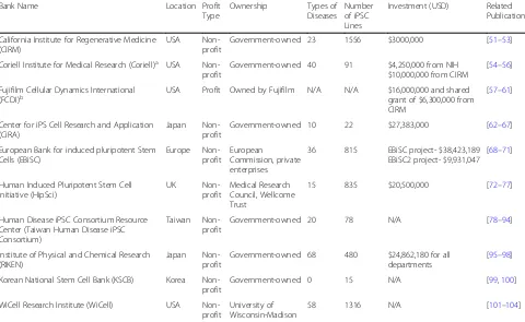
Table 2Brief overview of iPSC banks worldwide
Bank Name Location Profit
Type
Ownership Types of Diseases
Number of iPSC Lines
Investment (USD) Related Publication
California Institute for Regenerative Medicine (CIRM)
USA Non-profit
Government-owned 23 1556 $3000,000 [51–53]
Coriell Institute for Medical Research (Coriell)a USA Non-profit
Government-owned 40 91 $4,250,000 from NIH $10,000,000 from CIRM
[54–56]
Fujifilm Cellular Dynamics International
(FCDI)b USA Profit Owned by Fujifilm N/A N/A $16,000,000 and sharedgrant of $6,300,000 from CIRM
[57–61]
Center for iPS Cell Research and Application (CiRA)
Japan Non-profit
Government-owned 10 22 $27,383,000 [62–67]
European Bank for induced pluripotent Stem Cells (EBiSC)
Europe Non-profit
European
Commission, private enterprises
36 815 EBiSC project- $38,423,189 EBiSC2 project- $9,931,047
[68–71]
Human Induced Pluripotent Stem Cell Initiative (HipSci)
UK
Non-profit
Medical Research Council, Wellcome Trust
15 835 $20,500,000 [72–77]
Human Disease iPSC Consortium Resource Center (Taiwan Human Disease iPSC Consortium)
Taiwan Non-profit
Government-owned 20 78 N/A [78–94]
Institute of Physical and Chemical Research (RIKEN)
Japan Non-profit
Government-owned 68 480 $24,862,180 for all departments
[95–98]
Korean National Stem Cell Bank (KSCB) Korea Non-profit
Government-owned 0 15 N/A [99,100]
WiCell Research Institute (WiCell) USA Non-profit
University of Wisconsin-Madison
58 1316 N/A [101–104]
a
Coriell has its own iPSC depositories: (1) NIGMS Human Genetic Cell Repository (15 healthy donor-derived iPSC lines and 37 diseased iPSC lines); (2) NIA Aging Cell Repository (3 diseased iPSC lines); and (3) Allen Institute for Cell Science. By using CRISPR technology, the Allen Institute generates 36 fluorescent-tagged (EGFP/RFP) iPSC lines from one healthy donor, which produces a potent research tool by tagging different cellular organelles, proteins and compartments. Owing to Coriell’s expertise in cryopreservation and banking, Coriell was awarded with $10 million grant by CIRM to redeposit donor samples and iPSC lines from the Human iPSC Initiative project
b
and automation groups that are spread across European nations. High-standard SOPs for iPSC QA and QC are being established and shared by EBiSC [47]. Currently, the EBiSC offers 306 normal and 482 diseased iPSC lines, including 27 CRISPR-mediated isogenic controls now available to researchers worldwide [111]. In March
2019, EBiSC embarked on a second project “EBiSC2”,
with US $9.93 million in funding, aiming to provide: (1) a more complete catalog of CRISPR-mediated isogenic controls or gene-modified lines; (2) hiPSC-derived pro-genitor cells; and (3) ready-to-use screening platforms between control and diseased lines. In order to generate a large quantity while maintaining constant cell quality, automation of the pipeline is now underway. Users not only have access to iPSC lines but also stringent online filmed/documented protocols set up by the EBiSC.
Korean Society for Cell Biology (KSCB)
The KSCB is an organization for iPSC and ESC line banking and distribution that operates under the Korea National Institute of Health. Researchers can apply to access the 15 listed iPSC lines, most of which are from healthy donors’fibroblasts; however, there are a number of cell lines being developed using RNA-based gene de-livery to generate cytogenetic abnormalities. KSCB and its stem cell bank are completely government-owned and funded [112].
Human induced pluripotent stem cell initiative (HipSci)
Located in the UK and funded by the Medical Research Council/Wellcome, with a total of US $20.5 million in funding, HipSci has collected 835 donor samples, the ma-jority of which have a British background, including 15 disease lines [113, 114]. The organization heavily utilizes the Cytotune 2.0 Sendai Virus Kit to generate iPSC lines and collaborates with ECACC/EBiSC to deposit/distribute cells. The advantage of HipSci over other biobanks is their extensive effort in characterizing iPSC lines. Genetic and genomic assays (RNA seq/DNA methylation/whole gen-ome seq/exgen-ome seq), proteomic assays, and cellular phe-notyping assays are included in the pipeline. HipSci has 496 healthy donor-derived iPSC lines that can be used for identifying genetic variations that occur in the general population. Researchers can access these data online and apply for use; however, they currently do not offer cus-tomized iPSC generation [115].
RIKEN–BioResource research center (BRC)
To date, RIKEN BRC holds an iPSC bank with approxi-mately 480 normal iPSC lines and 68 unique diseased iPSC lines [116]. In addition to iPSC banking, RIKEN BRC is focusing on the development of iPSC characterization and iPSC-based drug discovery. With its collaboration with Kyoto University, they formed the CiRA in 2008,
which focuses on the iPS Cell Stock for Regenerative Medicine and aims to provide clinical grade iPSCs to in-dustry and research institutes [117].
Taiwan Human Disease iPSC Consortium
Taiwan Human Disease iPSC Consortium is the first, and the only iPSC resource center in Taiwan that aims to provide iPSC generation, characterization, and an iPSC banking service. The consortium was founded in
2015 under the Taiwan government’s National Research
Program for Biopharmaceuticals project. In 2017, the con-sortium was transferred into another program called the National Core Facility for Biopharmaceuticals. For the past three years, the consortium has received funding from the National Research Program for Biopharmaceuti-cals and the National Core Facility for BiopharmaceutiBiopharmaceuti-cals program which totals US $2.1 million. Blood or fibroblast samples are collected and sent to the iPSC cores to be generated into iPSC lines, which are subsequently sent to FIRDI for QC and iPSC banking [118]. To date, 78 Sendai virus reprogrammed iPSC lines have been generated by the Taiwan iPSC Consortium consisting of 11 normal and 67 diseased iPSC lines. As of February 2019, there have been 20 individual disease types banked in the Taiwan iPSC Consortium. Furthermore, out of the 78 iPSC lines, 57 are feeder-free iPSC lines and 21 are feeder-dependent iPSC lines, all of which are accessible to all researchers in Taiwan via the BCRC website.
WiCell
As a supporting organization of the University of Wisconsin-Madison, WiCell, established in 1999, is a non-profit organization focusing on the betterment of stem cell research. Starting with banking and distributing embryonic stem cell (ESC) lines, WiCell quickly expanded their collection into iPSC lines [119]. WiCell has gener-ated and characterized 1316 iPSC lines from donor blood with 58 individual disease types across the spectrum from sickle cell anemia to mental illness. These cell lines are readily available to both academic and industrial groups. WiCell offers services including cell line generation, mycoplasma detection, karyotyping, cell banking, and other services. Other than stem cell generation, WiCell also offers services in cell line banking, operating under good manufacturing practice conditions with modified iPSC lines and differentiated cell lines readily available for purchase [119].
Barriers in iPSC application
mutations can occur in the same gene and lead to differ-ent phenotypes in differdiffer-ent individuals. Also, genetic back-ground, epigenetic modifications, and variation amongst clones in iPSC lines can affect results observed by re-searchers. Thus, a large cohort of diseased iPSCs is needed to understand the underlying mechanism for each disease. To this end, projects for large-scale collection of iPSCs from normal and diseased individuals have been growing over the past ten years. The value of iPSC biobanks and resources are related to the information and QC that are provided to the users. This section aims to describe the hurdles faced in translating iPSC applications into the clinic. Although a few clinical studies based on iPSC deriv-atives are ongoing, QC, reproducibility, and immunogen-icity are the biggest barriers for iPSC utility.
Immunogenicity
The discovery of iPSC-based technology offers a promis-ing cell source for autologous cell transplantation for vari-ous degenerative diseases without side effects from immunosuppression and allograft rejection. In 2011, Zhao and colleagues reported that injection of iPSC-derived teratoma into syngeneic host mice resulted in immune re-jection. This study raises a concern regarding the use of autologous iPSC transplantation for cell therapy and the immunogenicity of undifferentiated iPSCs [120]. Almeida et al. tried to compare the immunogenicity of undifferenti-ated autologous iPSCs, iPSC derivatives, and syngeneic somatic cells after cell transplantation; they demonstrated that autologous iPSC derivatives could engraft into tissue without using immune suppression and elicited a tolero-genic immune response very similar to the syngeneic som-atic cell group. However, the autologous undifferentiated iPSC graft was rejected by the recipient with lymphocytic infiltration [121]. This work has proved that iPSC deriva-tives result in loss of immunogenicity. Moreover, Embrog et al. transplanted autologous iPSC-derived neural pro-genitor cells into the non-human primate brain and six months after transplantation, found no infiltration of mac-rophages and lymphocytes. This result suggests that the autologous iPSC-derived neural cell transplants were not rejected by the primate brain [122]. Another study showed that transplantation of autologous iPSC-derived dopamine
neurons into a non-human primate Parkinson’s disease
model for up to 2 years provided functional recovery and
immune tolerance without immunosuppression [123]. A
similar result was published in the first iPSC-based clinical trial in RIKEN in 2017 where the authors transplanted an autologous iPSC-derived retinal epithelial cell sheet into a patient with neovascular age-related macular degener-ation. The result indicated that the graft could survive more than two years after transplantation without im-mune suppression [11]. Together, these studies indicate
that iPSC-derived cells may provide a new source for cell therapy.
Timelines and costs
Although there are obvious advantages to using autolo-gous iPSC-based cell therapies, the pipeline of iPSC generation, characterization, and cell banking is a labor-intensive, highly time- and cost-consuming process. In general, it costs US $10,000–$25,000 to generate and val-idate a research grade iPSC line. The entire process re-quires between 6 to 9 months from patient recruitment to final characterization and requires further 3 to 6 months to produce large scale iPSC derivatives. Generating a clin-ical grade iPSC line costs approximately US $800,000 based on previously published reports [124, 125]. There-fore, to maximize the utility and efficiency of iPSCs and to significantly reduce the cost of generating an iPSC line, an alternative and practical strategy for personalized iPSC generation is to establish an allogenic iPSC resource for human leukocyte antigen (HLA)-matched tissue trans-plantation. Several similar projects have been started around the world since it has been proposed that 50 HLA
homozygous“super donors”could match over 90% of the
Japanese population [126]. A similar strategy, reported by Taylor et al., found that generating 150 selected HLA
homozygous donors’ iPSCs could match 93% of the UK
population [127].
Standardization
Variability within various iPSC lines and their derivatives remains a great concern when using iPSCs and their de-rivatives for disease modeling and cell therapy. Variabil-ity is often observed in iPSC differentiation potential, tumorigenicity, genome instability, epigenetic status, and maturation status within inter- and intra- iPSC lines when generated from different individuals and iPSC core
facilities. The successful generation of “comparable”
Genetic variations and stability
Recent studies of genetic and epigenetic variations in iPSCs raised concerns about safety in iPSC use. The presence of genetic variations in iPSCs includes gen-ome instability, single nucleotide variations, CNV, and loss of heterozygosity. These mutations can be intro-duced and accumulated in iPSCs from their parental cells, reprogramming process, and generated during
prolonged culture in vitro [129]. One safety concern
about genetic variations in iPSCs is the possibility of tumorigenicity. The first clinical iPSC trial which treated age-related macular degeneration with an au-tologous iPSC-derived retinal pigment epithelial cell
(RPE) sheet was conducted in 2014 in Japan [130];
however, Mandai et al. reported that three CNV were
found in the second patient’s iPSCs and iPSC-derived
RPE. Thus, the authors decided not to transplant the RPE sheet even if the iPSC-derived RPE passed the
tumorigenicity test [11] despite there being no human
iPSC-derivative clinical trials reporting the formation of neoplasia tissue after cell transplantation [11, 131,
132]. Moreover, it is known that various iPSC lines
have different differentiation efficiency [133, 134]. An-other concern for genetic and epigenetic variations among iPSCs is that variations may affect the iPSC differentiation potential and cause an unexpected phenotype of iPSC-derived cells [135–137]. The genetic variations in iPSCs may cause functional and safety consequences, thus, fur-ther studies and generation of a common iPSC-related mutation database and an established standard for screen-ing of genetic variation are required for genomic stability evaluation.
Interspecies chimerism
Currently, researchers are attempting to use human iPSCs to generate interspecies chimeras. They aim to improve in vivo research models by generating human organs and tissues in animals or by generating new man disease models. Wu et al. (2017) reported that hu-man iPSCs are capable of integrating into pig embryo [138]. However, there are still concerns in this field, for example, 1) for safety concern, the organ may be rejected by recipients even when receiving immuno-suppressants during the xenotransplantation process; 2) serious zoonotic risks and contamination from
ani-mal cells when creating human-aniani-mal chimeras [139];
3) the ethical issues, human-chimeric animals may have consciousness; 4) animal welfare issue, human cells may lead to unexpected suffer on chimeric animal
[140]. Even though there are some advantages for this
potential technique, the ethical issues for generating human-animal chimeras still requires further public discussion.
Conclusions
The discovery of iPSCs has not only expanded our knowledge of the cellular mechanisms involved in pluripotency and development but has also allowed the opportunity for enhanced, human-specific drug screen-ing and disease studies. These cells are becomscreen-ing ever more prominent and continue to play a vital role in bringing more relevant cell models into the lab. Fur-ther advancement in iPSC technology will highlight their role in regenerative medicine. However, the cost and time required for the generation iPSCs remain on-going roadblocks for many researchers. The continued development of iPSC banks provides a greater oppor-tunity for researchers to gain access to these valuable cells while at the same time beginning to standardize their quality and reliability.
Abbreviations
BCRC:Bioresource Collection and Research Center; BRC: BioResource Research Center; CiRA: Center for iPS Cell Research and Application; CIRM: California Institute for Regenerative Medicine; CNV: copy number variation; EBiSC: European Bank for induced pluripotent Stem Cells; ESC: embryonic stem cells; FIRDI: Food Industry Research and Development Institute; IBMS: Institute of Biomedical Sciences; IBMT: Fraunhofer-Institut für Biomedizinische Technik; iCeMS: Center for iPS Cell Research and Application; IMS: Information Management System; iPSC: induced pluripotent stem cell; KSCB: Korean Society for Cell Biology; QA: quality assurance; QC: quality control; RPE: retinal pigment epithelial cell; SNP: single nucleotide polymorphism
Acknowledgements
This study was supported by the Ministry of Science and Technology and the Academia Sinica. We thank our collaborators and their institutes, which are members of the Taiwan Human iPSC Consortium, for their support including Dr. Hong-Nerng Ho, Dr. Hsin-Fu Chen, and Dr. Hsiang-Po Huang at National Taiwan University Hospital; Dr. Shih-Hwa Chiou at Taipei Veterans General Hospital; Dr. Huai-En Lu and Dr. Shiaw-Min Hwang at the Food Industry Research and Development Institute; and Dr. B. Linju Yen at the National Health Research Institutes. We also thank the Taiwan Mouse Clinic and the National Center for Genome Medicine for technical support.
Authors’contributions
CYH: Manuscript outline, preparation of the draft manuscript; CLL, CYT, YTC and YCC: collected detailed information about the various iPSC banks and draft; MWN: English editing and introduction draft and suggestions; PCH: Design and coordination of the manuscript, reading and editing of the draft manuscript. All authors read and approved the final manuscript.
Funding
This work was supported by the Ministry of Science and Technology, Taiwan (MOST 108–2319-B-001-004, 108–2321-B-001-017, 108–3111-Y-001-053), the National Health Research Institutes grant EX108-10512SI and the Academia Sinica Program for Translational Innovation of Biopharmaceutical Development-Technology Supporting Platform Axis (AS-KPQ-106-TSPA), Thematic Research Program (AS-107-TP-L12) and Summit Research Program (MOST 107–0210-01-19-01),
Availability of data and materials
The information for normal/disease iPSC lines are available in various iPSC repositories.
Hyperlink for these repositories are listed below. CIRM:https://www.cirm.ca.gov
CiRA:https://www.cira.kyoto-u.ac.jp/e/
FCDI:https://fujifilmcdi.com/the-cirm-ipsc-bank/
EBiSC:http://www.ebisc.org/
Taiwan Human Disease iPSC Consortium:http://ipsc.ibms.sinica.edu.tw/index. html; https://catalog.bcrc.firdi.org.tw/Welcome
RIKEN:https://cell.brc.riken.jp/en/hps/patient_specific_ips
KSCB:http://www.cdc.go.kr
WiCell:https://www.wicell.org
Ethics approval and consent to participate
Not applicable.
Consent for publication
Not applicable.
Competing interests
The authors declare that they have no competing interests.
Author details
1Institute of Biomedical Sciences, Academia Sinica, Taipei, Taiwan.2Graduate Institute of Medical Genomics and Proteomics and Graduate Institute of Clinical Medicine, National Taiwan University College of Medicine, Taipei, Taiwan.3Cardiovascular Surgery Division, Department of Surgery, National Taiwan University Hospital, Taipei, Taiwan.
Received: 29 July 2019 Accepted: 9 October 2019
References
1. Takahashi K, Yamanaka S. Induction of pluripotent stem cells from mouse embryonic and adult fibroblast cultures by defined factors. Cell. 2006;126(4): 663–76.
2. Takahashi K, Tanabe K, Ohnuki M, Narita M, Ichisaka T, Tomoda K, Yamanaka S. Induction of pluripotent stem cells from adult human fibroblasts by defined factors. Cell. 2007;131(5):861–72.
3. Elitt MS, Barbar L, Tesar PJ: Drug screening for human genetic diseases using iPSC models. (1460–2083 (Electronic)).
4. Guengerich FP: Mechanisms of drug toxicity and relevance to pharmaceutical development. (1880–0920 (Electronic)).
5. Uhl EW, Warner NJ. Mouse models as predictors of human responses: evolutionary medicine. Curr Pathobiol Rep. 2015;3(3):219–23. 6. McNeish J, Gardner JP, Wainger BJ, Woolf CJ, Eggan K. From dish to
bedside: lessons learned while translating findings from a stem cell model of disease to a clinical trial. Cell Stem Cell. 2015;17(1):8–10.
7. Burbulla LF, Song P, Mazzulli JR, Zampese E, Wong YC, Jeon S, Santos DP, Blanz J, Obermaier CD, Strojny C, et al. Dopamine oxidation mediates mitochondrial and lysosomal dysfunction in Parkinson's disease. Science (New York, NY). 2017;357(6357):1255–61.
8. Ramsden CM, Nommiste B, RL A, Carr AF, Powner MB, JKS M, Chen LL, Muthiah MN, Webster AR, Moore AT, et al. Rescue of the MERTK phagocytic defect in a human iPSC disease model using translational read-through inducing drugs. Sci Rep. 2017;7(1):51.
9. Sa S, Gu M, Chappell J, Shao NY, Ameen M, Elliott KA, Li D, Grubert F, Li CG, Taylor S, et al. Induced pluripotent stem cell model of pulmonary arterial hypertension reveals novel gene expression and patient specificity. Am J Respir Crit Care Med. 2017;195(7):930–41.
10. Azuma K, Yamanaka S. Recent policies that support clinical application of induced pluripotent stem cell-based regenerative therapies. Regen Ther. 2016;4:36–47.
11. Mandai M, Watanabe A, Kurimoto Y, Hirami Y, Morinaga C, Daimon T, Fujihara M, Akimaru H, Sakai N, Shibata Y, et al. Autologous induced stem-cell-derived retinal cells for macular degeneration. New Engl J Med. 2017; 376(11):1038–46.
12. Takahashi J. Preparing for first human trial of induced pluripotent stem cell-derived cells for Parkinson's disease: an interview with Jun Takahashi. Regen Med. 2019;14(2):93–5.
13. First iPS Cell Trial for Heart Disease Raises Excitement, Concern [https:// www.the-scientist.com/news-opinion/first-ips-cell-trial-for-heart-disease-raises-excitement%2D%2Dconcern-64743].
14. Japan approves world-first trial using iPS cells to treat spinal cord injuries [https://www.japantimes.co.jp/news/2019/02/18/national/science-health/ japan-approves-world-first-trial-using-ips-stem-cells-treat-spinal-cord-injuries/ #.XXXZm6TVLIX].
15. Yu J, Vodyanik MA, Smuga-Otto K, Antosiewicz-Bourget J, Frane JL, Tian S, Nie J, Jonsdottir GA, Ruotti V, Stewart R, et al. Induced pluripotent stem cell lines derived from human somatic cells. Science (New York, NY). 2007; 318(5858):1917–20.
16. Haase A, Olmer R, Schwanke K, Wunderlich S, Merkert S, Hess C, Zweigerdt R, Gruh I, Meyer J, Wagner S, et al. Generation of induced pluripotent stem cells from human cord blood. Cell Stem Cell. 2009;5(4):434–41.
17. Loh YH, Hartung O, Li H, Guo CG, Sahalie JM, Manos PD, Urbach A, Heffner GC, Grskovic M, Vigneault F, et al. Reprogramming of T cells from human peripheral blood. Cell Stem Cell. 2010;7(1):15–9.
18. Bueno C, Sardina JL, Di Stefano B, Romero-Moya D, Munoz-Lopez A, Ariza L, Chillon MC, Balanzategui A, Castano J, Herreros A, et al. Reprogramming human B cells into induced pluripotent stem cells and its enhancement by C/EBPalpha. Leukemia. 2016;30(3):674–82.
19. Kim Y, Kang K, Lee SB, Seo D, Yoon S, Kim SJ, Jang K, Jung YK, Lee KG, Factor VM, et al. Small molecule-mediated reprogramming of human hepatocytes into bipotent progenitor cells. J Hepatol. 2019;70(1):97–107. 20. Yang W, Mills JA, Sullivan S, Liu Y, French DL, Gadue P: iPSC
Reprogramming from Human Peripheral Blood Using Sendai Virus Mediated Gene Transfer. In: StemBook.Cambridge (MA): Harvard Stem Cell Institute Copyright: (c) 2012 Wenli Yang, Jason A. Mills, Spencer Sullivan, Ying Liu, Deborah L. French, and Paul Gadue.; 2008.
21. Cheng YY, Yan YT, Lundy DJ, Lo AH, Wang YP, Ruan SC, Lin PJ, Hsieh PC. Reprogramming-derived gene cocktail increases cardiomyocyte proliferation for heart regeneration. EMBO Mol Med. 2017;9(2):251–64.
22. Rizzi R, Di Pasquale E, Portararo P, Papait R, Cattaneo P, Latronico MV, Altomare C, Sala L, Zaza A, Hirsch E, et al. Post-natal cardiomyocytes can generate iPS cells with an enhanced capacity toward cardiomyogenic re-differentation. Cell Death Differ. 2012;19(7):1162–74.
23. Merling RK, Sweeney CL, Choi U, De Ravin SS, Myers TG, Otaizo-Carrasquero F, Pan J, Linton G, Chen LF, Koontz S, et al. Transgene-free iPSCs generated from small volume peripheral blood nonmobilized CD34(+) cells. Blood. 2013;121(14):E98–E107.
24. Stadtfeld M, Nagaya M, Utikal J, Weir G, Hochedlinger K. Induced pluripotent stem cells generated without viral integration. Science (New York, NY). 2008;322(5903):945–\.
25. Fusaki N, Ban H, Nishiyama A, Saeki K, Hasegawa M. Efficient induction of transgene-free human pluripotent stem cells using a vector based on Sendai virus, an RNA virus that does not integrate into the host genome. Proc Jpn Acad Ser B, Phys Biol Sci. 2009;85(8):348–62.
26. Seki T, Yuasa S, Oda M, Egashira T, Yae K, Kusumoto D, Nakata H, Tohyama S, Hashimoto H, Kodaira M, et al. Generation of induced pluripotent stem cells from human terminally differentiated circulating T cells. Cell Stem Cell. 2010;7(1):11–4.
27. Okita K, Nakagawa M, Hyenjong H, Ichisaka T, Yamanaka S. Generation of mouse induced pluripotent stem cells without viral vectors. Science (New York, NY). 2008;322(5903):949–53.
28. Yu J, Hu K, Smuga-Otto K, Tian S, Stewart R, Slukvin II, Thomson JA, et al. Science (New York, NY). 2009;324(5928):797–801.
29. Okita K, Matsumura Y, Sato Y, Okada A, Morizane A, Okamoto S, Hong H, Nakagawa M, Tanabe K, Tezuka K, et al. A more efficient method to generate integration-free human iPS cells. Nat Methods. 2011;8(5): 409–12.
30. Kaji K, Norrby K, Paca A, Mileikovsky M, Mohseni P, Woltjen K. Virus-free induction of pluripotency and subsequent excision of reprogramming factors. Nature. 2009;458(7239):771–5.
31. Woltjen K, Michael IP, Mohseni P, Desai R, Mileikovsky M, Hamalainen R, Cowling R, Wang W, Liu P, Gertsenstein M, et al. PiggyBac transposition reprograms fibroblasts to induced pluripotent stem cells. Nature. 2009; 458(7239):766–70.
32. Yusa K, Rad R, Takeda J, Bradley A. Generation of transgene-free induced pluripotent mouse stem cells by the piggyBac transposon. Nat Methods. 2009;6(5):363–9.
33. Warren L, Manos PD, Ahfeldt T, Loh YH, Li H, Lau F, Ebina W, Mandal PK, Smith ZD, Meissner A, et al. Highly efficient reprogramming to pluripotency and directed differentiation of human cells with synthetic modified mRNA. Cell Stem Cell. 2010;7(5):618–30.
35. Li HO, Zhu YF, Asakawa M, Kuma H, Hirata T, Ueda Y, Lee YS, Fukumura M, Iida A, Kato A, et al. A cytoplasmic RNA vector derived from
nontransmissible Sendai virus with efficient gene transfer and expression. J Virol. 2000;74(14):6564–9.
36. Tokusumi T, Iida A, Hirata T, Kato A, Nagai Y, Hasegawa M. Recombinant Sendai viruses expressing different levels of a foreign reporter gene. Virus Res. 2002;86(1–2):33–8.
37. Inoue M, Tokusumi Y, Ban H, Kanaya T, Tokusumi T, Nagai Y, Iida A, Hasegawa M. Nontransmissible virus-like particle formation by F-deficient Sendai virus is temperature sensitive and reduced by mutations in M and HN proteins. J Virol. 2003;77(5):3238–46.
38. Shi G, Jin Y. Role of Oct4 in maintaining and regaining stem cell pluripotency. Stem Cell Res Ther. 2010;1(5):39.
39. Masui S, Nakatake Y, Toyooka Y, Shimosato D, Yagi R, Takahashi K, Okochi H, Okuda A, Matoba R, Sharov AA, et al. Pluripotency governed by Sox2 via regulation of Oct3/4 expression in mouse embryonic stem cells. Nat Cell Biol. 2007;9(6):625–35.
40. Nandan MO, Yang VW. The role of Kruppel-like factors in the
reprogramming of somatic cells to induced pluripotent stem cells. Histol Histopathol. 2009;24(10):1343–55.
41. Niwa H, Ogawa K, Shimosato D, Adachi K. A parallel circuit of LIF signalling pathways maintains pluripotency of mouse ES cells. Nature. 2009;460(7251): 118–22.
42. Shyh-Chang N, Daley GQ. Lin28: primal regulator of growth and metabolism in stem cells. Cell Stem Cell. 2013;12(4):395–406.
43. Scheper W, Copray S. The molecular mechanism of induced pluripotency: a two-stage switch. Stem Cell Rev. 2009;5(3):204–23.
44. Nakagawa M, Takizawa N, Narita M, Ichisaka T, Yamanaka S. Promotion of direct reprogramming by transformation-deficient Myc. Proc Natl Acad Sci U S A. 2010;107(32):14152–7.
45. Crook JM, Hei D, Stacey G. The international stem cell banking initiative (ISCBI): raising standards to bank on. In Vitro Cell Dev Biol Anim. 2010;46(3– 4):169–72.
46. European Bank for induced pluripotent Stem Cells [http://www.ebisc.org/]. 47. De Sousa PA, Steeg R, Wachter E, Bruce K, King J, Hoeve M, Khadun S,
McConnachie G, Holder J, Kurtz Aet al: Rapid establishment of the European Bank for induced pluripotent stem cells (EBiSC) - the hot start experience. Stem Cell Res 2017, 20(December 2015):105–114. 48. EBiSC Virtual Training Library [https://ebisc.org/trainings/].
49. EBiSC-Protocol for the Use of Induced Pluripotent Stem Cells [http://www. ebisc.org/files/Other-doc/EBiSC-User_Protocol-V2-2017-05].
50. The EBiSC Poster Book [ https://www.sigmaaldrich.com/content/dam/sigma-aldrich/docs/Sigma-Aldrich/General_Information/1/ebisc-poster-book.pdf]. 51. Lan F, Lee AS, Liang P, Sanchez-Freire V, Nguyen PK, Wang L, Han L, Yen M,
Wang Y, Sun N, et al. Abnormal calcium handling properties underlie familial hypertrophic cardiomyopathy pathology in patient-specific induced pluripotent stem cells. Cell Stem Cell. 2013;12(1):101–13.
52. de Almeida PE, Meyer EH, Kooreman NG, Diecke S, Dey D, Sanchez-Freire V, Hu S, Ebert A, Odegaard J, Mordwinkin NM, et al. Transplanted terminally differentiated induced pluripotent stem cells are accepted by immune mechanisms similar to self-tolerance. Nat Commun. 2014;5:3903.
53. Kooreman NG, Kim Y, de Almeida PE, Termglinchan V, Diecke S, Shao N-Y, Wei T-T, Yi H, Dey D, Nelakanti R, et al. Autologous iPSC-Based Vaccines Elicit Anti-tumor Responses In Vivo. Cell Stem Cell. 2018;22(4): 501–513.e507.
54. Zhang J, Lian Q, Zhu G, Zhou F, Sui L, Tan C, Mutalif RA, Navasankari R, Zhang Y, Tse HF, et al. A human iPSC model of Hutchinson Gilford Progeria reveals vascular smooth muscle and mesenchymal stem cell defects. Cell Stem Cell. 2011;8(1):31–45.
55. Kim E-M, Manzar G, Zavazava N. Human iPS cell-derived hematopoietic progenitor cells induce T-cell anergy in in vitro-generated alloreactive CD8(+) T cells. Blood. 2013;121(26):5167–75.
56. Ng R, Manring H, Papoutsidakis N, Albertelli T, Tsai N, See CJ, Li X, Park J, Stevens TL, Bobbili PJ, et al. Patient mutations linked to arrhythmogenic cardiomyopathy enhance calpain-mediated desmoplakin degradation. JCI Insight. 2019;5.
57. Diouf B, Crews KR, Lew G, Pei D, Cheng C, Bao J, Zheng JJ, Yang W, Fan Y, Wheeler HE, et al. Association of an inherited genetic variant with vincristine-related peripheral neuropathy in children with acute lymphoblastic leukemia. JAMA. 2015;313(8):815–23.
58. La Manno G, Gyllborg D, Codeluppi S, Nishimura K, Salto C, Zeisel A, Borm LE, Stott SRW, Toledo EM, Villaescusa JC, et al. Molecular Diversity of Midbrain Development in Mouse, Human, and Stem Cells. Cell. 2016;167(2): 566–580.e519.
59. Wakeman DR, Hiller BM, Marmion DJ, McMahon CW, Corbett GT, Mangan KP, Ma J, Little LE, Xie Z, Perez-Rosello T, et al. Cryopreservation maintains functionality of human iPSC dopamine neurons and rescues Parkinsonian phenotypes in vivo. Stem Cell Rep. 2017;9(1):149–61.
60. Collado MS, Cole BK, Figler RA, Lawson M, Manka D, Simmers MB, Hoang S, Serrano F, Blackman BR, Sinha S, et al. Exposure of induced pluripotent stem cell-derived vascular endothelial and smooth muscle cells in Coculture to hemodynamics induces primary vascular cell-like phenotypes. Stem Cells Transl Med. 2017;6(8):1673–83.
61. Grimm FA, Blanchette A, House JS, Ferguson K, Hsieh NH, Dalaijamts C, Wright AA, Anson B, Wright FA, Chiu WAet al: A human population-based organotypic in vitro model for cardiotoxicity screening. (1868-596X (Print)). 62. Takahashi K, Tanabe K, Ohnuki M, Narita M, Ichisaka T, Tomoda K, Yamanaka
S. Induction of pluripotent stem cells from adult human fibroblasts by defined factors. Cell. 2007;131(5):861–72.
63. Nakagawa M, Koyanagi M, Tanabe K, Takahashi K, Ichisaka T, Aoi T, Okita K, Mochiduki Y, Takizawa N, Yamanaka S. Generation of induced pluripotent stem cells without Myc from mouse and human fibroblasts. Nat Biotechnol. 2008;26(1):101–6.
64. Sugita S, Iwasaki Y, Makabe K, Kimura T, Futagami T, Suegami S, Takahashi M. Lack of T cell response to iPSC-derived retinal pigment epithelial cells from HLA homozygous donors. Stem Cell Rep. 2016;7(4):619–34. 65. Tateno H, Hiemori K, Hirayasu K, Sougawa N, Fukuda M, Warashina M,
Amano M, Funakoshi T, Sadamura Y, Miyagawa S, et al. Development of a practical sandwich assay to detect human pluripotent stem cells using cell culture media. Regen Ther. 2017;6:1–8.
66. Toyoda T, Kimura A, Tanaka H, Ameku T, Mima A, Hirose Y, Nakamura M, Watanabe A, Osafune K. Rho-associated kinases and non-muscle myosin IIs inhibit the differentiation of human iPSCs to pancreatic endoderm. Stem Cell Rep. 2017;9(2):419–28.
67. Minagawa A, Yoshikawa T, Yasukawa M, Hotta A, Kunitomo M, Iriguchi S, Takiguchi M, Kassai Y, Imai E, Yasui Y, et al. Enhancing T Cell Receptor Stability in Rejuvenated iPSC-Derived T Cells Improves Their Use in Cancer Immunotherapy. Cell Stem Cell. 2018;23(6):850–858.e854.
68. Cao L, McDonnell A, Nitzsche A, Alexandrou A, Saintot PP, Loucif AJ, Brown AR, Young G, Mis M, Randall A et al: Pharmacological reversal of a pain phenotype in iPSC-derived sensory neurons and patients with inherited erythromelalgia. (1946–6242 (Electronic)).
69. Fatima A, Ivanyuk D, Herms S, Heilmann-Heimbach S, O'Shea O, Chapman C, Izsvak Z, Farr M, Hescheler J, Saric T: Generation of human induced pluripotent stem cell line from a patient with a long QT syndrome type 2. (1876–7753 (Electronic)).
70. Koyuncu SA-Ohoo, Saez I, Lee HJ, Gutierrez-Garcia R, Pokrzywa W, Fatima A, Hoppe TA-Ohoo, Vilchez DA-Ohoo: The ubiquitin ligase UBR5 suppresses proteostasis collapse in pluripotent stem cells from Huntington's disease patients. (2041–1723 (Electronic)).
71. Schmid B, Prehn KR, Nimsanor N, Garcia BIA, Poulsen U, Jorring I, Rasmussen MA, Clausen C, Mau-Holzmann UA, Ramakrishna Set al: Generation of a set of isogenic, gene-edited iPSC lines homozygous for all main APOE variants and an APOE knock-out line. (1876–7753 (Electronic)).
72. Leha A, Moens N, Meleckyte R, Culley OJ, Gervasio MK, Kerz M, Reimer A, Cain SA, Streeter I, Folarin A, et al. A high-content platform to characterise human induced pluripotent stem cell lines. Methods. 2016;96:85–96. 73. Schwartzentruber J, Foskolou S, Kilpinen H, Rodrigues J, Alasoo K, Knights
AJ, Patel M, Goncalves A, Ferreira R, Benn CL, et al. Molecular and functional variation in iPSC-derived sensory neurons. bioRxiv. 2017:095943.
74. Kilpinen H, Goncalves A, Leha A, Afzal V, Alasoo K, Ashford S, Bala S, Bensaddek D, Casale FP, Culley OJ, et al. Common genetic variation drives molecular heterogeneity in human iPSCs. Nature. 2017;546:370. 75. Alasoo K, Rodrigues J, Mukhopadhyay S, Knights AJ, Mann AL, Kundu K,
Hale C, Dougan G, Gaffney DJ, Consortium H. Shared genetic effects on chromatin and gene expression indicate a role for enhancer priming in immune response. Nat Genet. 2018;50(3):424–31.
77. Barrell WB, Griffin JN, Harvey J-L, HC DD, Beales P, Grigoriadis AE, Liu KJ, Durbin R, Gaffney D, et al. Induction of Neural Crest Stem Cells From Bardet–Biedl Syndrome Patient Derived hiPSCs. Front Mol Neurosci. 2019; 12(139).https://doi.org/10.3389/fnmol.2019.00139.
78. Peng KY, Lee YW, Hsu PJ, Wang HH, Wang Y, Liou JY, Hsu SH, Wu KK, Yen BL. Human pluripotent stem cell (PSC)-derived mesenchymal stem cells (MSCs) show potent neurogenic capacity which is enhanced with cytoskeletal rearrangement. Oncotarget. 2016;7(28):43949–59. 79. Lin YT, Wang CK, Yang SC, Hsu SC, Lin H, Chang FP, Kuo TC, Shen CN,
Chiang PM, Hsiao M, et al. Elimination of undifferentiated human embryonic stem cells by cardiac glycosides. Sci Rep. 2017;7(1):5289.
80. Hsu YH, Wu YT, Huang CY, Ho MC, Cheng YC, Syu SH, Lu HE, Chen YC, Tsai CL, Lin HC, et al. Generation of an induced pluripotent stem cell line from a 39-year-old female patient with severe-to-profound non-syndromic sensorineural hearing loss and a A1555G mutation in the mitochondrial MTRNR1 gene. Stem Cell Res. 2017;25:245–9.
81. Huang CY, Ho MC, Lee JJ, Hwang DY, Ko HW, Cheng YC, Hsu YH, Lu HE, Chen HC, Hsieh PCH. Generation of induced pluripotent stem cells derived from an autosomal dominant polycystic kidney disease patient with a p. Ser1457fs mutation in PKD1. Stem Cell Res. 2017;24:139–43.
82. Ho MC, Huang CY, Lee JJ, Hsu SH, Cheng YC, Hsu YH, Hwang DY, Lu HE, Chen HC, Hsieh PCH. Generation of an induced pluripotent stem cell line, IBMS-iPSC-014-05, from a female autosomal dominant polycystic kidney disease patient carrying a common mutation of R803X in PKD2. Stem Cell Res. 2017;25:38–41.
83. Lee JJ, Ho MC, Huang CY, Wen CH, Cheng YC, Hsu YH, Hwang DY, Lu HE, Chen HC, Hsieh PCH. Induced pluripotent stem cells derived from an autosomal dominant polycystic kidney disease patient carrying a PKD1 Q533X mutation. Stem Cell Res. 2017;25:83–7.
84. Wu YT, Hsu YH, Huang CY, Ho MC, Cheng YC, Wen CH, Ko HW, Lu HE, Chen YC, Tsai CL, et al. Generation of an induced pluripotent stem cell (iPSC) line from a 40-year-old patient with the A8344G mutation of mitochondrial DNA and MERRF (myoclonic epilepsy with ragged red fibers) syndrome. Stem Cell Res. 2018;27:10–4.
85. Liu SP, Hsu YH, Huang CY, Ho MC, Cheng YC, Wen CH, Lu HE, Tsai CH, Shyu WC, Hsieh PCH. Generation of novel induced pluripotent stem cell (iPSC) line from a 16-year-old sialidosis patient with NEU-1 gene mutation. Stem Cell Res. 2018;28:39–43.
86. Lu HE, Yang YP, Chen YT, Wu YR, Wang CL, Tsai FT, Hwang DK, Lin TC, Chen SJ, Wang AG, et al. Generation of patient-specific induced pluripotent stem cells from Leber's hereditary optic neuropathy. Stem Cell Res. 2018;28:56–60. 87. Cheng YC, Huang CY, Ho MC, Hsu YH, Syu SH, Lu HE, Lin HI, Lin CH, Hsieh
PCH. Generation of 2 induced pluripotent stem cell lines derived from patients with Parkinson's disease carrying LRRK2 G2385R variant. Stem Cell Res. 2018;28:1–5.
88. Hsu WT, Huang CY, Yen CYT, Cheng AL, Hsieh PCH. The HER2 inhibitor lapatinib potentiates doxorubicin-induced cardiotoxicity through iNOS signaling. Theranostics. 2018;8(12):3176–88.
89. Hsu CC, Lu HE, Chuang JH, Ko YL, Tsai YC, Tai HY, Yarmishyn AA, Hwang DK, Wang ML, Yang YP, et al. Generation of induced pluripotent stem cells from a patient with BEST dystrophy carrying 11q12.3 (BEST1 (VMD2)) mutation. Stem Cell Res. 2018;29:134–8.
90. Peng CH, Huang KC, Lu HE, Syu SH, Yarmishyn AA, Lu JF, Buddhakosai W, Lin TC, Hsu CC, Hwang DK, et al. Generation of induced pluripotent stem cells from a patient with X-linked juvenile retinoschisis. Stem Cell Res. 2018; 29:152–6.
91. Wang LT, Jiang SS, Ting CH, Hsu PJ, Chang CC, Sytwu HK, Liu KJ, Yen BL. Differentiation of Mesenchymal stem cells from human induced pluripotent stem cells results in Downregulation of c-Myc and DNA replication pathways with immunomodulation toward CD4 and CD8 cells. Stem Cells. 2018;36(6):903–14.
92. Chan YH, Cheng YF, Chen YT, Huang CY, Lin CH, Hu CJ, Lu YC, Wu CC, Hsu CJ. Generation of induced pluripotent stem cells from a patient with hearing loss carrying GJB2 p.V37I mutation. Stem Cell Res. 2018;33:51–5. 93. Lee JJ, Cheng SJ, Huang CY, Chen CY, Feng L, Hwang DY, Kamp TJ, Chen
HC, Hsieh PCH. Primary cardiac manifestation of autosomal dominant polycystic kidney disease revealed by patient induced pluripotent stem cell-derived cardiomyocytes. EBioMedicine. 2019;40:675–84.
94. Choong OKCC, Zhang J, Lin JH, Lin PJ, Ruan SC, Kamp TJ, Hsieh PCH. Hypoxia-induced H19/YB-1 cascade modulates cardiac remodeling after infarction. Theranostics. 2019;9(22):6550–67.
95. Kanemura H, Go MJ, Shikamura M, Nishishita N, Sakai N, Kamao H, Mandai M, Morinaga C, Takahashi M, Kawamata S. Tumorigenicity studies of induced pluripotent stem cell (iPSC)-derived retinal pigment epithelium (RPE) for the treatment of age-related macular degeneration. PLoS One. 2014;9(1):e85336.
96. Kamao H, Mandai M, Okamoto S, Sakai N, Suga A, Sugita S, Kiryu J, Takahashi M. Characterization of human induced pluripotent stem cell-derived retinal pigment epithelium cell sheets aiming for clinical application. Stem Cell Rep. 2014;2(2):205–18.
97. Long C, Li H, Tiburcy M, Rodriguez-Caycedo C, Kyrychenko V, Zhou H, Zhang Y, Min Y-L, Shelton JM, Mammen PPA, et al. Correction of diverse muscular dystrophy mutations in human engineered heart muscle by single-site genome editing.Sci Adv. 2018;4(1):eaap9004.
98. Hayashi R, Ishikawa Y, Katayama T, Quantock AJ, Nishida K. CD200 facilitates the isolation of corneal epithelial cells derived from human pluripotent stem cells. Sci Rep. 2018;8(1):16550.
99. Choi HY, Kim S-J, Go GY, Kwon A, Im YS, Ha H-Y, Hong JT, Jung J-W, Koo SK. Generation of a human induced pluripotent stem cell line, KSCBi003-a, from human adipose tissue-derived mesenchymal stem cells using a
chromosomal integration-free system. Stem Cell Res. 2018;31:1–4. 100. Rim YA-O, Park N, Nam Y, Ham DS, Kim JW, Ha HY, Jung JW, Jung SM, Baek
IC, Kim SYet al: Recent progress of national banking project on homozygous HLA-typed induced pluripotent stem cells in South Korea. (1932–7005 (Electronic)).
101. Patel R, Page S, Al-Ahmad AJ. Isogenic blood–brain barrier models based on patient-derived stem cells display inter-individual differences in cell maturation and functionality. J Neurochem. 2017;142(1):74–88. 102. Lin Y-T, Wang C-K, Yang S-C, Hsu S-C, Lin H, Chang F-P, Kuo T-C, Shen C-N,
Chiang P-M, Hsiao M, et al. Elimination of undifferentiated human embryonic stem cells by cardiac glycosides. Sci Rep. 2017;7(1):5289. 103. Greuel S, Freyer N, Hanci G, Böhme M, Miki T, Werner J, Schubert F, Sittinger
M, Zeilinger K, Mandenius C-F. Online measurement of oxygen enables continuous noninvasive evaluation of human-induced pluripotent stem cell (hiPSC) culture in a perfused 3D hollow-fiber bioreactor. J Tissue Eng Regen Med. 2019;13(7):1203–16.
104. Greuel S, Hanci G, Böhme M, Miki T, Schubert F, Sittinger M, Mandenius C-F, Zeilinger K, Freyer N. Effect of inoculum density on human-induced pluripotent stem cell expansion in 3D bioreactors. Cell Prolif. 2019;52(4): e12604.
105. California Institute for Regenerative Medicine [https://www.cirm.ca.gov]. 106. CIRM iPSC Repository [https://www.cirm.ca.gov/researchers/ipsc-repository/
iPSC-collection-characteristics].
107. CIRM iPSC Bank [https://fujifilmcdi.com/the-cirm-ipsc-bank/].
108. CIRM-Stem Cell Agency Banks on $32 million New Approach to Advance Research [https://www.cirm.ca.gov/about-cirm/newsroom/press-releases/031 92013/stem-cell-agency-banks-32-million-new-approach-advance]. 109. Center for iPS Cell Research and Application KU: Establishment of the new
Center for iPSC Cell Research and Application. In.; 2010.
110. Center for iPS Cell Research and Application [https://www.cira.kyoto-u.ac.jp/e/]. 111. Schmid B, Prehn KR, Nimsanor N, Garcia BIA, Poulsen U, Jørring I,
Rasmussen MA, Clausen C, Mau-Holzmann UA, Ramakrishna S, et al. Generation of a set of isogenic, gene-edited iPSC lines homozygous for all main APOE variants and an APOE Knock-out line. Stem Cell Res. 2019;34(June 2017):101349.
112. Korean Society for Cell Biology [http://www.cdc.go.kr].
113. Human Induced Pluripotent Stem Cell Initiative [http://www.hipsci.org/]. 114. Kilpinen H, Goncalves A, Leha A, Afzal V, Alasoo K, Ashford S, Bala S,
Bensaddek D, Casale FP, Culley OJ, et al. Common genetic variation drives molecular heterogeneity in human iPSCs. Nature. 2017;546(7658):370–5. 115. Streeter I, Harrison PW, Faulconbridge A, Consortium THS, Flicek P,
Parkinson H, Clarke L. The humaninduced pluripotent stem cell initiative -data resources for cellular genetics. Nucleic Acids Res. 2017;45(D1):D691–7. 116. RIKEN Patient-Specific iPSC [https://cell.brc.riken.jp/en/hps/patient_
specific_ips].
117. RIKEN Tsukuba Branch Annual Report Pamphlet [http://www.riken.jp/~/ media/riken/pr/publications/pamphlets/brc-en.pdf].
118. Taiwan Human Disease iPSC Service Consortium [http://ipsc.ibms.sinica.edu. tw/index_e.html].
119. WiCell [https://www.wicell.org].