University of New Orleans University of New Orleans
ScholarWorks@UNO
ScholarWorks@UNO
University of New Orleans Theses and
Dissertations Dissertations and Theses
Summer 8-4-2011
Synthesis of chiral intermediates by deritivization of
Synthesis of chiral intermediates by deritivization of
monosaccharides
monosaccharides
Sanjeeva Dodlapati
University of New Orleans, sodlapa@my.uno.edu
Follow this and additional works at: https://scholarworks.uno.edu/td
Part of the Chemistry Commons
Recommended Citation Recommended Citation
Dodlapati, Sanjeeva, "Synthesis of chiral intermediates by deritivization of monosaccharides" (2011). University of New Orleans Theses and Dissertations. 457.
https://scholarworks.uno.edu/td/457
This Thesis-Restricted is protected by copyright and/or related rights. It has been brought to you by
ScholarWorks@UNO with permission from the rights-holder(s). You are free to use this Thesis-Restricted in any way that is permitted by the copyright and related rights legislation that applies to your use. For other uses you need to obtain permission from the rights-holder(s) directly, unless additional rights are indicated by a Creative Commons license in the record and/or on the work itself.
Synthesis of chiral intermediates by derivatization of
monosaccharaides
A Thesis
Submitted to the Graduate Faculty of the
University of New Orleans
In partial fulfillment of the
Requirements for the degree of
Master of Science
In
Chemistry
By
Sanjeeva Reddy Dodlapati
August 2011
Dedicated to:
My mother, Suguna
My father, Saidi Reddy
My uncle, Madhava Reddy
Acknowledgements:
I would like to thank my advisor, Dr. Guijun Wang, for her guidance and support for
the past five semesters at the University of New Orleans. Also, I would like to thank my MS.
committee members, Dr. Mark L. Trudell and Dr. Branko Jursic for their precious time. I
would also like to thank current and former group members: Dr. Kristopher Williams,
Navneet Goyal, Michael St. Martin, Hao Yong, Bhargav Parikh and Hariprasad Reddy
Mangunuru. Thank you to the Department of Chemistry for supporting me with teaching
assistance ship. Thank you to Anthony Delucca and group of USDA, New Orleans. The
research was supported in part by AHA SDG 043085N.
Table of Contents
List of Schemes………... v
List of figures ………... vi
Abstract ………... v iii Chapter 1: Literature review on bicyclic amino acids………... 1
Introduction………... ... 1
Bicyclic amino acids in peptidomimetics……… 2
Bicyclic amino acids in drug design and development………... 9
Aeruginosins………... 16
Synthesis of bicyclic amino acid core structures of aeruginosins……… ... 23
Synthesis of other bicyclic amino acids……… ... 33
Reference………... 40
Chapter 2: Synthetic process towards bicyclic amino acids………... 48
Introduction………... ... 48
Results and discussion………... ... 57
Experimental section………... ... 62
Reference………... 70
Vita ………... 72
List of schemes
Scheme 1.1 ………... 25
Scheme 1.2 ………... 26
Scheme 1.3 ………... 27
Scheme 1.4 ………... 28
Scheme 1.5 ………... 28
Scheme 1.6 ………... 30
Scheme 1.7 ………... 32
Scheme 1.8 ………... 34
Scheme 1.9 ………... 35
Scheme 1.10 ………... 36
Scheme 1.11 ………... 36
Scheme 1.12 ………... 37
Scheme 1.13 ………... 38
Scheme 2.1 ………... 53
Scheme 2.2 ………... 56
Scheme 2.3 ………... 58
Scheme 2.4 ………... 59
Scheme 2.5 ………... 60
Scheme 2.6 ………... 61
Scheme 2.7 ………... 61
List of figures
Figure 1.1 ………... 3
Figure 1.2 ………... 4
Figure 1.3 ………... 5
Figure 1.4 ………... 5
Figure 1.5 ………... 6
Figure 1.6 ………... 6
Figure 1.7 ………... 7
Figure 1.8 ………... 8
Figure 1.9 ………... 9
Figure 1.10 ………... 11
Figure 1.11 ………... 12
Figure 1.12 ………... 14
Figure 1.13 ………... 15
Figure 1.14 ………... 17
Figure 1.15 ………... 18
Figure 1.16 ………... 19
Figure 1.17 ………... 20
Figure 1.18 ………... 21
Figure 1.19 ………... 22
Figure 1.20 ………... 30
Figure 2.1 ………... 48
Figure 2.3 ………... 50
Figure 2.4 ………... 51
Figure 2.5 ………... 55
Figure 2.6 ………... 57
Abstract
Conformationally constrained bicyclic amino acids are invaluable in the synthesis of
natural products and peptidomimetics. Aeruginosins contain novel bicyclic amino acid, 2-‐
carboxy-‐6-‐hydroxyl octahydrindole (Choi) as the core structure. Aeruginosins are tetra
peptide serine protease inhibitors isolated from marine sponges and cyanobacterial water
blooms. Rigid bicyclic amino acid (Choi) is an essential core structure, which strongly
influence biological activity of aeruginosin family members. Aeruginosins showed
promising inhibitory activity against thrombin, trypsin, and factor VIIa. Thrombin
and factor VIIa play a major role in blood clotting cascade; excessive coagulation lead to
thrombosis and other cardiovascular diseases. Several research groups have reported
a number of synthetic aeruginosin analogs. In this thesis, some of the synthetic
methodologies of bicyclic amino acid core of aeruginosins are presented. Importance of
bicyclic amino acids in peptidomimetic synthesis and drug designing is presented. Mainly,
syntheses of ring oxygenated Choi analogs starting from glucose and mannose are
presented.
Key words:
Bicyclic amino acid,
2-‐carboxy-‐6-‐hydroxyl octahydrindole (L-‐Choi), ring
oxygenated carboxy octahydrindole (O-‐Choi), aeruginosins, thrombin and
Chapter 1: Literature review on bicyclic amino acids
Introduction:
Conformationally constrained bicyclic compounds have been invaluable in
medicinal chemistry. Plenty of biologically significant natural products and their synthetic
analogs possess various bicyclic scaffolds as the core structure. Especially, bicyclic amino
acids are prominent building blocks in the synthesis of peptidomimetics and natural
product analogs. Aeruginosin family members are natural product
tetra peptides comprised of 2-‐carboxy-‐6-‐hydroxyoctahydrindole (Choi) or analogs of Choi.
Promising anti-‐thrombin activity of aeruginosin family members made them attractive new
class of small molecule anticoagulant drug targets. Several research groups have been
pursuing synthesis of aeruginosin analogs and their core structure; some of
the significant efforts will be discussed in this chapter. Conformationally constrained
bicyclic amino acids provide many advantages such as preventing metabolic degradation of
small molecule peptides. Hence bicyclic amino acids have been continuously receiving
considerable attention from synthetic community. However, it is not possible to discuss all
classes of bicyclic amino acids, mainly octahydrindole derivatives and azabicyclo amino
acid analogs will be discussed.
Bicyclic amino acids in peptidomimetics
Peptidomimetic compounds are molecules whose essential elements
(pharmacophores) mimic a natural peptide or protein in three-‐dimensional space and
which retain the ability to interact with the biological target and produce the same
biological effect. Peptide motifs are present in many biologically active molecules.1-‐3 Drug
design often involves the examination of protein-‐protein interactions associated
with disease, followed by the design of small molecules that can bind to one of the
interacting proteins. Often, the biological activity of proteins stems from a small-‐localized
region of a protein surface created by secondary structural elements such as beta strand.
Despite their remarkable activities in enzymatic or receptor-‐based assays, peptides suffer
from a number of disadvantages that compromise their use as drugs.4-‐6 Linear peptide
molecules possess greater flexibility to adopt random conformations and compromise their
biological activity. Peptide molecules are prone to rapid metabolic degradation; they are
rapidly excreted through the liver and kidney, which contribute to their exclusion as drug
substance in clinical practice.
A synthetic peptidomimetic that is not subject to cleavage while maintaining the
biological activity of the original peptide may have distinct advantages as a potential drug
substance. Conformationally pre-‐organized or fixed into a shape that is recognized by a
receptor can have higher affinity for that receptor, also have reduced entropy. Successful
chemical modification approaches are involved in the restriction of conformations by
blocks.7, 8 Naturally occurring amino acids in a potential inhibitor molecule can be replaced
by conformationally constrained monocyclic and bicyclic unnatural amino acids to avoid
metabolic degradation and maintain its biological activity. It may also exhibit
enhanced potency over their parent sequences, and more importantly, it avoids undesired
side effects from byproducts of peptide degradation.9-‐11
Figure 1.1. Possible conformations of bicyclo (3.3.0)octane
Bicyclic skeletons like bicyclo(3.3.0)octane (Fig. 1) is interesting because it has a
rigid ring junction as well as conformationally flexible ends.12 Depending upon the nature
of substituents, they can adopt any one of the possible conformations in a given
circumstances. Sugar derived amino acids with 5,5-‐ and 6,5-‐ bicyclic scaffolds have been
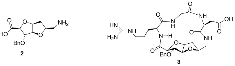
reported as β-‐turn inducers.13, 14 For example, as shown below, bicyclic sugar derived
amino acid (SAA) 2 was incorporated to induce the bioactive conformation in small cyclic
RGD (Arg-‐Gly-‐Asp) peptide analog 3, which acts as selective antagonist of
αvβ3 integrins expressed on GM 7373 cells.15
Figure 1.2. Cyclic peptide 3 consists sugar amino acid derivative bicyclic scaffold 2 as a
core structure
Integrins are a large family of α/β heterodimeric transmembrane receptors
comprise of glycoproteins that attach cells to extracellular matrix (ECM) proteins. Primary
functions of integrins are attachment of the cell to the extracellular matrix, and signal
transduction from the ECM to the cell. Many cells have multiple types of integrins on their
surface, which involve in, along with fundamental cellular processes, various disease
states such as tumors, immune, and inflammatory disorders. Constrained RGD analogs,
with rigid bicyclic amino acid as an inducer of the desired conformation, have been
reported as inhibitors of integrin receptors. Peptidomimetic RGD analog 4 with
pyrroloazepinone core structure is a potent nonselective inhibitor of αvβ3 (IC50 = 3.7 nM,
Ki = 3nM) and αvβ5 (IC50 = 1.4 nM).16
αvβ3/αvβ5 inhibitor IC50 = 3.7/1.4 nM αvβ5 inhibitor IC50 = 4.1 nM
Figure 1.3. Cyclic peptidomimetics 4 and 5, αvβ5 inhibitors
Subsequent replacement of the pyrroloazepinone by (3R, 6R, 9S) 3-‐benzyl-‐
indolizidine-‐2-‐one provided αvβ5 inhibitor 5 (IC50 = 4.1 nM).17 Constrained
azabicycloalkanone amino acids such as 6 -13 have been reported as useful in
the synthesis of peptidomimetics to induce beta turns.18
Figure 1.4. Various conformationally constrained azabicycloalkanone amino acids,
A cyclic vinyl amide scaffold 16 has been designed to mimic the segment Phe43-‐
Leu44 of CD4 15. CD4 is a co-‐receptor of a T-‐cell receptor, which is responsible
for recognition of antigens bound to major histocompatibility complex (MHC) molecules.19
Its main function is to amplify the signal generated by the TCR by recruiting the enzyme
known as tyrosine kinase lck, which is essential for activating many molecules involved in
the signaling cascade of an activated T cell.
Figure 1.5. Mimicry of the Phe43-‐Leu44 segment of CD4 by compound 16
The bold line indicates the region of mimicry where constraints are made from a single
side.
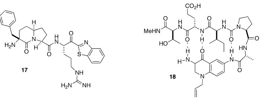
Bicyclic compound 17 is an example of beta strand mimetic. X-‐ray crystal structure
of thrombin complex with compound 17 revealed an anti-‐parallel beta strand hydrogen
bonds between the carbonyl oxygen and the amino nitrogen atom of bicyclic scaffold and
the active site Ser214-‐Gly216 of thrombin’s S2 subunit.20 Hetero bicyclic compounds have
also been used as β-‐strand inducers. In tripeptide derivative of the bicyclic 3,6-‐
diaminoquinolone 18, hydrogen bonding scaffold induced a rigid β-‐strand conformation in
attached amino acids.21 A hydrogen atom was required on the amino group at position 3 of
the quinone scaffold for the formation of one of the necessary hydrogen bonds of the β-‐
strand conformation.
Figure 1.7. a) Schematic representation of anti-‐parallel β-‐strand hydrogen bonding
between Aeruginosin 98B (19) and trypsin. b) Schematic presentation of the hydrogen
bonding between 20-22 and thrombin.
Octahydrindole scaffold in the natural product tetrapeptide orientates its
pharmacophores in proximity to the active site of protease enzyme trypsin. X-‐ray crystal
bonding pattern that is illustrative of anti-‐parallel beta strand binding.22 Compounds 20-22
have diazabicyclic scaffold also show extended H-‐bonding with the active site of
thrombin.23 Short peptide compounds 23-27, which contain 5,6-‐fused bicyclic amino acids
with multiple hetero atoms as core structure, have been
reported as excellent peptidomimetic inhibitors of serine proteases: trypsin, and thrombin
in the nano-‐molar range.24-‐28
Figure 1.8. Compounds contain 5,6-‐fused bicyclic amino acid core structures with
Bicyclic amino acids in drug design and development
Broad range of biological activities of bicyclic amino acids is being explored. Various
bicyclic amino acids and their derivatives showed biological activity against inflammatory
disorders, autoimmune disorders, cell proliferative disorders, epilepsy, faintness attacks,
hypokinesia, cranial disorders, neurodegenerative disorders, depression, anxiety, panic,
pain, arthritis, neuropathological, and sleep disorders. Bicyclic amino acids have also been
used as core structures of drug molecules. For example, ACE inhibitors (26-29)
Trandolapril and Prindopril contain the bicyclic octahydrindole 2-‐carboxylic acid as a core
structure, Quinapril contains tetrahydroisoquinolin-‐3-‐carboxylic acid, and Ramipril has a
5,5 fused bicyclo amino acid.
Figure 1.9. Some of the drug molecules that have constrained bicyclic amino acid as core
structure
Azabicycloalkane amino acid analogs 30-39 have been reported as angiotensin
converting enzyme (ACE) and neutral endopeptidase (NEP) inhibitors. ACE is
an exopeptidase that catalyzes the conversion of angiotensin I to angiotensin II, a potent
vasoconstrictor; also, degrades bradykinin, a potent vasodilator. These enzymes play
an important role in blood pressure regulation, body fluid homeostasis, and cell growth.
Hence inhibition of these enzymes is a main goal in the treatment of conditions such as
heart failure, high-‐blood pressure, diabetic neuropathy, and type-‐2 diabetes mellitus.
Analogs of 32 (35-38), which have oxygen or methylene group in place of sulfur also 5
membered ring in 36 and 38, showed equivalent potency to 32 against ACE and NEP.
Among all these azabicycloalkane amino acid analogs, compound 30 showed
higher potency against both ACE and NEP. Compound 39 showed decent potency against
ACE, but its activity against NEP is not that impressive.
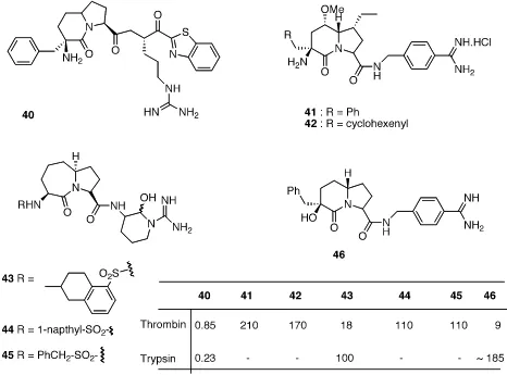
Azabicycloalkane amino acids 40- 46, diazabicyclo alkane amino acids 47- 54,
and thia-‐aza bicyclo alkane amino acids 55- 58 showed inhibitory activity against serine
protease thrombin. Compound 40 showed decent activity against both thrombin and
trypsin, with Ki = 0.85 nM and 0.23 nM respectively, but obviously selectivity was
a big concern. Compound 43 with tetrahydro napthyl sulfate as R group showed 6 fold
higher potency against thrombin compared to its counter parts 44 and 45.33-‐35 Compound
46 showed significant potency against thrombin with Ki = 9 nM, also exhibited 20-‐fold
selectivity over trypsin.36 Since 41 and 46 are slightly different, regarding substituents on
bicyclic scaffold, it would be interesting to know the comparative selectivity in favor of
Figure 1.10. Bicyclic amino acid ACE and NEP inhibitors
Figure 1.11. Azabicyclo amino acid derivative protease inhibitors
Compounds 47 and 48 showed good potency against both thrombin and trypsin, but
failed to show any selectivity.33 Compounds 49 – 52 and 58 showed decent inhibitory
activity against thrombin, but biological activity data against trypsin is not available.37-‐44, 46
Compounds 55 and 56, which differ with respect to the stereochemistry at C6
potency.45 Conformationally constrained azabicycloalkane amino acids 59- 63
exhibited nanomolar range inhibitory activity against caspases1 and caspase3. 6-‐aza-‐
piperdinoazipinone-‐aspartate-‐α-‐aldehydes 59 and 61 are the most potent among all with
Ki = 1nM for Caspase1.47, 48 Compound 61 inhibited both Caspase1 and Caspase3 with Ki =
1nM and 10 nM respectively.49 Compound 62 selectively inhibited caspase1 with IC50 = 36
nM, and compound 63 selectively inhibited caspase3 with IC50 = 18 nM. Caspase1 belongs
to a family of cystine proteases that always cleave peptide bonds following an aspartic acid
residue. Caspase3 has been identified as a mediator of apoptosis in mammalian cells. Over
expression of both caspase1 and 3 would kill cells indiscriminately. Inhibitors
of caspases have been useful in the treatment of diseases such as rheumatoid arthritic,
Alzheimer’s disease, amyotrophic lateral sclerosis (ALS), and Parkinson’s disease.
Figure 1.13. Various GPCR ligands
Various constrained unnatural bicyclic amino acid analogs have been explored as G
protein-‐coupled receptor (GPCR) ligands. G protein-‐coupled receptors comprised of
a large protein family of trance membrane proteins that receive chemical signals from
GPC receptors involve in several diseases, and have become targets for almost 30% of
the medicinal drugs that are currently being used in the treatment of various diseases.
When a ligand binds to the GPCR, it causes a conformational change in the GPCR, and
activates G protein. Further effect depends on the G protein that involved in signal
transduction or disease. Bicyclic scaffolds 64 and 65 act as TRH-‐R1/2 partial agonists with
Ki 1.4 μM and 1.5 μM, respectively.50, 51 Compound 66 has shown antagonist activity
against the Cholecystokinin (CCK1) receptor. Dihydroindolizidinone tripeptide mimic 67
acted as a specific neurokinin (NK1) receptor antagonist with Ki = 79 nM.54, 55
Aeruginosins
The novel bicyclic amino acid 2-‐carboxy-‐6-‐hydroxyocta-‐hydrindole (Choi)
is important motif commonly shared among aeruginosins. Aeruginosins are linear tetra-‐
peptides isolated from microcystic aeruginosa, which is the most common toxic cyano
bacterium in eutrophic fresh water.37, 57 Cyanobacteria are prokaryotic photosynthetic
microorganisms that are a rich source of a variety of structurally novel bioactive nitrogen
compounds. Among the wide range of serine protease inhibitors found in cyanobacteria,
aeruginosin 298 A, the first member of the family, was isolated by Murakama and co-‐
workers in 1994.56 In the span of a decade, continuous search for biologically active natural
products resulted in isolation of 20 new aeruginosins that are structurally closely related.
They were isolated from geographically different locations from different sources having
no clear relationship to the Microcystis waterbloom.37, 56-‐66 Regardless of diverse origin, all
carboxyperhydrindole as the core structure. With few exceptions, they exhibited varying
degree of inhibitory activity against one or more serine proteases. Almost all aeruginosins
showed in vitro activity mainly against serine proteases: Thrombin, Trypsin, and Factor
VIIa. Some of the aeruginosins have also been evaluated against other enzymes, such as
plasmin, chemotrypsin, elastase, and cystine protease papain. For simplification,
generalized structure of aeruginosins virtually can be divided into four common portions:
a C-‐terminal guanidine containing group P1, a 2-‐carboxyperhydrindole core P2, a bulky
hydrophobic amino acid P3 and an N-‐terminal hydroxy or acidic group P4.
Figure 1.14. Generalized structure of the aeruginosins
Figure 1.15. Natural product aeruginosins
Aeruginosin 98A (69), aeruginosin 98B (70), aeruginosin 98C (71), and
aeruginosin 101 (72) have three common subunits: P1 basic guanidine group, O-‐sulfate L-‐
Choi as P2, and D-‐allo-‐Ile as P3 subunit. The fourth subunit is D-‐Hpla with
different substituents on the aromatic ring. They showed significant potency against
thrombin, but they did not show any selectivity to thrombin over trypsin. Where as,
aeruginosin 98A and aeruginosin 98B showed moderate selectivity in favor of trypsin.
Presence of halo substituents on the aromatic ring of D-‐Hpla seems to be causing loss of
selectivity.
Figure 1.16. Aeruginosins that differ at P3 resudue
Compounds 73-77 contain argal group in cyclic form (6 membered) as P1 moiety,
and O-‐sulfated D-‐Hpla P4 residues as common subunits. Aeruginosins 89A and 89B have
D-‐Leu, while other three compounds: aeruginosins 102A, 102B, and 103A have D-‐Tyr as
ether, all other four compounds showed excellent activity against both thrombin and
trypsin. Above details indicate that modifications in P3-‐P4 portion don’t have much impact
on potency.
Figure 1.17. Aeruginosin 298 B, aeruginosin EI461, and dysinosins with cyclic ariginine
analog.
Aeruginosin 298B and aeruginosin EI461, without P1 subunit, lost their biological
activity. It clearly indicates that P1 basic unit with guanidine group is essential for activity.
P1 basic subunit and O-‐Me-‐O-‐SO3-‐ D-‐lysinic acid influencing biological activity in
Chloro substituent in Chlorodysinosin A appears to be responsible for high activity.
Aeruginosin 205 B has 2-‐chloro substituted D-‐Leu showed significant potency. Hanessian
and group have explained this phenomenon as chlorine effect.
Figure 1.19. Schematic representation of thrombin active site bind to aeruginosin 298A
X-‐ray crystal structures of the enzyme in complex with several aeruginosin family
members provided valuable information about enzyme–inhibitor interactions.56 Most of
the aeruginosin family members interacted with the active site of thrombin enzyme in a
similar way and revealed the most important regions of the thrombin active site for
inhibition. As shown in the above figure subsites S1, S2, and S3 are crucial for the biological
activity. The S1 subsite, also known as the specificity pocket, can recognize and engage in
ionic interactions with inhibitors containing C-‐terminal guanidine group P1. Carboxy
hydrindole core structure P2 residue fits into S2 packet. Hydrophobic amino acid residue
P3 and hydroxyphenyllactic acid (Hpla) P4 interact with S3 subsite.
So far, X-‐ray crystal structures of thrombin enzyme complex with Aeruginosin 298A,
dysinosin A, oscillarin, and chlorodysinosin A have been reported. All of them revealed
similar binding modes in the active site, with slight variations in orienting polar
substituents.68-‐71 there are no hydrogen bonds between the hydroxy groups of the
octahydrindole and thrombin active site is evident. Therefore, we can conclude that
hydroxy groups on bicyclic core structure have no influence on biological activity. Sandler
and co-‐workers have reported X-‐ray crystal structure of trypsin complex with aeruginosin
98B. They speculated that the selectivity of 6-‐O-‐ sulfate aeruginosin 98B to trypsin over
thrombin might be due to the sulfate group that was projected into the hydrophobic pocket
of thrombin active site. Whereas in case of trypsin the sulfate group of aeruginosin 98B was
projected in to the solution present out side the active site.72 Bicyclic amino acid
core structure is essential to bring rigidity and stability to aeruginosins.
Synthesis of bicyclic amino acid core structure of aeruginosins:
Thrombosis and related complications are major causes of potentially fatal
cardiovascular and cerebrovascular disease throughout the world.73 Current
anticoagulation therapies, such as the administration of heparins and coumarins, are
limited by narrow therapeutic windows, severe side effects, and/or the need for
parenteral administration.74 Intensive efforts are being made to develop new
anticoagulants relying on direct inhibition of coagulation enzymes.75-‐78
The central role of thrombin in the blood coagulation cascade has made it an
attractive target for the development of antithrombotic drugs.79-‐84 Thus, aeruginosins,
which exhibited inhibitory potency against blood coagulation factors, have become
attractive small-‐molecule targets in the search for new anticoagulants. For some of the
members of aeruginosin family, stereochemistry assignments were not fully established.
Several members were revised, and still being revised. A structural revision of aeruginosin
205B has been reported in 2010.92
Synthesis of constrained bicyclic amino acid core structure, L-‐Choi, achieved by
Bonjoch et al. Absolute stereochemistry of L-‐Choi (104) has also been established as (2S,
3aS, 6R, 7aS)-‐6-‐hydroxy octahydrindole-‐2-‐carboxylic acid. The azabicyclic core unit
synthesis commenced from aromatic amino acid 92. Birch reduction followed by acidic
cleavage of the enol ether, and Michael-‐type addition of the pendant
amine nucleophile provided a mixture of isomers 94 and 95. After benzylation of acid and
amine groups, benzyl ester was converted to methyl ester on treatment with methanol in
the presence of acid resin. On treatment with concentrated acid, 98a would be converted to
more stable and desired bicyclic intermediate 98b. Benzyl deprotection and acetyl
protection of secondary amine followed by reduction of keto function group yielded
desired bicyclic core structure 104 (scheme 1.1).85
Scheme 1.1. Synthesis of methyl 6-‐hydroxy-‐cis-‐octahydrindole-‐2-‐carboxylate derivatives
by Bonjoch et al
Scheme 1.2. Synthesis of aeruginosin 298A by Bonjoch et al
After establishing the stereochemistry of bicyclic core structure, total synthesis of
aeruginosin 298A was pursued starting from keto intermediate 98b.86,87 For the
convenience, benzyl protection was removed, and Boc protection was installed for
secondary amine. Various reduction conditions were attempted to establish optimizing
reduction conditions. Bulky reducing regent L-‐selectride gave desired 6R stereochemistry
OH resulted in dipeptide 107. Following the same protocol, coupling of protected hydroxy
phenyllactic acid with intermediate 107 resulted in tripeptide 108. Hydrolysis of methyl
ester, and coupling with L-‐Arg (NO2)-‐OMe.HCl followed by removal of the protecting
groups by hydrolysis afforded aeruginosin 298A.
Scheme 1.3. Synthesis of bicyclic core structure by Shibasaki et al
In 2004, Shibasaki and group reported a versatile synthetic process for aeruginosin
298A as well as several analogs. Stereochemistry of all the centers was controlled by
catalytic asymmetric phase-‐transfer reaction promoted by two-‐center asymmetric catalysts
112a & 112b. Asymmetric epoxidation promoted by a Lanthanide-‐BINOL complex.89-‐90
Synthetic strategy for bicyclic amino acid core structure 114 and then aeruginosin
298A was commenced from compound 111. Reaction between enolate of 110 and allyl
(80% yield and 88% ee). Ketal deprotection and subsequent intramolecular Michael-‐
type addition afforded bicyclic undesired ketone 98a and desired ketone 98b in 2:1 ratio.
Then, acid treatment of 98a, following the method previously reported by Bonjoch and co-‐
workers, yielded 98b in 78% yield.
Scheme 1.4. Synthesis of 119 by Shibasaki et al
Allylglycine precursor 116 was obtained from compound 111 and allyl bromide
115 by utilizing asymmetric phase-‐transfer alkylation in the presence of 112b. Functional
group deprotection, Boc protection, hydroxylation by asymmetric hydroboration followed
by oxidation yielded 117, which further transformed into guanidine intermediate 118.
Peptide coupling with 114 followed by Boc removal afforded dipeptide L-‐Choi-‐Argal
intermediate 119 in 72% yield. Asymmetric epoxidation of 120 gave 121, and then
converted to amide 122 on coupling with D-‐4, 5-‐dehydroleucine-‐OtBu. Reduction of the
double bond by catalytic hydrogenation, protection of alcohol, and cleavage of the t-‐butyl
ester gave Hpla-‐Leu fragment 123. Coupling with 119 in the presence of HATU yielded 124
and followed by methyl ester removal afforded aeruginosin 298A.
Figure 1.20. Some of the synthetic aeruginosin analogs
Total synthesis of other members of aeruginosin family also being pursued by
several research groups. Hanessian and co-‐workers via ring-‐closing metathesis achieved
first synthesis of a potent thrombin and factor VIIa inhibitor dysinosin A. This method
Choi and Adc structural motifs. Allylglutamate precursor 129, which is readily available
from L-‐glutamic acid in high yields, converted to pyroglutamate 130, followed by reduction
of the lactam function group with super-‐hydride (LiBHEt3) and O-‐acetylation of
the intermediate provided 131 in 85% yield. Alkylation of the N-‐
acyl iminium ion intermediate was achieved in the presence of allylbutyl stannane and
BF3.Et2O to give 132, and then treatment with Grubb’s second-‐generation catalyst in
refluxing dichloromethane yielded bicyclic alkene 133. Trans dihydroxylation and
protecting group manipulation gave 5-‐hydroxy L-‐Choi derivative 135 in 44% overall yield
from 129.
Scheme 1.6. Synthesis of Choi analog 135
So far, limited number of total syntheses of natural product aeruginosins has been
achieved. Quite a few numbers of synthetic aeruginosin analogs were reported.
Considerable amount of variations made to P1, P3 and P4 residues, but very limited
number of efforts have been made to synthesize aeruginosin analogs, with various
synthetic octahydrindole analogs as a core structure. Compounds 125, 126, and 127 are
the representative members of aeruginosin analogs with different bicyclic scaffold. Among
all the natural product aeruginosins and their synthetic analogs, compound 125 is the most
potent thrombin inhibitor reported to date. Several research groups have also reported the
synthesis of various Choi analogs and their improved synthetic methodologies. Some of the
significant methodologies have been discussed below.
Tandem catalysis has been utilized to perform Diels-‐Alder reaction between
bromoacrolein and furan followed by Mukaiyama Aldol reaction affording bromo
intermediate 139, with decent stereo selectivity (dr. 5:1, ee. 86:14).91-‐93 Then treated 139
with potassium tertiary butoxide to obtain intermediate 140. Compound 140 was
treated with m-‐CPBA to form epoxide, then H2/Pd reducing conditions were employed to
reduce α, β-‐ unsaturation, followed by K2CO3 treatment provided lactone intermediate
141.
Scheme 1.7. Synthesis of bicyclic core structure 149 by Carreira et al
Azalactone 143 was obtained by treating 141 with bulky base and 142. Lactone of
143 was opened, and the resulting azide was reduced to amine and protected using CbzCl.
Epoxide was opened selectively using low-‐valent Ti reagents followed by resulting 2o
alcohol was protected selectively as acetate, then 3o alcohol was removed by following the
previously reported procedure. Deprotection of Cbz group followed by TMSOTf mediated
nucleophilic opening yielded dihydroxy octahydrindole 149, the core structure of
MicrocineSF608.93
Synthesis of other bicyclic amino acids
Many other bicyclic scaffolds that closely resemble Choi analogs, like tetrahydroxy
octahydrindole 155, consist in natural product alkaloids. Some of the hydroxy indolizidine
alkaloids noteworthy to mention are: lentiginosine, an amyloglucosidase inhibitor;
swainsonine, an α-‐ mannosidase inhibitor; castanospermine, an α-‐ glucosidase inhibitor.
Synthesis of tetrahydroxy octahydrindole 155 and other prominent bicyclic amino acid
analogs, mainly azabicyclo amino acids and bicyclic scaffolds with more than
one heteroatom, have been discussed below.94
Scheme 1.8. Synthesis of tetrahydroxy octahydrindole 155
Azabicyclo amino acid analogs 165-168 can be obtained from a common starting
material pyroglutamate 156. DIBAL-‐H reduction of 156, followed by reaction with
allyltrimethylsilane provides intermediate 158. Terminal hydroxylation using 9-‐BBN and
Dess-‐Martine periodinine oxidation of resulting primary hydroxy group gives aldehyde
intermediate 161. Reduction of unsaturation of 164 followed by saponification and peptide
coupling afford azabicyclo amino acids 165 and 166.95 Following the similar reaction
conditions, 167 and 168 can also be obtained from their respective starting materials 159
and 160.96 Another azabicyclo amino acid 174, with aryl substitution, obtained from 160
in 8 steps starting from 160, as shown in scheme 10.97
Scheme 1.9. Synthesis of various azabicyclo amino acids 165-168 starting from 156
Scheme 1.10. Synthesis of 174 from proline derivative 160
Scheme 1.11. Synthesis of 175 from proline derivative 179
Scheme 1.12. Synthesis of azabicyclo amino acids 192- 194 from proline via ring cyclo
alkene metathesis
Synthesis of bicyclic amino acid 179 from keto diester 175 in 6 steps has been
reported. Protection of keto function group using ethylene glycol followed by reduction of
ester function groups in the presence of mild reducing agent DIBAL-‐H at low
temperatures afford dialdehyde intermediate 176, then let it react with 2 equivalents of
162 and undergo reduction to give 178. Deprotection of the amine group and keto group
followed by cyclization under mild basic conditions yielded 179.98
Bicyclic amino acids 192-194 can be obtained from proline derivative 180. Cis-‐vinyl
proline (5R) 182 was synthesized by oxidation of 180 and addition of bis (trimethylsilyl)
acetylene followed by lindar’s reduction provides 182. Diene 188 was obtained from
compound 182 by treating with compound 183. Then, ring closing metathesis
conditions were employed to compound 188 to yield bicyclic scaffold 192. Following
similar reaction conditions, azabicyclic amino acid analogs 193 and 194 have been
synthesized from compound 182, as shown in scheme 12.98
Azabicyclo compounds 203- 206 have been obtained from a homo allyl proline
derivative 195, which is a homologue of compound 182.98, 99 Similar reaction conditions as
used in scheme 1.12 were employed to compound 195 to obtain corresponding products
199, 200, 201, and 202. Finally, ring closing metathesis of dienes 199-202 using reagent
190 yields their corresponding bicyclic scaffolds 203- 206, as shown in scheme 13.
Synthesis of hetero bicyclic amino acid analogs oxa-‐quinolidine ester 209 and aza-‐
quinolidine ester 211 were obtained from Boc-‐serinyl-‐allylglycne methyl ester 207. Thia-‐
quinolinidine ester 214 was obtained form S-‐(trityl)-‐N-‐(Boc)cysteinyl-‐allylglycine methyl
ester 212 in excellent yield.100
Reference:
1. Lam, K. S.; Salmon, S. E.; Hersh.; Evan, M.; Hurby; Victor, J. Nature. 1991, 354, 82-‐84.
2. Kiso, Yoshiak, Trends Pharmacol. Sci. 1995, 16, 67.
3. Singh, Y.; Dolphin, G, T.; Razkin, J.; Dumy, P. Chem Bio Chem. 2006, 7, 1298-‐1314.
4. West, M. L.; Fairlie, D. P. Trends Pharmacol. Sci. 1995, 16, 67.
5. Hirschmann, R. Angew. Chem., Int. Ed. Engl. 1991, 30, 1278.
6. Smith, A. B., III; Hirschmann, R.; Pasternak, A.; Akaishi, R.; Guzman, M. C.; Jones, D. R.;
Keenan, T. P.; Sprengeler, P. A.; Darke, P. L.; Emini, E. A.; Holloway, M. K.; Schleif, W.
A. J. Med. Chem. 1994, 37, 215.
7. Fairlie, D. P.; Abbenante, G.; March, D. R. Curr. Med. Chem. 1995, 2, 654.
8. Abbenante, G.; March, D. R.; Bergman, D. A.; Hunt, P. A.; Garnham, B.; Dancer, R. J.;
Martin, J. L.; Fairlie, D. P. J. Am. Chem. Soc. 1995, 117, 10220.
9. Trabocchi, A.; Scarpi, D.; Guarna. Amino Acids 2008, 34, 1–24.
10. Cowell, S. M.; Lee, Y. S.; Cain, J, P.; Hruby, V. J. Curr. Med. Chem. 2004, 11, 2785–2798.
11. Hanessian, S.; McNaughton-‐ Smith, G.; Lombart, H.-‐G.; Lubell, W. D. Tetrahedron
1997, 53, 12789–12854.
12. Sung, J. Y.; Kyung, S. J.; Hogyu, H.; Nakcheol, J. Tetrahedron Letters, 2006, 47, 7389-‐
7393.
13. Subasinghe, N. L.; Bontems, R. J.; McIntee, E. ; Mishra, R. K.; Johnson, R. L. J. Med.
Chem., 1993, 36, 2356.
14. Subasinghe, N. L.; Khalil, E. M.; Johnson, R. L. Tetrahedron Lett. 1997, 38, 1317.
15. Francesco, P.; Roberta, B.; Enrico, C.; Luca, D. G.; Barbara, F.; Marco, P.; Elena, T.;
16. Belvisi, L.; Caporale, A.; Colombo, M.; Manzoni, L.; Potenza, D.; Scolastico, C.;
Castorina, M.; Cati, M.; Giannini, G.; Pisano, C. Helv Chem Act 2002, 85, 4353– 4368.
17. Belvisi, L.; Bernardi, A.; Checchia, A.; Manzoni, L.; Potenza, D.; Scolastico, C.;
Castorina, M.; Cupelli, A.; Giannini, G.; Carminati, P.; Pisano, C. Org Lett 2001, 3,
1001–1004.
18. Josef, V.; Hongchang, Q.; Victor, J. H. Chemical Biology 2008, 12, 292-‐296.
19. Boumendjel, A.; Roberts, J. C.; Hu, E.; Pallai, P. V.; Rebek, J., Jr. J. Org. Chem. 1996, 61,
4434.
20. St. Charles, R.; Matthews, J. H.; Zhang, E.; Tulinsky, A. J. Med. Chem. 1999, 42, 1376.
21. Michne, W. F.; Schroeder, J. D. Int. J. Pept. Protein Res. 1996, 47, 2.
22. Sandler, B.; Murakami, M.; Clardy, J. J. Am. Chem. Soc. 1998, 120, 595.
23. Boatman, P. D.; Ogbu, C. O.; Eguchi, M.; Kim, H.-‐O.; Nakanishi, H.; Cao, B.; Shea, J. P.;
Kahn, M. J. Med. Chem. 1999, 42, 1367.
24. Fuchi, N.; Doi, T.; Cao, B.; Kahn, M.; Takahashi, T. Synlett 2002, 285.
25. Fuchi, N.; Doi, T.; Harada, T.; Urban, J.; Cao, B.; Kahn, M.; Takahashi, T. Tetrahedron
Lett. 2001, 42, 1305.
26. Ogbu, C. O.; Kim, H.-‐O.; Blaskovich, M. A. Chem. Abstr 2003, 139, 197496; PCT Int.
Appl., Molecumetics Ltd., USA, WO 20030821, 2003.
27. Ogbu, C. O.; Qabar, M. N.; Boatman, P. D.; Urban, J.; Meara, J. P.; Ferguson, M. D.;
Tulinsky, J.; Lum, C.; Babu, S.; Blaskovich, M. A.; Nakanishi, H.; Ruan, F.; Cao, B.;
Minarik, R.; Little, T.; Nelson, S.; Nguyen, M.; Gall, A.; Kahn, M. Bioorg. Med. Chem.
28. Boatman, P. D.; Urban, J.; Nguyen, M.; Qabar, M.; Kahn, M. Bioorg. Med. Chem. Lett.
2003, 13, 1445.
29. Robl, J. A.; Simpkins, L. M.; Asaad, M. M. Bioorg Med Chem Lett 2000, 10, 257–260.
30. Bohacek, R.; DeLombaert, S.; McMartin, C.; Priestle, J.; Guter, M. J Am Chem Soc 1996,
118, 8231–8249.
31. Robl, J. A.; Sun, C. Q.; Stevenson, J.; Ryono, D. E.; Simpkins, L. M.; Cimarusti, M. P.;
Dejnaka, T.; Slusarchyk, W. A.; Chao, S.; Stratton, L.; Misra, R. N.; Bednarz, M. S.;
Asaad, M. M.; Cheung, H. S.; Abboa-‐Offei, B. E.; Smith, P. L.; Mathers, P. D.; Fox, M.;
Schaeffer, T. R.; Seymour, A. A.; Trippodo, N. C. J Med Chem 1997, 40, 1570 –1577.
32. St Charles, R.; Matthews, J. H.; Zhang, E.; Tulinsky, A. J Med Chem 1999, 42, 1376–
1383.
33. Hanessian, S.; Therrien, E.; Granberg, K.; Nilsson, I. Bioorg Med Chem Lett 2002, 12,
2907–2911.
34. Salimbeni, A.; Paleari, F.; Canevotti, R.; Criscuoli, M.; Lippi, A.; Angiolini, M.; Belvisi, L;
Scolastio, C.; Columbo, L. Bioorg Med Chem Lett 1997, 7, 2205– 2210.
35. Hanessian, S.; Balaux, E.; Musil, D.; Olsson, L.L.; Nilsson, I. Bio- org. Med. Chem. Lett.,
2000, 10, 243.
36. Guijun, W.; Navneet, G. Cardiovascular & Hematological Agents in Medicinal
Chemistry 2009, 7, 147-‐165.
37. Leblond, L.; Grouix, B.; Boudreau, C.; Yang, Q.; Siddiqui, M.A.; Winocour, P. D.
Thromb. Res., 2000, 100, 195.
38. Lévesque, S.; St-‐Denis, Y .; Bachand, B.; Préville, P .; Leblond, L.; Winocour, P.D.;