R E S E A R C H
Open Access
Bench Validation of a Compact Low-Flow
CO
2
Removal Device
Alexandra G. May
1,2, R. Garrett Jeffries
2,3, Brian J. Frankowski
2, Greg W. Burgreen
4and William J. Federspiel
1,2,3,5** Correspondence:wfedersp@pitt. edu
1Department of Chemical and
Petroleum Engineering, University of Pittsburgh, Pittsburgh, USA 2
McGowan Institute for
Regenerative Medicine, University of Pittsburgh, 3025 East Carson Street, Suite 226, Pittsburgh, PA 15203, USA
Full list of author information is available at the end of the article
Abstract
Background:There is increasing evidence demonstrating the value of partial extracorporeal CO2removal (ECCO2R) for the treatment of hypercapnia in patients with acute exacerbations of chronic obstructive pulmonary disease and acute respiratory distress syndrome. Mechanical ventilation has traditionally been used to treat hypercapnia in these patients, however, it has been well-established that aggressive ventilator settings can lead to ventilator-induced lung injury. ECCO2R removes CO2 independently of the lungs and has been used to permit lung protective ventilation to prevent ventilator-induced lung injury, prevent intubation, and aid in ventilator weaning. The Low-Flow Pittsburgh Ambulatory Lung (LF-PAL) is a low-flow ECCO2R device that integrates the fiber bundle (0.65 m2) and centrifugal pump into a compact unit to permit patient ambulation.
Methods:A blood analog was used to evaluate the performance of the pump at various impeller rotation rates. In vitro CO2removal tested under normocapnic conditions and 6-h hemolysis testing were completed using bovine blood. Computational fluid dynamics and a mass-transfer model were also used to evaluate the performance of the LF-PAL.
Results: The integrated pump was able to generate flows up to 700 mL/min against the Hemolung 15.5 Fr dual lumen catheter. The maximum vCO2of 105 mL/min was achieved at a blood flow rate of 700 mL/min. The therapeutic index of hemolysis was 0.080 g/(100 min). The normalized index of hemolysis was 0.158 g/(100 L). Conclusions:The LF-PAL met pumping, CO2removal, and hemolysis design targets and has the potential to enable ambulation while on ECCO2R.
Keywords:Extracorporeal CO2removal, Artificial lung, Acute respiratory distress syndrome, COPD
Background
Mechanical ventilation is commonly used to help normalize arterial blood gases in patients with acute hypercapnia but can also contribute to ventilator-induced lung injury (VILI). VILI results from over-distension of the lung, barotrauma,
and alveolar damage caused by high volume ventilation [1, 2]. Extracorporeal
CO2 removal (ECCO2R) provides a minimally invasive option to remove CO2
in-dependently of the lungs and allow lung rest. ECCO2R has been used in patients
with acute exacerbations of chronic obstructive pulmonary disease (ae-COPD) to avoid invasive mechanical ventilation, avoid intubation, or assist in extubation
and in ventilator weaning [3–5]. In patients with moderate acute respiratory
distress syndrome (ARDS), ECCO2R has been used in conjunction with lung
pro-tective or ultra-propro-tective ventilator settings (tidal volume less than 6 mL/kg [6]
or 3 mL/kg [7], respectively) to reduce VILI and correct acidosis [7, 8].
Contemporary ECCO2R devices use simplified designs, biocompatible coatings, and
polymethylpentene fibers to reduce adverse events [9]. Dual lumen catheters permit
single site veno-venous (vv) cannulation and obviate the need for, and risks of, arterio-venous (av) cannulation. A 2016 epidemiological study shows that the trend is
toward vv cannulation [10]. The recent focus has been on improving the gas exchange
efficiency of ECCO2R devices. Active mixing, blood acidification, electrodialysis, and carbonic anhydrase immobilization to the fiber surface are being explored in an effort to reduce fiber surface area and further lower blood flow rates [11–14].
ECCO2R utilizes similar principles as extracorporeal membrane oxygenation
(ECMO), but with the main goal of removing CO2 in patients with otherwise
suf-ficient oxygenation and at a fraction of ECMO blood flow rates. Lower blood
flow rates are usable in ECCO2R due to the linear slope of the CO2 dissociation
curve within the physiological pCO2 range. Thus, the amount of CO2 available
per volume of blood decreases linearly with decreasing pCO2. Comparatively, the
sigmoidal oxy-hemoglobin dissociation curve plateaus at pO2 values above
100 mmHg thereby limiting the amount of O2 that can be transferred to the
blood [9]. Clinically used ECCO2R blood flow rates vary from 180 to 1700 mL/
min [15] and are classified as either low-flow (< 1 L/min) or mid-flow (1–2 L/
min) with ECMO considered high-flow.
Proponents of mid-flow ECCO2R contend that higher blood flow rates are
quired to decrease the likelihood of thrombus formation and to attain the
re-quired CO2 removal rates. Both of these concerns stem from the velocity of the
blood through the device. Research has shown that regions of a device with
low-blood velocity are prone to thrombus formations [16], and that increasing
the velocity of the blood past the fibers increases the gas exchange efficiency
[17]. There are ways, however, to engineer an ECCO2R device with increased
blood velocity independent of bulk blood flow and permit low-flow ECCO2R. The
Hemolung RAS and the ultra-low flow ECCO2R device (ULFED) each use active
mixing technology to increase the blood velocity at the fiber surface while still
removing a clinically significant amount of CO2 [12, 18]. The Hemolung RAS
de-vice has been successfully used at blood flow rates below 500 mL/min to correct
hypercapnia in patients [19–21]. The Low-Flow Pittsburgh Ambulatory Lung
(LF-PAL) evaluated in this manuscript operates in the low-flow region and uses a
narrow bundle cross sectional area to increase blood velocity past the fibers [17].
Here, the performance of the LF-PAL as a low-flow ECCO2R device is evaluated
through bench studies. The LF-PAL utilizes a 0.65 m2 bundle integrated with
cen-trifugal pump into a highly compact device aimed at increasing patient mobility.
The CO2 removal performance of the LF-PAL was modeled and then measured in
vitro at blood flow rates up to 700 mL/min. Additionally, the hydrodynamic per-formance of the LF-PAL and the resistance of the Hemolung 15.5 Fr catheter were measured and used to determine the anticipated operating conditions. Lastly,
in vitro hemolysis was evaluated in the 0.65 m2 LF-PAL and compared to two
Methods
Device description
The LF-PAL incorporates the hollow fiber membrane (HFM) bundle into a highly com-pact integrated pump-lung. The centrifugal pump drives blood flow from the patient, through the HFM bundle, and back to the patient via a dual lumen catheter located in the jugular vein. The impeller is magnetically coupled to, and driven by, an external motor. The device utilizes a 0.65 m2cylindrical, stacked-type HFM bundle with a diam-eter of 1.75 in. The bundle is manufactured from polymethypentene fiber sheets
(OXY-PLUS, Membrana, Wuppertal, Germany) [17]. This prototype device weighs 1850 g
and is intended to have the option to be worn by the patient. The specific design and
manufacturing details of the LF-PAL devices have been previously published [22]. The
device has previously been evaluated for high-flow adult oxygenation [22], but not for
low-flow CO2removal.
CO2removal model
The CO2removal model was based on a previously published mass transfer correlation
and assumes radially uniform flow through the bundle [17,23]. Briefly, the overall CO2 mass balance is
Qb
dCCO2 dz ¼πR
2
kavΔPCO2 ð1Þ
where Qbis the blood flow rate, CCO2 is the total concentration of CO2,z is the axial coordinate, Ris the bundle radius, k is the mass transfer coefficient,av is the surface area by volume ratio, and ΔPCO2 is the CO2pressure gradient between the sweep gas and blood. The averagePCO2in the sweep gas was assumed to be 4 mmHg and is based on a previously calculated average of the inlet and outlet sweep gasPCO2[23].
A fit of the CO2dissociation curve allows forCCO2 to be written as a function of par-tial pressure [23].
CCO2¼qP t
CO2 ð2Þ
whereqandtare regression parameters equal to 0.128 and 0.369, respectively. A previ-ously developed [23] mass transfer correlation relating the Sherwood (Sh), Reynolds (Re), and the Schmidt (Sc), numbers was used:
Sh¼0:54 Re0:42Sc1=3 ð3Þ
The Sherwood number is defined as Sh¼kCO2df
αCO2Df, wherekCO2is the mass transfer co-efficient, dfis fiber diameter,αCO2is the solubility of CO2in blood, andDfis the facili-tated diffusivity. The Reynolds number is defined as Re¼φρavμ [24], where ρ is the
fluid density, vis the superficial fluid velocity, φ is the cylindrical particle correction factor,ais the surface area per volume of the fiber bundle, andμis fluid viscosity. The
Schmidt number is defined as Sc¼ vb
Deff where vbis kinematic viscosity, andDeffis the
effective diffusivity which takes in to account chemically bound CO2. The importance
solver built into MATLAB (MathWorks, Natick, MA) and based on the Runge-Kutta method was used to solve the differential equation formed by Eqs.1–3.
Computational fluid dynamics (CFD) was used to analyze the hydraulic and hemodynamic aspects of the LF-PAL device as well as to ensure radially uniform flow through the fiber bundle as assumed by the mass transfer model. Blood flow velocities and pressures within the LF-PAL were modeled via laminar CFD analysis performed using ANSYS Fluent v17 (ANSYS, Canonsburg, PA). Blood was treated as a
homoge-neous incompressible fluid of density 988 kg/m3and constant viscosity of 3.4 cP. The
fiber bundle was modeled as porous media with a uniform viscous resistance [23] of
1e9 m−2and a fluid porosity [25] of 0.58. The CFD mesh consisted of 5.7 M tetrahedral cells, and rotor motion was handled using a frozen relative motion frame of reference.
In vitro gas exchange
Gas exchange was performed in bovine blood collected from a local slaughterhouse
and adhered to the ISO7199 standard [26]. The blood was filtered (40μm Pall
Biomed-ical, Inc., Fajardo, PR), heparinized (30 U/mL), and treated with gentamicin (0.1 mg/ mL). Blood was diluted to a hemoglobin of 12 ± 1 g/dL with phosphate-buffered saline. The test circuit (Fig. 1) consisted of a LF-PAL device, two 6-L compliant blood reser-voirs and an Affinity oxygenator (Medtronic, Minneapolis, MN). The reservoir bags were submerged in a water bath to maintain blood temperature at 37 ± 1 °C. The blood was recirculated through a single reservoir while the Affinity oxygenator was used to balance the blood gases to venous conditions. Once the blood was conditioned, clamps were used to divert blood flow through the LF-PAL and into the empty, second reser-voir. Blood gas measurements were taken before and after the LF-PAL and analyzed by a Rapidpoint 405 blood gas analyzer (Siemens, Deerfield, IL).
Blood flow rates ranged from 250 to 700 mL/min and were measured by an ultra-sonic flow probe (Tranultra-sonic Systems Inc., Ithaca, NY). Hoffman clamps were used to simulate the resistance of the Hemolung 15.5 Fr dual lumen catheter (ALung
Fig. 1Schematic of the single pass in vitro CO2removal loop. Clamps are used to allow the reservoir filled
with venous-conditioned blood to flow through the device and in to the empty second reservoir. Once the first reservoir empties, gases to the de-oxygenator are turned on and the blood is reconditioned to
Technologies, Pittsburgh, PA). The pressure across the device was monitored with a differential fluid pressure transducer (PX771-025DI; Omega Engineering, Inc.,
Stam-ford, CT). The normocapnic condition was tested at an inlet pCO2 of 45 ± 5 mmHg
and sO2of 65 ± 5%. The gas exchange rate at each flow rate was measured in triplicate. The oxygen sweep gas flow rate was controlled by a gas flow controller (Fathom
Technologies, Georgetown, TX) and ranged from 9 to 19.5 L/min. A WMA-4 CO2
analyzer (PP Systems, Amesbury, MA) measured the CO2concentration in the sweep
gas (FCO2) exiting the LF-PAL. Steady state was achieved when the CO2concentration in the sweep gas changed by less than 10 ppm. CO2removal rate (vCO2) was calculated according to Eq.4and normalized to an inlet pCO2of 45 mmHg (vCO2*) according to Eq.5[27].
vCO2¼QSGFCO2 ð4Þ
vCO2¼vCO2
45 mmHg
PInletCO2
ð5Þ
where QSGis sweep gas flow rate,FCO2is the concentration of CO2in the sweep gas,
andPInlet
CO2 is the inlet blood pCO2.
Hydrodynamic performance
The hydrodynamic performance of the 0.65 m2LF-PAL was evaluated using an 8.5 g/L
solution of carboxymethylcelluose sodium salt (CMC) (Sigma Aldrich, St. Louis, MO) as the working fluid. The viscosity of the CMC solution at 37 °C was 3.5 cP and verified using a capillary viscometer (Cannon Instrument Company, State College, PA). The LF-PAL was connected to a reservoir submerged in a 37 °C water bath. The rotation rate of the impeller was varied between 800 and 2000 RPM. Hoffman clamps placed at the inlet and outlet to the LF-PAL were used to vary the flow rate between 0 and 1.4 L/ min. Pressure was measured at the inlet and outlet to the device using a differential fluid pressure transducer (PX771-025DI; Omega Engineering, Inc., Stamford, CT).
The anticipated catheter for use with the LF-PAL is the Hemolung 15.5 Fr dual lumen catheter. The catheter was inserted into a 1600-mL reservoir bag, and pressure was measured at the inflow and outflow ports of the catheter using a differential fluid pressure transducer. Pressure within the reservoir was assumed to be spatially uniform so that the resistance of the catheter may be calculated as the pressure difference be-tween the inflow and outflow tubing connection ports of the catheter. Flow was driven by a Biomedicus BP-80 pump (Medtronic, Minneapolis, MN) and ranged between 100 and 900 mL/min.
In vitro hemolysis
Bovine blood was collected and prepared as in the gas exchange experiments. The hemolysis test circuit consisted of the LF-PAL and the Hemolung 15.5 Fr dual lumen femoral catheter in order to reflect clinical use. The LF-PAL was tested at 500 mL/min. The control circuit replaced the LF-PAL with a PediMag pump (Thoratec, Pleasan-ton, CA) and Minimax PLUS Hollow Fiber Oxygenator (Medtronic, Minneapolis, MN) and was run in parallel with the test circuit. The control circuit was operated at
operated at 500 mL/min [28]. Hoffman clamps were used to simulate inclusion of a 12 Fr arterial cannula (Medtronic Bio-Medicus Cannula #96820-012) and 14 Fr venous
cannula (Medtronic Bio-Medicus Cannula #96830-014) [29, 30]. Both circuits
con-tained an 800-mL compliant blood reservoir (Medtronic; Minneapolis, MN) submerged in a water bath to maintain blood temperature at 37 ± 2 °C.
Blood samples from each circuit were taken every 30 min over a 6-h period to meas-ure plasma free hemoglobin (PfHb), hematocrit, and hemoglobin. Details of the sam-pling and PfHb measurement methods, calculation of the normalized index of hemolysis (NIH), and therapeutic index of hemolysis (TIH) have been previously pub-lished [12,17]. Three independent trials were conducted for each circuit. The results of a second control (Medtronic Biomedicus BP-50, Minimax, and Medtronic Bio-Medicus Cannulas) are also included and methods have been previously described by our group [12]. This second control, BP-50 control, was operated at the blood flow rate required
for the Minimax to meet our 70 mL/min CO2removal target.
Statistics
Calculations for statistical comparisons were completed using SPSS (IBM, Armonk, NY). A one-way ANOVA with Tukey HSD post hoc analysis was used to compare the
mean TIH values and the mean NIH values. Levene’s test was used to test for
homo-geneity of variances. The effect of device type was considered significant. Comparisons between the three TIH and NIH of the devices were considered significant atp< 0.05.
Results
Model and in vitro CO2removal
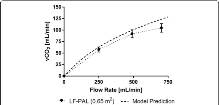
In vitro gas transfer results and model predictions of the 0.65-m2 bundle are
shown in Fig. 2. The CO2 removal rate increased with increasing blood flow rate.
The maximum CO2 removal rate of the LF-PAL was 105 ± 9.2 mL/min at a blood
flow rate of 703 mL/min. The model predicted CO2 removal rates were within
7.7–15.4% of the experimental values.
Pump requirements
The pressure generated by the 0.65 m2LF-PAL device is shown in Fig. 3. The pressure
requirements for operation with the 15.5 Fr dual lumen catheter are also shown in
Fig.3. The 0.65 m2LF-PAL reached the required flow rate range of 250 to 700 mL/min
at impeller rotation rates between 800 and 1800 RPM.
Typical CFD results shown in Fig. 4 for the 0.65 m2LF-PAL demonstrate adequate
In vitro hemolysis
Table1 provides the TIH and NIH values for the 0.65 m2LF-PAL and control circuits.
The rate of PfHb increase over time was linear (R2> 0.90) for the LF-PAL and control circuit. The TIH of the LF-PAL (0.08 ± 0.017 g/100 min), the Pedimag control (0.043 ± 0.0004 g/100 min), and the BP-50 control (0.123 ± 0.013 g/100 min) all significantly dif-fered from one another (p< 0.05). The NIH of the LF-PAL (0.158 ± 0.034 g/100 L), the Pedimag control (0.029 ± 0.003 g/100 L), and the BP-50 control (0.105 ± 0.012 g/100 L) all significantly differed from one another (p< 0.05).
Discussion
Clinical evidence demonstrates that ECCO2R can prevent the need for intubation, allow for protective and ultra-protective lung ventilation, and aid in weaning patients from mechanical ventilation [6,31]. Low-flow ECCO2R devices aim to provide minimally in-vasive, complementary treatment options for ae-COPD patients and patients with moderate ARDS requiring mechanical ventilation. This manuscript details the in vitro
and computational characterization of the LF-PAL for ECCO2R. The LF-PAL is an
inte-grated pump-lung designed to allow simplified patient ambulation while on ECCO2R.
Fig. 2In vitro CO2removal data of the LF-PAL. The model predicted CO2removal rate is also plotted. The
maximum CO2removal rate for the LF-PAL was 105 mL/min. The model predicted the performance
between 7.7–15.4% of the in vitro results
The LF-PAL removed up to 105 mL/min of CO2thereby exceeding the 70 mL/min tar-get and had acceptable hemolysis.
ECCO2R has been applied to a variety of clinical applications including weaning pa-tients from mechanical ventilation, avoiding intubation, and permitting lung protective ventilation [3–5,7,8]. As a result of the range of clinical applications, the CO2removal rate required for ECCO2R is not well defined. Additionally, CO2removal is dependent on the total CO2content of the blood. Hence, the degree of hypercapnia will affect the CO2removal rate at a given blood flow rate. The CO2removal rate, however, will pro-portionally increase with increases in pCO2. The CO2removal target must therefore be reported as a percentage of the rate of metabolically produced CO2or the pCO2of the blood entering the device must be specified. Trahanas et al. provide a review of studies
since 2009 of ECCO2R used in hypercapnic COPD patients and CO2 removal rates
ranged from 80 to 160 mL/min [32]. From this, the authors proposed that an
ambula-tory ECCO2R device must remove at least half of the metabolic CO2. Under
normocap-nic conditions, this would be approximately 100 mL/min. Commercial ECCO2R
devices report removal rates of 20–40% of the metabolically produced CO2 [33–35].
Under normocapnic conditions, these rates are equal to 40–80 mL/min. Based on this
data, we set 70 mL/min as the minimum target CO2removal rate for the LF-PAL at
normocapnia. The LF-PAL exceeded this target at low-flow ECCO2R blood flow rates.
Recent, on-going, and upcoming ECCO2R clinical trials (XTRAVENT [7], REST
(NCT02654327), SUPERNOVA (NCT02282657), and VENT-AVOID (NCT03255057))
should provide a more defined CO2removal target for devices in development.
Concerns with low-flow ECCO2R, compared to mid-flow, include inadequate CO2
re-moval and thrombus formation resulting from low-velocity regions within the device [16]. To mitigate both of these effects, the LF-PAL uses a fiber bundle with a narrow cross sectional area to increase local blood velocities and achieve clinically significant
Fig. 4CFD analysis results for 850 RPM and 0.25 L/min showing (a) predicted pressure (mmHg) throughout the device, (b) fluid velocity (m/s) through the fiber bundle, and (c) near wall velocity magnitudes (m/s) on the device surfaces
Table 1In vitro hemolysis of the LF-PAL
Device Flow Rate NIH TIH
[mL/min] [g/100 L] [g/100 min]
LF-PAL (0.65 m2) 500 0.158 ± 0.034† 0.080 ± 0.017‡
Pedimag control 1500 0.029 ± 0.003† 0.043 ± 0.004‡
BP-50 control 1250 0.105 ± 0.012† 0.123 ± 0.013‡
CO2 removal. In addition, CFD results of the LF-PAL demonstrate uniform blood velocity through the bundle. Other low-flow devices use active mixing technology to
increase local blood velocity in an effort to achieve required CO2 removal rates. The
Hemolung incorporates a rotating core [18], and the ULFED uses rotating impellers
[12]. The drawback is that too high of an increase in blood velocity may detrimentally increase hemolysis. The Hemolung, however, has been used in humans without causing clinically significant hemolysis, though no in vitro hemolysis data are available for com-parison [20].
At least one center has begun ambulating ECCO2R patients to reduce muscle
decon-ditioning and allow for greater physical therapy [3]. A compact device which does not require a saline infusion or vacuum pump, such as the LF-PAL, that could also be worn
would simplify ambulation. Current, clinically used ECCO2R devices are portable, but
are not designed to be worn by the patient [36–40]. The jugular cannulation and cart-ridge design of the Hemolung RAS permits ambulation. The device, however, must res-ide on the roller cart, which houses the required saline infusion and gas sres-ide vacuum pump and does not provide the option to be worn by the patient. The arterio-venous
CO2removal (AVCO2R) device is also under development as a wearable ECCO2R
de-vice [41]. The av-cannulation of the AVCO2R, however, relies on the patient’s
cardio-vascular system to drive blood flow. The CO2removal rate is therefore dependent on
the cardiac function of the patient [42]. The vv-cannulation and pump-driven blood
flow of the LF-PAL allows the clinician greater control of the extracorporeal blood flow
and, in turn, the CO2removal rate. The compact design and dual lumen cannulation of
the LF-PAL lends itself to ambulation.
In this study the hemolysis of the LF-PAL was only evaluated at 500 mL/min. Shear stress within the circuit will increase as blood flow increases and likely result in ele-vated hemolysis at higher blood flow rates. Thus, when the LF-PAL is operated at the maximum blood flow rate, 700 mL/min, the TIH of the LF-PAL will likely increase, as would the TIH of the control circuit when operated at a higher blood flow rate. A limi-tation to the TIH is the lack of an established threshold value correlated to clinically significant hemolysis in vivo. Thus, in vitro studies are limited to a comparative assess-ment between two circuits. Future in vivo studies will thoroughly evaluate if the hemolysis generated by the LF-PAL is clinically significant in addition to any effect the device may have on platelet activation or end organ function.
Conclusion
Evidence demonstrating the benefits of partial CO2 removal by ECCO2R systems in
conjunction with non-invasive ventilation or lung protective ventilation continues to
grow. The LF-PAL provides the CO2 removal benefits of low-flow ECCO2R in a
compact design. Future work will focus on 7-day in vivo studies to further characterize the LF-PAL performance and the effect of the device on the cardiopulmonary system.
Abbreviations
ae-COPD:Acute exacerbation of chronic obstructive pulmonary disease; ARDS: Acute respiratory distress syndrome; av: Arterio-venous; CFD: Computational fluid dynamics; ECCO2R: Extracorporeal CO2removal; ECMO: Extracorporeal
Acknowledgements
Editorial assistance was provided by Ryan Orizondo, PhD.
Funding
This work was supported by National Institutes of Health grants R01HL117637 and R01HL135482 and the McGowan Institute for Regenerative Medicine. Funding for AGM and RGJ was partially provided by an NIH training grant (T32 HL076124) for the University of Pittsburgh Cardiovascular Bioengineering Training Program.
Availability of data and materials
All data used to develop the conclusions of this study are included within the manuscript. No additional data was submitted.
Authors’contributions
AGM conducted experiments and wrote manuscript. GWB completed CFD analysis. All authors contributed to analysis and interpretation of results and approved the final manuscript.
Ethics approval and consent to participate Not applicable
Consent for publication Not applicable
Competing interests
WJF is the head of the scientific advisory board and an equity holder in ALung Technologies.
Publisher’s Note
Springer Nature remains neutral with regard to jurisdictional claims in published maps and institutional affiliations.
Author details
1
Department of Chemical and Petroleum Engineering, University of Pittsburgh, Pittsburgh, USA.2McGowan Institute for Regenerative Medicine, University of Pittsburgh, 3025 East Carson Street, Suite 226, Pittsburgh, PA 15203, USA. 3
Department of Bioengineering, University of Pittsburgh, Pittsburgh, USA.4Computational Fluid Dynamics Group, Center for Advanced Vehicular Systems, Mississippi State University, Mississippi State, MS, USA.5Department of Critical Care Medicine, University of Pittsburgh Medical Center, Pittsburgh, USA.
Received: 24 April 2018 Accepted: 7 September 2018
References
1. Ricard J-D, Dreyfuss D, Saumon G (2003) Ventilator-induced lung injury. Eur Respir J 22:2s–9s
2. Slutsky A, Ranieri V (2013) Ventilator-induced lung injury. N Engl J Med 369:2126–2136.https://doi.org/10.1056/ NEJMra1208707
3. Abrams D, Brenner K, Burkart K et al (2013) Pilot study of extracorporeal carbon dioxide removal to facilitate extubation and ambulation in exacerbations of chronic obstructive pulmonary disease. Ann Am Thorac Soc 10:307–314 4. Bonin F, Sommerwerck U, Lund L, Teschler H (2013) Avoidance of intubation during acute exacerbation of chronic
obstructive pulmonary disease for a lung transplant candidate using extracorporeal carbon dioxide removal with the Hemolung. J Thorac Cardiovasc Surg 145:e43–e44
5. Kluge S, Braune S, Engel M et al (2012) Avoiding invasive mechanical ventilation by extracorporeal carbon dioxide removal in patients failing noninvasive ventilation. Intensive Care Med 38:1632–1639
6. Terragni P, Del Sorbo L, Mascia L et al (2009) Tidal volume lower than 6 ml/kg enhances lung protection. Anesthesiology 111:826–835
7. Bein T, Weber-Carstens S, Goldmann A et al (2013) Lower tidal volume strategy (~3 ml/kg) combined with extracorporeal CO2 removal versus“conventional”protective ventilation (6 ml/kg) in severe ARDS. Intensive Care Med 39:847–856
8. Zimmerman M, Bein T, Arlt M et al (2009) Pumpless extracorporeal interventional lung assist in patients with acute respiratory distress syndrome: a prospective pilot study. Crit Care 13:R10
9. Lund L, Federspiel W (2013) Removing extra CO2 in copd patients. Curr Respir Care Rep 2:131–138
10. Karagiannidis C, Brodie D, Strassmann S et al (2016) Extracorporeal membrane oxygenation: evolving epidemiology and mortality. Intensive Care Med 42:889–896
11. Arazawa D, Kimmel J, Federspiel W (2015) Kinetics of CO2 exchange with carbonic anhydrase immobilized on fiber membranes in artificial lungs. J Mater Sci Mater Med 26:1–8
12. Jeffries R, Lund L, Frankowski B, Federspiel W (2017) An extracorporeal carbon dioxide removal device operating at hemodialysis blood flowrates. Intensive Care Med Exp 5:41
13. Zanella A, Castagna L, Salerno D et al (2015) Respiratory electrodialysis: a novel, highly efficient extracorporeal CO2 removal technique. Am J Respir Crit Care Med 192:719–726
14. Zanella A, Mangili P, Giani M et al (2014) Extracorporeal carbon dioxide removal through ventilation of acidified dialysate: an experimental study. J Heart Lung Trans 33:536–541
15. Abrams D, Roncon-Albuquerque R, Brodie D (2015) What’s new in extracorporeal carbon dioxide removal for COPD? Intensive Care Med 41:906–908
17. Madhani S, Frankowski B, Federspiel W (2017) Fiber bundle design for an integrated wearable artificial lung. ASAIO J.
https://doi.org/10.1097/MAT.0000000000000542
18. ALung Technologies, Inc (2015) How activmix technology enhances CO2 removal in the hemolung RAS. HL-PL-0321_RA 19. Akkanti B, Rajagopal K, Patel K et al (2017) Low-flow extracorporeal carbon dioxide removal using the Hemolung
Respiratory Dialysis system® to facilitate lung-protective mechanical ventilation in acute respiratory distress syndrome. J Extracorpor Technol 49:112–114
20. Burki N, Mani R, Herth F et al (2013) A novel extracorporeal CO2 removal system: results of a pilot study of hypercapnic respiratory failure in patients with COPD. Chest 143:678–686
21. Parilla F, Bergesio L, Aguirre-Bermeo H et al (2015) Ultra-low tidal volumes and extracorporeal carbon dioxide removal (Hemolung® RAS) in ards patients. a clinical feasibility study. Intensive Care Med Exp 3:A7
22. Madhani S, Frankowski B, Burgreen G et al (2017) In vitro and in vivo evaluation of a novel integrated wearable artificial lung. J Heart Lung Transplant 36:806-811
23. Svitek RG, Federspiel WJ (2008) A mathematical model to predict co2 removal in hollow fiber membrane oxygenators. Ann Biomed Eng 36:992–1103
24. Hines A, Maddox R (1985) Mass transfer: fundamentals and applications. Prentice Hall, Englewood Cliffs 25. Pacella H, Eash H, Frankowski B, Federspiel W (2011) Darcy permeability of hollow fiber bundles used in blood
oxygenation devices. J Membr Sci 382:238–242
26. American National Standard (2009) Cardiovascular Implants and Artificial Organs - Blood-Gas Exchangers (Oxygenators) 7199 27. Svitek R, Frankowski B, Federspiel W (2007) Evaluation of a pumping assist lung that uses a rotating fiber bundle. ASAIO
J 51:773–778
28. Medtronic (2008) Medtronic minimax plus, instructions for use
29. Svitek R, Smith D, Magovern J (2007) In vitro evaluation of the tandem heart pediatric centrifugal pump. ASAIO J 53: 747–753
30. Medtronic (2017) Find your ideal cannulae
31. Combes A, Pesenti A, Ranieri V (2017) Is extracorporeal circulation the future of acute respiratory distress syndrome management? Am J Respir Crit Care Med 195:1161–1170
32. Trahanas J, Lynch W, Bartlett R (2016) Extracorporeal support for chronic obstructive pulmonary disease: a bright future. J Intensive Care Med.https://doi.org/10.1177/0885066616663119
33. ALung Technologies, Inc (2015)“Low-flow”versus“mid-flow”extracorporeal CO2 removal: a review of clinical performance and device efficiency
34. Ruberto F, Pugliese F, D’Alio A et al (2009) Extracorporeal removal CO2 using a venovenous, low-flow system (Decapsmart) in a lung transplanted patient: a case report. Transplant Proc 41:1412–1414
35. Terragni P, Birocco A, Faggiano C, Ranieri V (2010) Extracorporeal CO2 removal. Contrib Nephrol 165:185–196 36. ALung Technologies, Inc (2012) Hemolung RAS: the first fully-integrated respiratory dialysis system 37. Maquet Getinge Group (2010) Pump assisted lung protection cardiohelp system
38. Maquet Getinge Group (2015) HLS Set Advanced
39. Novalung A Xenios Company (2015) iLA activve system platform custom-tailored extrapulmonary lung support 40. Seiler F, Trudzinski F, Hennemann T et al (2017) The Homburg lung: efficacy and safety of a minimal-invasive
pump-driven device for veno-venous extracorporeal carbon dioxide removal. ASAIO J 63:659–665
41. Wang D, Lick S, Campbell K et al (2005) Development of ambulatory arterio-venous carbon dioxide removal (AVCO2R): the downsized gas exchanger prototype for ambulation removes enough CO2 with low blood resistance. ASAIO J 51: 385–389