Introduction
Stem cells are a quintessential key to proper behavior of homeostatic processes. Th ey are often thought of as the solution to a wide range of human conditions, with the ability to rescue malfunctioning or non-functioning organs and tissues. However, there is increasing evidence that stem cells can play a central role in disease. Most recently, stem cells have been implicated in cancer after not responding to homeostasic controls, such as
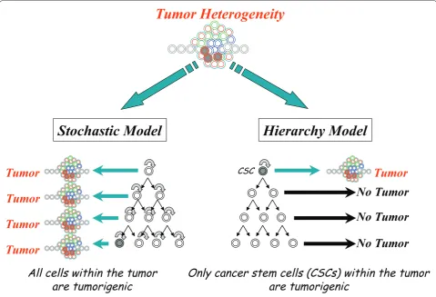
proliferation and diff erentiation [1]. Th ere are currently two models for the cellular origin of cancer and their lines are becoming blurred as research in the area con-tinues (Figure 1). Th e hierarchy model identifi es a limited number of tumor cells, called cancer stem cells (CSCs), capable of initiating a heterogenous tumor, whereas the stochastic model describes a probability that specifi c events in a tumor cell population have the potential to transform any tumor cell into a tumor-initiating cell [2,3]. Cancer is the second leading cause of death in the US, and colorectal cancer is the third most common cancer among men and women and accounts for 10% of all new cancers [4]. Colorectal cancer has been suggested to follow a hierarchy or CSC model, being initiated by a CSC [5], although not all CSCs are derived from stem cells. It is important to note that the CSC hypothesis is still a model for how cancer arises and provides resistance
to therapy. Th is model is supported by experimental
evidence [2,6-8] but will need further experimental support, particularly in the context of human cancer. Further more, a report by Quintana and colleagues [9] in human melanoma suggests the possibility that not all cancers follow a CSC model. With a severely immuno-compromised mouse, 25% of human tumor cells were tumorigenic, suggesting that tumorigenic cells are more common in some human cancers than previously thought [9,10] and may correspond to what is expected in a stochastic model more than in a hierarchy model. However, the impact and interpretation of studies such as these are still up for discussion [8]. Th is review will focus on the most recent evidence for the existence of CSCs and their implication in tumorigenesis, metastases, recurrence and therapy resistance using colon cancer as a model system.
Obedient versus defi ant stem cells
Self-control and homeostasis
A myriad of cells contribute to the normal function and maintenance of adult tissues. Some cells, such as goblet cells that produce mucus in the colon, play functional roles in specifi c tissues. Th ese altruistic cells are terminally diff erentiated and will die serving the tissue. Other rare and undiff erentiated cells, called stem cells,
Abstract
Stem cells maintain homeostasis in adult tissues via self-renewal and generation of terminally
diff erentiated cells. Alterations in this intricate balance can result in disease. It has become increasingly evident that cancer can be initiated at the level of stem cells. Therefore, understanding what causes stem cells to become cancerous may lead to new therapeutic approaches. Multiple signaling pathways ultimately aff ect stem cell survival and proliferation, thus maintaining homeostasis in the gut. Changes in these pathways could perturb normal stem cell behavior, leading to cancerous stem cells. In addition, cancerous stem cells show resistance to current therapies and may lead to a dangerous selection process resulting in recurrence and metastasis. Genomic instability, the driving force of mutation and resistance, may give cancerous stem cells an adaptive advantage, especially when subjected to cancer therapies. Targeting the unique characteristics of cancerous stem cells to promote either terminal diff erentiation or destruction would eff ectively eradicate cancer and improve patient care and survival.
© 2010 BioMed Central Ltd
Cancerous stem cells: deviant stem cells with
cancer-causing misbehavior
Julie M Chandler* and Eric Lagasse*
R E V I E W
*Correspondence: juc24@pitt.edu or lagasse@pitt.edu
McGowan Institute for Regenerative Medicine, Department of Pathology, University of Pittsburgh Medical School, 450 Technology Drive, Suite 300, Pittsburgh, PA 15219, USA
are responsible for replenishing the pool of diff erentiated cells while maintaining an adequate supply of themselves through the process of self-renewal. Stem cells can divide asymmetrically to produce one daughter cell that is more committed to a specifi c cell lineage, a transit-amplifying (TA) cell, and one that retains stem-ness. TA cells have a limited life span and self-renewal potential while re-populating the diff erentiated cells of the tissue.
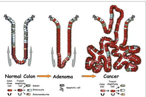
Th e colon is organized in this hierarchical fashion and its epithelium is renewed every 5 days in humans [11]. In this very dynamic process lies a complex collection of epithelial cell lineages along with an intricate set of molecular mechanisms to maintain order. To preserve tissue function, the colon is structurally organized in an elegant network of invaginations, termed crypts, which aid in the absorption of water and vitamins. Stem cells of the colon are located at the base of these crypts and produce epithelial cells that are committed to three diff erent cell lineages. Th ese diff erentiated cells are the absorptive enterocytes, mucus-secreting goblet cells, and hormonal enteroendocrine cells that will migrate up the crypt wall to form the colon (Figure 2).
Defi ance and altered management
Since one of the hallmarks of stem cells is their ability to self-renew and diff erentiate, investigators were prompted to explore the similarities and diff erences that exist between normal stem cell maintenance of tissues and organs and the uncontrolled proliferation of cancer [2]. In essence, tumors can be viewed as small aberrant organs containing a hierarchy of progenitor cells and diff erentiated cells (Figure 2, for example). Albeit dys-func tional when compared to physiologically dys-functioning organs, they maintain their own abnormal proliferation and survival mechanisms [7]. If tumors are indeed aberrant organs, then there is a program by which the system is controlled, however loosely it might be. Th ere-fore, in the cellular hierarchy of a tumor, there is a diff erentiation mechanism from tumor stem cell to tumor progenitor cell to mature tumor cell which ends in apoptosis and turnover [6]. Abnormal cellular behavior in this tightly controlled system can occur via genetic alterations, such as tumor suppressor loss or gene destabilization, which result in incremental neoplastic gains and disruption of the homeo static system [12].
Figure 1. Model for cellular origin of cancer.Two models are proposed to explain the cellular heterogeneity in cancer: the stochastic model and the hierarchy model. In the stochastic model, every tumor cell can stochastically generate a tumor. In the hierarchy model, only the cancer stem cells (CSCs) will generate tumors.
Stochastic Model
Hierarchy Model
Tumor Heterogeneity
Tumor
Tumor
Tumor
Tumor Tumor
No Tumor
No Tumor
No Tumor
All cells within the tumor
are tumorigenic
Only cancer stem cells (CSCs) within the tumor
are tumorigenic
With these alterations, resulting in a loosely controlled system, cancer will eventually prevail. If the alterations can be restricted and coerced to force cells to terminally diff erentiate and die, the invading neoplastic tissue will cease to exist.
To determine what makes a normal stem cell mis-behave, it must fi rst be determined what makes them behave normally. Wnt signaling is one of the driving forces in crypt formation and maintenance of the colon [13]. When Wnt proteins bind Frizzled/LRP recep tors, the canonical Wnt pathway ensues. Th is involves the disruption of the destruction complex that sequesters beta-catenin in the cytoplasm with axin, adenomatous polyposis coli (APC), and glycogen synthase kinase 3 (GSK3) beta. Th is complex marks beta-catenin for degra-da tion and leads to a decrease in Wnt targeted transcrip-tion. When the complex is disrupted, beta-catenin can enter the nucleus and, in concert with transcription factor 4 (TCF4), eff ect transcription of Wnt targets [14]. Th e dysregulation of the Wnt pathway and its role in self-renewal as well as diff erentiation have been shown to be required for the development of cancer [13]. Deviance in any of these factors will tip the balance toward cancer.
Furthermore, recent in vivo studies in rodents have indi-cated that colonic crypts are derived from Lgr5+
(leucine-rich repeat-containing G protein-coupled receptor 5-positive) crypt base columnar cells. Lineage-tracing assays using Lgr5-lacZ mice indicated that the rare crypt base columnar cells represent the stem cells of the colon [15]. Th e Lgr5 gene is a Wnt target and is repressed upon inhibition of the pathway [15]. Th erefore, if aberrantly active, the Wnt pathway could result in an expanded progenitor population and colon cancer (Figure 1).
One of the most commonly mutated genes in human colorectal cancer is Apc, which has been implicated in both the sporadic and inherited forms of this cancer [16]. As discussed above, this is one of the players involved in the destruction of beta-catenin. Upon disruption of this protein interaction, beta-catenin is free to roam into the nucleus, where it upsets gene transcription via inter-actions with transcription factors and subsequently disrupts homeostasis. Th is concept was nicely
demon-strated with Lgr5+ colon stem cells when Lgr5-EGFP
mice with fl oxed Apc were treated with tamoxifen. Th e
resulting deletion of the Apc gene in the stem cell
compartment supported the idea that stem cell-specifi c
Figure 2. Model for colon cancer initiated by stem cells.Colon stem cells are located at the base of the crypt in normal colon and will diff erentiate while moving up the crypt in about 5 days. Adenoma will develop upon deregulation of stem cell homeostasis. Upon further neoplastic injuries, stem cells will transform into cancerous stem cells (CSCs) with some limited ability to diff erentiate.
Apoptotic cell Colon
Stem Cell
Transit Amplifying
Cell Goblet Enterocyte
Enteroendocrine
Colon CSC
Transit Amplifying
Cell
loss of Apc will result in a progressive neoplasia and eventually cancer [17]. Since some of these transformed cells had retained their Lgr5 expression, Lgr5+ tumor
cells could also be considered true CSCs and the Lgr5 epitope as a possible CSC marker (Figure 2).
Parallel to the studies investigating Lgr5 and its role as a stem cell marker of the intestine was the investigation of Bmi1. Bmi1 is a transcriptional receptor in the polycomb group (PcG) gene family and is involved in stem cell maintenance, proliferation, diff erentiation to more committed cells, malignant transformation, and biologic aggressiveness of human carcinomas [18,19]. It is a member of the PcG proteins and, more specifi cally, the polycomb-repressing complex 1 (PRC1), indicating a role in maintaining chromatin silencing [20]. As expected, any factor involved in gene silencing has the capacity to exponentially misbehave. If the genes aff ected are involved in maintaining control, their silencing could perturb the structured environment of a tissue. Bmi1 transcriptionally regulates the INK4a locus encoding p16INK4a and p19ARF tumor suppressors [21]. By
suppres-sing a tumor suppressor, a double-negative results in a positive for cancer growth as well as for the expansion of cells with stem cell properties. Th erefore, Bmi1 became an interesting endeavor for the cancer stem cell biologist. As with Lgr5, a lacZ reporter assay identifi ed Bmi1 as the source for all diff erentiated cells of the mouse intestine. In addition, its ablation led to crypt loss and adenoma formation upon stable beta-catenin expression in these cells [22]. Th e relation of Bmi1 to cancer was further enhanced by expression profi les, which showed that increased Bmi1 expression correlates with poor clinical survival in patients with colon cancer [23].
Bmi1 also gives a good example of how epigenetics can infl uence the initiation or propagation of cancer or both. Epigenetic modifi cations can result in altered signaling and function, thus contributing to the formation or progression of cancer or both [24]. Being a PcG protein, Bmi1 coordinates the regulation of histone modifi cation and methylation, adding a layer of epigenetic modifi -cation to its already existing regulatory role. As seen in breast cancer, Bmi1 overexpression can block terminal diff erentiation, leading to an expansion of cells capable of self-renewing [19]. Normally, Bmi1 is lowly expressed in diff erentiated cells and highly expressed in stem cells. Th e increased expression of PcG proteins in metastatic breast and prostate cancer occurs in a pool of cancer cells with stem cell-like properties, indicating that this alteration may occur in the CSC population [25].
Although the studies on Lgr5 and Bmi1 suggest that the transformation of stem cells from obedient to deviant cells may lead to tumorigenesis, complete molecular mechanisms have yet to be resolved. Understanding the steps by which stem cell homeostasis is lost and cancer is
initiated would pave the way for possible cancer treatments. Th e Hedgehog-Gli (HH-Gli) signaling path-way is proposed as a mechanism through which normal stem cell maintenance can be dysregulated, leading to colon cancer [26]. In an active HH pathway, Patched is blocked, releasing Smoothened to activate Gli. Although colon cancers and their stem cells of all stages have active HH-Gli signaling, an increase in HH-Gli signaling is seen in metastases. Th is may indicate that while APC/beta-catenin signaling disruption may be the fi rst of many hits toward colon cancer, HH-Gli may be an added push toward deviance [27].
In addition to these pathways, other signaling mecha-nisms involved in normal stem cell function and homeostasis may contribute to the misbehavior evident in cancer and its stem cells. Th ese include the phospha-tase and tensin homolog (PTEN) and Notch pathways [28]. PTEN is a tumor suppressor whereas the Notch pathway regulates cell fate decisions [29,30]. PTEN not only has been implicated in the signaling of CSCs themselves but also has been found in stromal cells of the
surrounding tumor microenvironment. Th e genetic
inactivation of Pten in stromal fi broblasts was shown to accelerate the initiation, progression, and malignant transformation of mammary epithelial tumors in mice, a phenotype that was correlated with a specifi c PTEN signature in patients with breast cancer [31].
Identifying the culprit in human cancer
Unfortunately, identifying CSCs in human cancers has remained challenging. CD133, a cell surface marker for many normal stem cells, has been thought of as a CSC marker in human cancers, and particularly in human colon cancer, with some controversy about whether it marks the true stem cell population. Using the AC133 antibody to identify CD133 expression, Ricci-Vitiani and colleagues [32] and O’Brien and colleagues [33] both
showed in 2007 that it is the CD133+ population of
human colon cancer cells which initiates the tumor whereas the CD133− population does not. It seems that
such a strong correlation would evoke some sort of function of CD133 to the cancer-initiating cells. However, upon siRNA (short interfering RNA) knockdown, there was no impact on proliferation, migration, colony formation, or invasion, indicating that while CD133 may serve as a prognostic marker of colon cancer and its stem cells, it is not a feasible target to eradicate the trouble-some cells [34]. Importantly, the CD133+ popu lation of
anti body used to identify CD133, clone AC133, recognizes a glycosylated epitope on the human CD133 antigen. Furthermore, AC133 is not present on all colon cancers and is lost upon diff erentiation whereas CD133 is not [35-37].
Th e epithelial cell adhesion molecule (EpCAM) is
another claimed CSC marker that has been implicated in stem cell signaling via the Wnt pathway [38]. While EpCAM presence is abundant in the membrane of normal epithelial tissues, it is prone to cleavage in cancer tissue, making it diffi cult to use as a target [39]. CD44 is also described as a molecule whose splicing variants were shown to be diff erentially expressed between normal stem cells and CSCs [40]. In combi nation with CD133, CD44 and CD166 may provide a better way to identify the CSC population. However, this makes therapeutic targeting a challenge because of the heterogenous expres-sion of these markers. Colorectal carcinomas assayed for expression of these markers found that CD133 held the strongest single-marker adverse correlation with patient survival. While CD166 and CD44 alone may have little or no correlation with survival, their combined analysis with CD133 may allow the separation of low-, intermediate-, and high-risk colorectal cancers [41].
Control or destroy?
So how do we control the CSCs? To date, colon cancer treatments have often resulted in recurrence and meta-stases. One reason may be that CSCs are more resistant to chemotherapy treatments than their more diff eren-tiated progeny are, allowing recurrence and metastases. Th erefore, therapies that target either the terminal diff erentiation or destruction of CSCs must be developed [28,42,43]. Th ere have been great eff orts to treat acute promyelocytic leukemia with all-trans retinoic acid (ATRA), which acts to diff erentiate leukemic pro myelo-cytes to normal, terminally diff erentiated neutrophils [44]. It is thought that such a diff erentiation therapy may be benefi cial in the emerging population of solid cancers that arise from malfunc tioning CSCs. Th e diff erentiation theory would cause CSCs to lose their dangerous edge: the ability to self-renew [7]. Another approach would be to destroy CSCs directly by harnessing the pathways used for CSC mainte nance and survival [45,46].
What do we know?
A recent study of colon cancer examined FOLFOX (folinic acid, 5-fl uorouracil plus oxaliplatin), a widely used treatment for colon cancer [47]. In that study, initial treatment of colon cancer cells enriched the population for CD133+, CD44+, and/or CD166+ cells with increased
levels of epidermal growth factor receptor-positive (EGFR+). Upon subsequent treatment of the surviving
cells (the CSCs) with curcumin or curcumin plus
FOLFOX, a reduction in CSCs was seen. Although the mechanism is not fully understood, it was shown to increase methylation of the EGFR promoter. Th is hyper-methylation, via changes in the level of DNA methyl-transferase 1, decreases the expression of EGFR, stabiliz-ing the chromatin and preventstabiliz-ing the bindstabiliz-ing of trans-cription factors [47]. While this may be a desired eff ect, care must be taken to investigate possible detri mental eff ects of this type of treatment. Hyper methy lation is also known to inactivate the transcription factor p16INK4a [47].
As discussed previously, increased Bmi1 expression leads to inactivation of p16INK4a. Th erefore, a therapy that may
promote hypermethylation and gene silencing may cause additional imbalances in the already mischievous CSCs.
Recent drug screenings revealed salinomycin as a specifi c inhibitor of CSCs. Th is study focused on breast
cancer and used CD44high/CD24low as the molecular
profi le of CSCs. Th ey found that the number of these cells decreased with salinomycin treatment as compared with a current treatment of paclitaxel. Surprisingly, paclitaxel actually increased the number of CSCs. In addition, there was a decrease in tumor sphere formation as well as decreased metastases over paclitaxel [48]. Although this study used breast cancer as a model, colon cancer has been shown to be resistant to paclitaxel. Modifi cations that include the use of a mitogen-activated protein kinase (MAPK) inhibitor in combination with paclitaxel to enhance apoptosis of colon cancer cells have been suggested [49]. In light of the results in breast cancer, it may be benefi cial to investigate the selection of colon CSCs by treatment with paclitaxel. Such a treatment may give a survival advantage that results in a more invasive and aggressive cancer. Th is is especially true given that salinomycin has been shown to induce apoptosis as well as overcome apoptotic resistance in breast cancer cells [50]. Th ere is a strong connection between colon and breast cancer because they are both epithelial cancers that resemble the tissue of origin, even at metastatic sites.
results were found with small-molecule and RNA knock-down studies showing that the inhibition of Notch leads to increased apoptotsis, decreased self-renewal, and increased secretory cell lineage diff erentiation of CSCs in colon cancer [54]. In yet another study focusing on the Notch pathway, inhibition of Delta-like ligand 4 (DLL4) inhibited the expression of Notch targets and reduced tumor-initiating cell frequency [55]. Th ese targeted therapies may enhance the effi cacy of chemo therapeutic drugs when used in sequence or combination [46]. However, one must realize that these are mecha nisms shared by normal intestinal stem cells and may lead to toxicity.
While there are treatments that seem to reduce the number of CSCs, two problems remain. Just reducing the population of misbehaving cells does not solve the problem. As soon as the treatment ceases, these cells can again fl ourish. Unfortunately, thus far, there has been no demonstration that targeting CSCs will improve the outcome of patients with cancer.
Peer pressure: the mastermind
If stem cells are indeed the culprit behind colon cancer and alterations in their behavior are the driving force, what causes the alterations? Th e answer may be genomic instability (GI), which could explain why colon cancer remains such a diffi cult disease to treat despite the breadth of knowledge obtained over recent years. GI occurring in stem cells may be one of the initiating peer pressure events that drive normal stem cells to go awry and become CSCs [56-58]. If it is not an initiating event, GI may be the result of other alterations, thus steering the stem cell population into cancer. Chromosomal aberrations and DNA repair mutations aff ecting migra-tion and proliferamigra-tion in these cells lead to metastases and cancers that are more aggressive [59]. In addition to carcinogenesis and disease progression, GI may be responsible for resistance to current therapies making colon cancer such a devastating disease [60]. As men-tioned previously, current therapies may eff ectively treat the bulk tumor but, in eff ect, could select for the more tumorigenic CSCs. If this is the cell population that harbors GI, these cells are more likely to evade the toxic eff ects of cancer drugs by adaptations that only their instability has allowed. In essence, natural selection selects not only the fi ttest organisms but also the fi ttest cells [5,36]. After the selective therapy has occurred, the population of the fi ttest cells, which includes those with enhanced abilities to migrate and proliferate, expands. Even with a change in treatment regimen, GI continues to perpetuate the peer pressure to become adaptively deviant, resulting in a cancer without restraint.
Two types of GI are present in colon cancer: micro-satellite instability (MSI) and chromosomal instability (CIN). MSI refers to genetic or epigenetic alterations or
both in DNA mismatch repair mechanisms, including MSH2 and MLH1, which can lead to loose control of DNA metabolism and cell cycle control and result in carcinogenesis [56,57]. Th is results in a nucleotide mutation rate that is two to three orders of magnitude greater than that of normal cells [56]. MSI in tumors may
determine site-related diff erences when comparing
proximal and distal colon tumors; therefore, MSI-bearing tumors are unique and may be indicative of a class of tumors more prone to genetic alterations and adaptations [61]. Surprisingly, MSI tumors are associated with a better prognosis and are less prone to metastasis than their microsatellite-stable (MSS) counterparts [62]. Th e mechanism behind this phenomenon in MSI involves a mutation in transforming growth factor-beta receptor 2 and the resulting inability to undergo epithelial-mesen-chymal-transition (EMT) as seen with MSS tumors [63]. EMT is often associated with the ability of cancers to metastasize and therefore leads to a poorer prognosis [64]. Such results indicate that GI may not always support cancer progression and the use of MSI status for prognosis or therapy response or both has the potential to improve patient care [58,65]. In addition, the information obtained from MSI tumors may unveil ways to harness MSS tumors.
While colon cancers with MSI make up only a small portion of cases, most of the remaining colon cancers exhibit CIN involving an increased tendency for gain or loss of chromosomes in part or whole [66]. While seemingly minor mutations resulting from MSI have been linked to colon cancer, CIN has been linked to the previously mentioned Apc mutations commonly seen in colon cancer. Prior to the beta-catenin dysregulation that results in increased self-renewal, altered APC-mediated microtubule regulation may be the fi rst point of deviance. Th e mitotic infi delity at this point places CIN earlier in carcinogenesis progression that may determine the future of a particular colon cancer and has been correlated with poor prognosis [67,68]. As a consequence of APC deletion, the calcium gradient present in the crypts of the colon is perturbed [69]. Normally, Ca+2
concentrations increase as cells migrate up the crypt. Th is is paralleled by an increased expression of the calcium-sensing receptors (CaSRs) and consequential E-cadherin expression. Th is normal process drives terminal diff erentiation and apoptosis at the crypt mucosal surface. In cases in which APC is lost, an expansion of TA cells below the Ca+2 gradient
change leads to a pool of proliferating cells that cannot express CaSR, thus perpetuating or even accelerating the development of disease [70,71]. While it seems as though colon cancer will prevail, targeting its GI could be an Achilles heel for treatment.
may provide CSCs with the ability to evade treatment [68]. If CSCs can be targeted by approaches that are more specifi c, the fact that GI is also present in these CSCs will make them moving targets [60]. As a consequence, both the specifi c targeting of CSCs and the inhibition of GI appear to be necessary for a successful approach to eradicating cancer in patients.
Conclusions
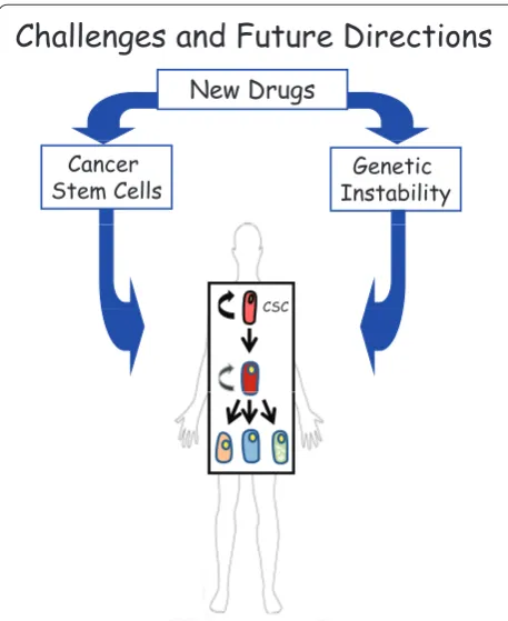
In colon cancer, like many other cancers, it seems that Mother Nature used two of its best creations, stem cells and GI, to produce tumor cells with survival skills unmatched by any other cells. Pathways such as Wnt, Notch, and HH may be potential therapeutic targets to force control of CSCs, but owing to GI, these approaches may subsequently fi nd resistance. Th erefore, treatments that are specifi c to both CSCs and GI may be the only solution to this diffi cult problem (Figure 3). Only when all CSCs are eradicated via either destruction or terminal diff eren tiation will cancer be truly cured. However, much still needs to be understood about normal stem cell versus CSC function and maintenance before eff ective treat ments can be generated for such defi ant cellular mechanisms.
Abbreviations
APC, adenomatous polyposis coli; CaSR, calcium-sensing receptor; CIN, chromosomal instability; CSC, cancer stem cell or cancerous stem cell; EGFR, epidermal growth factor receptor; EMT, epithelial-mesenchymal-transition; EpCAM, epithelial cell adhesion molecule; FOLFOX, folinic acid, 5-fl uorouracil plus oxaliplatin; GI, genomic instability; HH, Hedgehog; Lgr5, leucine-rich repeat-containing G protein-coupled receptor 5; MSI, microsatellite instability; MSS, microsatellite-stable; PcG, polycomb group; PTEN, phosphatase and tensin homolog; TA, transit amplifying.
Competing interests
The authors declare that they have no competing interests.
Acknowledgments
This work was supported by the Jeannik M Littlefi eld-American Association for Cancer Research (EL) and Cellular Approaches to Tissue Engineering and Regeneration-National Institutes of Health grant support 2 T32 EB001026-06) (JC). The authors thank Lindsey Boone and Aaron DeWard for their comments.
Published: 20 May 2010
References
1. Buick RN, Pollak MN: Perspectives on clonogenic tumor cells, stem cells, and oncogenes.Cancer Res 1984, 44:4909-4918.
2. Reya T, Morrison SJ, Clarke MF, Weissman IL: Stem cells, cancer, and cancer stem cells.Nature 2001, 414:105-111.
3. Dick JE: Looking ahead in cancer stem cell research.Nat Biotechnol 2009, 27:44-46.
4. Jemal A, Siegel R, Ward E, Hao Y, Xu J, Thun MJ: Cancer statistics, 2009.
CA Cancer J Clin 2009, 59:225-249.
5. Odoux C, Fohrer H, Hoppo T, Guzik L, Stolz DB, Lewis DW, Gollin SM, Gamblin TC, Geller DA, Lagasse E: A stochastic model for cancer stem cell origin in metastatic colon cancer.Cancer Res 2008, 68:6932-6941.
6. Al-Hajj M, Clarke MF: Self-renewal and solid tumor stem cells.Oncogene
2004, 23:7274-7282.
7. Sell S: Stem cell origin of cancer and diff erentiation therapy.Crit Rev Oncol Hematol 2004, 51:1-28.
8. Shackleton M, Quintana E, Fearon ER, Morrison SJ: Heterogeneity in cancer: cancer stem cells versus clonal evolution.Cell 2009, 138:822-829. 9. Quintana E, Shackleton M, Sabel MS, Fullen DR, Johnson TM, Morrison SJ:
Effi cient tumour formation by single human melanoma cells.Nature 2008, 456:593-598.
10. Schatton T, Murphy GF, Frank NY, Yamaura K, Waaga-Gasser AM, Gasser M, Zhan Q, Jordan S, Duncan LM, Weishaupt C, Fuhlbrigge RC, Kupper TS, Sayegh MH, Frank MH: Identifi cation of cells initiating human melanomas.
Nature 2008, 451:345-349.
11. Potten CS, Kellett M, Roberts SA, Rew DA, Wilson GD: Measurement of in vivo
proliferation in human colorectal mucosa using bromodeoxyuridine.Gut
1992, 33:71-78.
12. Vogelstein B, Kinzler KW: Cancer genes and the pathways they control.
Nat Med 2004, 10:789-799.
13. Korinek V, Barker N, Moerer P, van Donselaar E, Huls G, Peters PJ, Clevers H: Depletion of epithelial stem-cell compartments in the small intestine of mice lacking Tcf-4.Nat Genet 1998, 19:379-383.
14. Clevers H: Wnt/beta-catenin signaling in development and disease.Cell
2006, 127:469-480.
15. Barker N, van Es JH, Kuipers J, Kujala P, van den Born M, Cozijnsen M, Haegebarth A, Korving J, Begthel H, Peters PJ, Clevers H: Identifi cation of stem cells in small intestine and colon by marker gene Lgr5.Nature 2007, 449:1003-1007.
16. Kwong LN, Dove WF: APC and its modifi ers in colon cancer.Adv Exp Med Biol
2009, 656:85-106.
17. Barker N, Ridgway RA, van Es JH, van de Wetering M, Begthel H, van den Born M, Danenberg E, Clarke AR, Sansom OJ, Clevers H: Crypt stem cells as the cells-of-origin of intestinal cancer.Nature 2009, 457:608-611.
18. Park IK, Morrison SJ, Clarke MF: Bmi1, stem cells, and senescence regulation.
J Clin Invest 2004, 113:175-179.
19. Pietersen AM, Evers B, Prasad AA, Tanger E, Cornelissen-Steijger P, Jonkers J, van Lohuizen M: Bmi1 regulates stem cells and proliferation and diff erentiation of committed cells in mammary epithelium.Curr Biol 2008, 18:1094-1099.
Figure 3. Potential approach to cure cancer. A combined approach to target both the tumor-initiating cell (cancer stem cell or CSC) and its driving force (genomic instability) is proposed for the best outcome in patients with cancer.
Challenges and Future Directions
New Drugs
Genetic Instability Cancer
Stem Cells
20. Valk-Lingbeek ME, Bruggeman SW, van Lohuizen M: Stem cells and cancer; the polycomb connection.Cell 2004, 118:409-418.
21. Jacobs JJ, Kieboom K, Marino S, DePinho RA, van Lohuizen M: The oncogene and Polycomb-group gene bmi-1 regulates cell proliferation and senescence through the ink4a locus.Nature 1999, 397:164-168. 22. Sangiorgi E, Capecchi MR: Bmi1 is expressed in vivo in intestinal stem cells.
Nat Genet 2008, 40:915-920.
23. Du J, Li Y, Li J, Zheng J: Polycomb group protein Bmi1 expression in colon cancers predicts the survival.Med Oncol 2009 Dec 1. [Epub ahead of print]. 24. Feinberg AP: Phenotypic plasticity and the epigenetics of human disease.
Nature 2007, 447:433-440.
25. Mathews LA, Crea F, Farrar WL: Epigenetic gene regulation in stem cells and correlation to cancer.Diff erentiation 2009, 78:1-17.
26. Gulino A, Ferretti E, De Smaele E: Hedgehog signalling in colon cancer and stem cells.EMBO Mol Med 2009, 1:300-302.
27. Varnat F, Duquet A, Malerba M, Zbinden M, Mas C, Gervaz P, Ruiz i Altaba A: Human colon cancer epithelial cells harbour active HEDGEHOG-GLI signalling that is essential for tumour growth, recurrence, metastasis and stem cell survival and expansion.EMBO Mol Med 2009, 1:338-351. 28. Massard C, Deutsch E, Soria JC: Tumour stem cell-targeted treatment:
elimination or diff erentiation.Ann Oncol 2006, 17:1620-1624.
29. Myers MP, Pass I, Batty IH, Van der Kaay J, Stolarov JP, Hemmings BA, Wigler MH, Downes CP, Tonks NK: The lipid phosphatase activity of PTEN is critical for its tumor supressor function.Proc Natl Acad Sci U S A 1998,
95:13513-13518.
30. Artavanis-Tsakonas S, Rand MD, Lake RJ: Notch signaling: cell fate control and signal integration in development.Science 1999, 284:770-776. 31. Trimboli AJ, Cantemir-Stone CZ, Li F, Wallace JA, Merchant A, Creasap N,
Thompson JC, Caserta E, Wang H, Chong JL, Naidu S, Wei G, Sharma SM, Stephens JA, Fernandez SA, Gurcan MN, Weinstein MB, Barsky SH, Yee L, Rosol TJ, Stromberg PC, Robinson ML, Pepin F, Hallett M, Park M, Ostrowski MC, Leone G: Pten in stromal fi broblasts suppresses mammary epithelial tumours.Nature 2009, 461:1084-1091.
32. Ricci-Vitiani L, Lombardi DG, Pilozzi E, Biff oni M, Todaro M, Peschle C, De Maria R: Identifi cation and expansion of human colon-cancer-initiating cells.
Nature 2007, 445:111-115.
33. O’Brien CA, Pollett A, Gallinger S, Dick JE: A human colon cancer cell capable of initiating tumour growth in immunodefi cient mice.Nature 2007, 445:106-110. 34. Horst D, Scheel SK, Liebmann S, Neumann J, Maatz S, Kirchner T, Jung A: The cancer stem cell marker CD133 has high prognostic impact but unknown functional relevance for the metastasis of human colon cancer.J Pathol
2009, 219:427-434.
35. Kemper K, Sprick MR, de Bree M, Scopelliti A, Vermeulen L, Hoek M, Zeilstra J, Pals ST, Mehmet H, Stassi G, Medema JP: The AC133 epitope, but not the CD133 protein, is lost upon cancer stem cell diff erentiation.Cancer Res
2010, 70:719-729.
36. Lagasse E: Cancer stem cells with genetic instability: the best vehicle with the best engine for cancer.Gene Ther 2008, 15:136-142.
37. Taieb N, Maresca M, Guo XJ, Garmy N, Fantini J, Yahi N: The fi rst extracellular domain of the tumour stem cell marker CD133 contains an antigenic ganglioside-binding motif.Cancer Lett 2009, 278:164-173.
38. Munz M, Baeuerle PA, Gires O: The emerging role of EpCAM in cancer and stem cell signaling.Cancer Res 2009, 69:5627-5629.
39. Maetzel D, Denzel S, Mack B, Canis M, Went P, Benk M, Kieu C, Papior P, Baeuerle PA, Munz M, Gires O: Nuclear signalling by tumour-associated antigen EpCAM.Nat Cell Biol 2009, 11:162-171.
40. Miletti-Gonzalez KE, Chen S, Muthukumaran N, Saglimbeni GN, Wu X, Yang J, Apolito K, Shih WJ, Hait WN, Rodriguez-Rodriguez L: The CD44 receptor interacts with P-glycoprotein to promote cell migration and invasion in cancer.Cancer Res 2005, 65:6660-6667.
41. Horst D, Kriegl L, Engel J, Kirchner T, Jung A: Prognostic signifi cance of the cancer stem cell markers CD133, CD44, and CD166 in colorectal cancer.
Cancer Invest 2009, 27:844-850.
42. Pierce GB: The cancer cell and its control by the embryo. Rous-Whipple Award lecture.Am J Pathol 1983, 113:117-124.
43. Spira AI, Carducci MA: Diff erentiation therapy.Curr Opin Pharmacol 2003, 3:338-343.
44. Wang ZY, Chen Z: Acute promyelocytic leukemia: from highly fatal to highly curable.Blood 2008, 111:2505-2515.
45. Gupta R, Vyas P, Enver T: Molecular targeting of cancer stem cells.Cell Stem Cell 2009, 5:125-126.
46. Thenappan A, Li Y, Shetty K, Johnson L, Reddy EP, Mishra L: New Therapeutics Targeting Colon Cancer Stem Cells.Curr Colorectal Cancer Rep 2009, 5:209. 47. Yu Y, Kanwar SS, Patel BB, Nautiyal J, Sarkar FH, Majumdar AP: Elimination of
Colon Cancer Stem-Like Cells by the Combination of Curcumin and FOLFOX.Transl Oncol 2009, 2:321-328.
48. Gupta PB, Onder TT, Jiang G, Tao K, Kuperwasser C, Weinberg RA, Lander ES: Identifi cation of selective inhibitors of cancer stem cells by high-throughput screening.Cell 2009, 138:645-659.
49. Xu R, Sato N, Yanai K, Akiyoshi T, Nagai S, Wada J, Koga K, Mibu R, Nakamura M, Katano M: Enhancement of paclitaxel-induced apoptosis by inhibition of mitogen-activated protein kinase pathway in colon cancer cells.
Anticancer Res 2009, 29:261-270.
50. Fuchs D, Heinold A, Opelz G, Daniel V, Naujokat C: Salinomycin induces apoptosis and overcomes apoptosis resistance in human cancer cells.
Biochem Biophys Res Commun 2009, 390:743-749.
51. van den Brink GR, Bleuming SA, Hardwick JC, Schepman BL, Off erhaus GJ, Keller JJ, Nielsen C, Gaffi eld W, van Deventer SJ, Roberts DJ, Peppelenbosch MP: Indian Hedgehog is an antagonist of Wnt signaling in colonic epithelial cell diff erentiation.Nat Genet 2004, 36:277-282.
52. Cheng T: Cell cycle inhibitors in normal and tumor stem cells.Oncogene
2004, 23:7256-7266.
53. van Es JH, van Gijn ME, Riccio O, van den Born M, Vooijs M, Begthel H, Cozijnsen M, Robine S, Winton DJ, Radtke F, Clevers H: Notch/gamma-secretase inhibition turns proliferative cells in intestinal crypts and adenomas into goblet cells.Nature 2005, 435:959-963.
54. Sikandar SS, Pate KT, Anderson S, Dizon D, Edwards RA, Waterman ML, Lipkin SM: NOTCH signaling is required for formation and self-renewal of tumor-initiating cells and for repression of secretory cell diff erentiation in colon cancer.Cancer Res 2010, 70:1469-1478.
55. Hoey T, Yen WC, Axelrod F, Basi J, Donigian L, Dylla S, Fitch-Bruhns M, Lazetic S, Park IK, Sato A, Satyal S, Wang X, Clarke MF, Lewicki J, Gurney A: DLL4 blockade inhibits tumor growth and reduces tumor-initiating cell frequency.Cell Stem Cell 2009, 5:168-177.
56. Boyer JC, Umar A, Risinger JI, Lipford JR, Kane M, Yin S, Barrett JC, Kolodner RD, Kunkel TA: Microsatellite instability, mismatch repair defi ciency, and genetic defects in human cancer cell lines.Cancer Res 1995, 55:6063-6070. 57. Loeb LA: A mutator phenotype in cancer.Cancer Res 2001, 61:3230-3239. 58. Vilar E, Gruber SB: Microsatellite instability in colorectal cancer-the stable
evidence.Nat Rev Clin Oncol 2010, 7:153-162.
59. Zeitoun G: [Cellular and molecular deregulations driving the metastatic phenotype].Med Sci (Paris) 2009, 25 Spec No 1:29-32.
60. Greaves M: Cancer stem cells: back to Darwin?Semin Cancer Biol 2010 Mar 30. [Epub ahead of print].
61. Zhao Y, Oki E, Ando K, Morita M, Kakeji Y, Maehara Y: The impact of a high-frequency microsatellite instability phenotype on the tumor location-related genetic diff erences in colorectal cancer.Cancer Genet Cytogenet, 196:133-139. 62. Gryfe R, Kim H, Hsieh ET, Aronson MD, Holowaty EJ, Bull SB, Redston M,
Gallinger S: Tumor microsatellite instability and clinical outcome in young patients with colorectal cancer.N Engl J Med 2000, 342:69-77.
63. Pino MS, Kikuchi H, Zeng M, Herraiz M-T, Sperduti I, Berger D, Park D-y, Iafrate AJ, Zukerberg LR, Chung DC: Epithelial to mesenchymal transition is impaired in colon cancer cells with microsatellite instability.
Gastroenterology 2010, 138:1406-1417.
64. Mani SA, Guo W, Liao MJ, Eaton EN, Ayyanan A, Zhou AY, Brooks M, Reinhard F, Zhang CC, Shipitsin M, Campbell LL, Polyak K, Brisken C, Yang J, Weinberg RA: The epithelial-mesenchymal transition generates cells with properties of stem cells.Cell 2008, 133:704-715.
65. De Grassi A, Segala C, Iannelli F, Volorio S, Bertario L, Radice P, Bernard L, Ciccarelli FD: Ultradeep sequencing of a human ultraconserved region reveals somatic and constitutional genomic instability.PLoS Biol 2010, 8:e1000275.
66. Lengauer C, Kinzler KW, Vogelstein B: Genetic instabilities in human cancers.
Nature 1998, 396:643-649.
67. Caldwell CM, Kaplan KB: The role of APC in mitosis and in chromosome instability.Adv Exp Med Biol 2009, 656:51-64.
68. Thompson SL, Bakhoum SF, Compton DA: Mechanisms of chromosomal instability.Curr Biol 2010, 20:R285-295.
69. Sansom OJ, Reed KR, Hayes AJ, Ireland H, Brinkmann H, Newton IP, Batlle E, Simon-Assmann P, Clevers H, Nathke IS, Clarke AR, Winton DJ: Loss of Apc in vivo immediately perturbs Wnt signaling, diff erentiation, and migration.
70. Whitfi eld JF: Calcium, calcium-sensing receptor and colon cancer.Cancer Lett 2009, 275:9-16.
71. Chakrabarty S, Wang H, Canaff L, Hendy GN, Appelman H, Varani J: Calcium sensing receptor in human colon carcinoma: interaction with Ca(2+) and 1,25-dihydroxyvitamin D(3).Cancer Res 2005, 65:493-498.
doi:10.1186/scrt13
Cite this article as: Chandler JM, Lagasse E: Cancerous stem cells: deviant stem cells with cancer-causing misbehavior.Stem Cell Research & Therapy