C A S E R E P O R T
Open Access
A case report of ventricular dysfunction post
pericardiocentesis: stress cardiomyopathy or
pericardial decompression syndrome?
Chadi Ayoub
1,2†, Michael Chang
1,2†and Leonard Kritharides
1,2*Abstract
We report a case of transient biventricular dysfunction post therapeutic pericardiocentesis, with classic features of stress cardiomyopathy (SCM). In our patient, the clinical and echocardiographic features were more in keeping with Takotsubo-type SCM than pericardial decompression syndrome (PDS). Our case is instructive in challenging our understanding of the aetiology of LV dysfunction complicating pericardiocentesis, and in highlighting the importance of careful clinical evaluation (altered heart rate and dyspnoea) in suspecting acute LV dysfunction after initial clinical improvement with pericardial aspiration.
Keywords:Pericardiocentesis, Stress cardiomyopathy, Pericardial decompression syndrome, Ventricular dysfunction, Echocardiography
Background
We report a case of reversible biventricular dysfunction following successful pericardiocentesis with classic fea-tures of stress or “Takotsubo” cardiomyopathy (SCM). Reports of SCM after pericardiocentesis are rare [1], as distinct from so-called pericardial decompression syndrome (PDS) which encompasses a spectrum of features of cardiac decompensation after large volume pericardiocentesis, including pulmonary oedema, adult respiratory distress syndrome, severe bi-ventricular failure and cardiogenic shock [2]. Our case is instruct-ive in challenging our understanding of the aetiology of LV dysfunction complicating pericardiocentesis, and in highlighting the importance of careful clinical ob-servations (heart rate and dyspnoea) in suspecting acute LV dysfunction after initial clinical improvement with pericardiocentesis.
Case report
A 62-year-old male presented with progressive dyspnoea for 10 days. He had a background of stage IV metastatic
non-small lung carcinoma treated for 6 months with non-cardiotoxic chemotherapy (carboplatin and gem-citabine), and recently commenced on target therapy (Erlotinib). Clinical examination revealed signs con-sistent with cardiac tamponade, including significant pulsus paradoxus, tachycardia (heart rate 101), tachyp-nea (respiratory rate 25), elevated jugular venous pres-sure and muffled heart sounds. He was normotensive at 130/90mmHg. The patient was extremely anxious and spontaneously expressed concern about his imminent death.
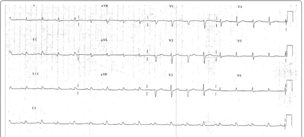
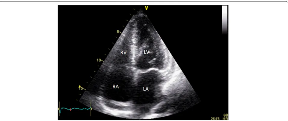
His electrocardiogram (ECG) (Fig. 1) demonstrated electrical alternans and bedside transthoracic echocar-diography (TTE) revealed a large pericardial effusion with features of cardiac tamponade, including diastolic compression of both right atrium and ventricle (Fig. 2, Additional file 1: Video 1 and Additional file 2: Video 2) and large mitral inflow variation (Fig. 3). Urgent peri-cardiocentesis was performed with a restricted aspir-ation of only 600 ml drained initially over the first hour, and a total drainage of 1.8 l of heavily blood-stained pericardial fluid over 36 h. During initial aspiration of pericardial fluid there was immediate symptomatic re-lief and haemodynamic improvement (heart rate [HR] decreased to 80/min, respiratory rate [RR] decreased to 15 breaths/min and BP increased to 150/70 mmHg). * Correspondence:leonard.kritharides@sydney.edu.au
†Equal contributors 1
Department of Cardiology, Concord Repatriation General Hospital, Concord 2139, NSW, Australia
2
The University of Sydney, Sydney, NSW, Australia
CARDIOVASCULAR ULTRASOUND
© 2015 Ayoub et al. This is an Open Access article distributed under the terms of the Creative Commons Attribution License (http://creativecommons.org/licenses/by/4.0), which permits unrestricted use, distribution, and reproduction in any medium, provided the original work is properly credited. The Creative Commons Public Domain Dedication waiver (http:// creativecommons.org/publicdomain/zero/1.0/) applies to the data made available in this article, unless otherwise stated.
Ayoubet al. Cardiovascular Ultrasound (2015) 13:32
Overnight (9 h post procedure) the patient developed chest discomfort, dyspnea, tachycardia (HR 110) and tachypnoea (RR 24). TTE the next morning showed no re-accumulation of pericardial fluid, but detected new severe impairment in function of both ventricles, with akinesis of the apex and peri-apical region (Figs. 4 and 5, Additional file 3: Video 3 and Additional file 4: Video 4). Biomarkers demonstrated a rise in highly sensitive tropo-nin from 8 to 224ng/L, but creatitropo-nine kinase did not rise significantly (107 to 116U/L). ECG after chest pain
demonstrated resolution of the electrical alternans, with new loss of R waves in the anterior leads (Fig. 6).
Based on a presumptive diagnosis of SCM, angiotensin converting enzyme inhibitor and long acting beta-blocker were commenced, chemotherapy withheld and the patient discharged for early clinical and echocar-diographic review. Serial follow up TTEs showed normalization of bi-ventricular function after two weeks (Figs. 7 and 8, Additional file 5: Video 5 and Additional file 6: Video 6), and restoration of R waves on subsequent Fig. 1ECG on first presentation with tamponade demonstrating reduced voltage and electrical alternans
Fig. 2Apical four chamber view on initial presenation demonstating large pericardial effusion with tamponade causing compression of right heart chambers (red arrows). LV function was normal prior to pericardiocentesis
ECGs (Fig. 9). Subsequent computed tomography examin-ation showed normal coronary arteries with a calcium score of zero and no evidence of LAD laceration or dissection.
The patient presented three months later with re-accumulation of pericardial effusion and tamponade. Therapeutic pericardiocentesis was performed with 500 ml of blood stained pericardial fluid drained im-mediately, with 1.9 L in total over 36 h. On this pres-entation he was relaxed and well adjusted in regards to his diagnosis. No LV dysfunction was detected on serial follow-up echocardiograms after the second peri-cardiocentesis (Fig. 10).
Discussion
Our patient developed biventricular apical dysfunction following successful and judicious pericardiocentesis, with features typical of stress or “Takotsubo cardio-myopathy”. The case is instructive for its comparison with PDS and the clinical pattern of initial improve-ment followed by deterioration respectively due to pericardial aspiration and myocardial pathology.
In light of the timing of onset of biventricular impair-ment immediately post procedure PDS is an important differential diagnosis. Other differentials such as lacer-ation to the ventricle or left anterior descending (LAD) coronary artery were clinically unlikely. The former was Fig. 3Transmitral inflow traces showing signicicant respiratory phase variation, consistant with tamponade on first presentation
Fig. 4Parasternal long view post pericardiocentesis demonstrating apical ballooning (red arrows) as a result of apical and peri-apical akinesis
excluded by the absence of new pericardial bleed post procedure. Laceration of the LAD was also clinically unlikely given relatively small rise in cardiac enzymes and absence of large infarct, the presence of concurrent RV dysfunction, spontaneous recovery of ventricular function in a short period of time; additionally CT scan showed no evidence of haematoma or injury to the LAD.
Accordingly, we reviewed the literature describing SCM and PDS. Whereas SCM has been rarely reported after pericardiocentesis, much has been published on
PDS. The incidence of PDS or new left or right sys-tolic dysfunction has been reported to range from 5 % to 36 % of patients post pericardiocentesis [3, 4], espe-cially after malignant pericardial effusions. Although the first case report of PDS in 1983 noted APO with preserved LV function [5], most subsequent reports describe severe impairment of left, right or bi- ven-tricular function, which may be segmental or global (Tables 1 and 2).
A number of mechanisms have been proposed to ex-plain the pathogenesis of LV systolic dysfunction in PDS. Fig. 5Apical four chamber view post pericardiocentesis demonstrating apical ballooning (red arrows) as a result of apical and peri-apical akinesis
Fig. 6ECG after the chest discomfort following pericardiocentesis showing resolution of electrical alternans, and loss of R waves in V1 and V2
Acute withdrawal of exaggerated sympathetic drive dur-ing relief of tamponade may trigger paradoxical haemo-dynamic instability [5]. Mechanical, inter-ventricular volume mismatch may also contribute, with sudden re-lief of pericardial constraint leading to abrupt, dispro-portionate increase in RV volume and a paradoxical rise in pulmonary artery pressure, resulting in raised LV end diastolic pressure and transient left heart failure [5–9]. Others have proposed myocardial stunning from coron-ary perfusion mismatch with acute distension of cardiac chambers after decompression [6, 10, 11]. Taken to-gether, it is likely that a combination of hormonal and
mechanical pathophysiologic mechanisms contribute to LV dysfunction and the final clinical sequelae in PDS.
The classic echocardiographic feature in SCM is tran-sient LV apical ballooning, although other segmental patterns have been described [12, 13]. A stressor leading to sympathetic overdrive and excessive catecholamine release is the currently accepted trigger in the development of SCM [12]. The catecholamine surge precipitates 1) ‘peripheral arterial vasospasm leading to increased after-load and transient increase in LV end-systolic pressure’, 2) ‘acute multiple coronary artery vasospasm leading to myocardial ischaemia’, and 3) direct catecholamine-Fig. 7Is an apical four chamber view 2 weeks post pericardiocentesis and development of LV dysfunction showing resolution of the apical ballooning in systole with normal LV systolic function
Fig. 8Parasternal long view in systole (2 weeks post pericardiocentesis and development of LV dysfunction), showing resolution of both akinesis in the mid septum and apical ballooning (apex not well visulised here)
Fig. 9ECG 2 months post event demonstrating resolution of ischemic changes in ECG in Figure 6
Fig. 10Time line of clinical events
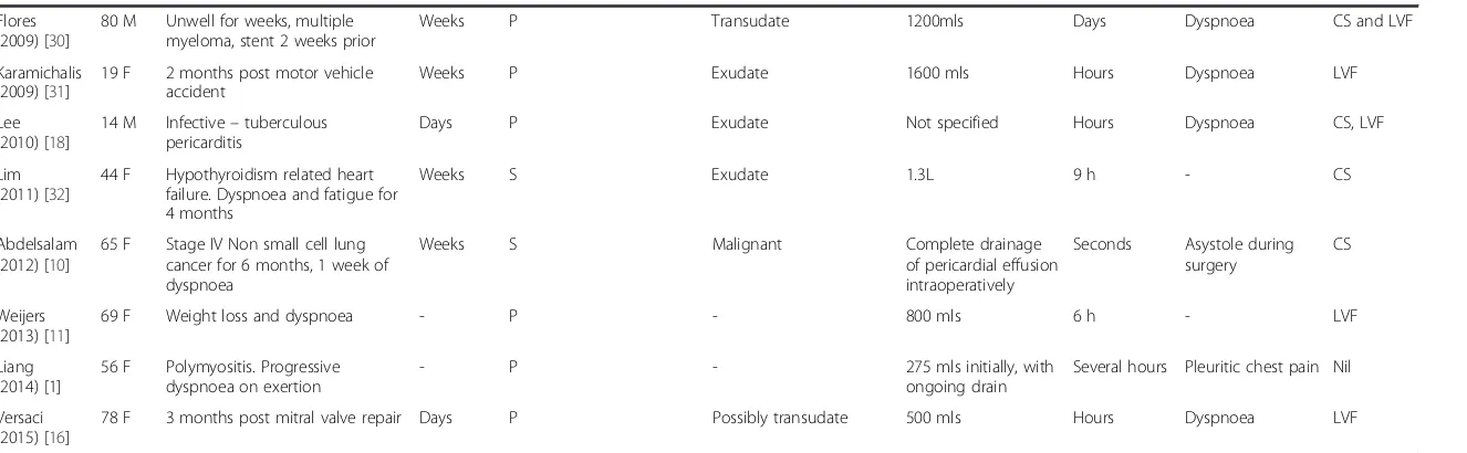
Table 1Summary of reported cases of LVF post pericardiocentesis: Clinical characteristics
Report Age/
Gender
Clinical Scenario Chronicity
of effusion
Type of pericardi-ocentesis Nature of pericardial fluid Fluid drained Time to onset of symptoms
Symptom Signs
VanDyke (1983) [5]
42 M Unwell for 10 days Days P Exudate (malignant) 680 mls Minutes Dyspnoea LVF
Shenoy (1984) [22]
57 M Recent myocardial infarction Days P Transudate 1000 mls Minutes Dyspnoea LVF
Glasser (1988) [23]
33 M Respiratory tract infection 3 months prior, history of Down’s and Ventricular Septal Defect
Weeks S Transudate 2000 mls Minutes Dyspnoea LVF
Downey (1991) [24]
50 M Traumatic (3 weeks post motor vehicle accident)
Weeks P Not specified 450 mls then 1500
mls
Minutes Dyspnoea LVF
Wolfe (1993) [19]
46 F 2 weeks, history of breast cancer prior
Weeks P Exudate 650 mls Weeks Dyspnoea LVF
Wolfe (1993) [19]
50 F 2 weeks, history of breast cancer prior
Weeks P Exudate 650 mls Weeks Dyspnoea LVF
Hamaya (1993) [25]
16 F Unwell, lymphoma with pericardial effusion for 3 years
Months P Not specified 700 mls Weeks Dyspnoea CS, and no
APO
Braverman (1994) [26]
27 F Unwell for 3 weeks (Atrial Septal Defect closure 13 years prior)
Weeks P then S Transudate 500 mls then 100 mls Days Dyspnoea, pleuritic
chest pain
LVF, RVF, CS
Anguera (1996) [27]
68 F History of bowel cancer, anorexia and dyspnoea for 1 month
Weeks P Malignant 800 mls Minutes - CS
Sunday (1999) [8]
60 F 3 days of dyspnoea, lung cancer with pericardial involvement
Days S Exudate 700 mls Minutes Dyspnoea CS, LVF
Chamoun (2003) [6]
36 F 2 months post Mitral valve replacement and Tricuspid repair
Days P Exudate 1070 mls Hours Dyspnoea CS, LVF
Chamoun (2003) [6]
46 F Metastatic cancer Weeks P Exudate 1000 mls Hours Dyspnoea CS, LVF
Geffroy (2004) [7]
53 M 1 month post chemotherapy for cancer
Weeks S Exudate 1500 mls Not specified Dyspnoea, hypoxia CS, LVF, RVF
Ligero (2006) [20]
41 F Lung cancer with hepatic metastases
Days P Exudate 1000 mls Hours Dyspnoea LVF, RHF
Bernal (2007) [28]
45 F Acute myeloid leukemia Days P Exudate 500 mls Hours Dyspnoea CS, LVF
Dosios (2007) [9]
66 F Hematoma, 10 day history of dyspnoea
Days S Exudate 500 mls initially Hours - CS
Sevimli (2008) [17]
42 F Infective - tuberculous pericarditis
Days S Exudate 500 mls Hours Dyspnoea CS and LVF
Khalili (2008) [29]
32 F 2 months post aortic and mitral valve replacement surgery
Weeks P Transudate 1000 mls Hours Dyspnoea CS
Table 1Summary of reported cases of LVF post pericardiocentesis: Clinical characteristics(Continued)
Flores (2009) [30]
80 M Unwell for weeks, multiple myeloma, stent 2 weeks prior
Weeks P Transudate 1200mls Days Dyspnoea CS and LVF
Karamichalis (2009) [31]
19 F 2 months post motor vehicle accident
Weeks P Exudate 1600 mls Hours Dyspnoea LVF
Lee (2010) [18]
14 M Infective–tuberculous pericarditis
Days P Exudate Not specified Hours Dyspnoea CS, LVF
Lim (2011) [32]
44 F Hypothyroidism related heart failure. Dyspnoea and fatigue for 4 months
Weeks S Exudate 1.3L 9 h - CS
Abdelsalam (2012) [10]
65 F Stage IV Non small cell lung cancer for 6 months, 1 week of dyspnoea
Weeks S Malignant Complete drainage
of pericardial effusion intraoperatively
Seconds Asystole during surgery
CS
Weijers (2013) [11]
69 F Weight loss and dyspnoea - P - 800 mls 6 h - LVF
Liang (2014) [1]
56 F Polymyositis. Progressive dyspnoea on exertion
- P - 275 mls initially, with
ongoing drain
Several hours Pleuritic chest pain Nil
Versaci (2015) [16]
78 F 3 months post mitral valve repair Days P Possibly transudate 500 mls Hours Dyspnoea LVF
Abbreviations:Ppercutaneous,Ssurgical,CScardiogenic shock (hypotension, tachycardia),LVFLeft heart failure,RVFright heart failure
Ayoub
et
al.
Cardiova
scular
Ultrasound
(2015) 13:32
Page
8
of
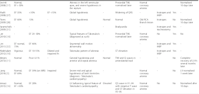
Table 2Summary of reported cases of LVF post pericardiocentesis: Electrocardiographic, biochemical, echocardiographic and outcome parameters
Report LV function pre tap
LV function post tap
RV function post tap
Regional wall motion abnormality
Bio marker ECG Coronary artery
imaging
Inotrope, IABP or Intubation
Death LV recovery
VanDyke (1983) [5]
Normal Normal (EF 67%) - Nil Normal Normal - Intubation No Normal LV
Shenoy (1984) [22]
- Mild LV
impairment
Normal Septal hypokinesis Normal T wave abnormality
and ST elevation V5-6
- - No Normalised few
days later
Glasser (1987) [23]
- Pulmonary
capillary wedge pressure normal
Normal (RVP increased)
- - - - Intubation No Clinical
improvement
Downey (1991) [24]
- Inferred to be
normal
Normal - - Normal - No No Normal LV
Wolfe (1993) [19]
Normal, EF > 50%
EF 30% - Severe global hypokinesis of LV - - - - No Normalised after
7 days
Wolfe (1993) [19]
Normal, EF > 50%
EF 25% - Antero-apical akinesis and
apical dyskinesis
- - - Normalised after
2 weeks
Hamaya (1993) [25]
Normal - - Not provided Normal ST elevation - Inotropes and
intubation
No
-Braverman (1994) [26]
EF 20% EF 20% EF <15% Not provided - - - EF 45% in 9 days
then normalised after a few weeks
Anguera (1996) [27]
- Mildly impaired.
Normal capillary wedge pressure
Severely dilated and severely impaired contractility, EF <15%
Paradoxical septal motion - - Normal
coronary arteries
Inotropes No Complete
recovery of biventricular fn after 10 days
Sunday (1999) [8]
EF 65% EF 30% Severely impaired
contractility
Global hypokinesis - - - Intubation Yes No
Chamoun (2003) [6]
Normal, EF > 50%
EF 20% - Regional wall motion
abnormality
- SR Normal
coronary arteries
Inotropes and IABP
No Normalised 2 weeks later
Chamoun (2003) [6]
Normal, EF > 50%
EF 20% - Akinesis of mid anterior wall
and septum /dilatation of LV
- SR - No No Normalised
2 weeks later
Geffroy (2004) [7]
Normal, EF > 50%
EF >50% EF <15% Akinetic and dilated RV Elevated Old RBBB Normal
coronary arteries Inotropes and intubation Yes -Ligero (2006) [20]
Normal, EF 75%
EF 25% Severe impairment Akinesis of anterior, septum and apex
Normal CK Normal Normal
coronary arteries
Inotropes No Normalised 10 days later
Bernal (2007) [28]
Normal, EF 60-65%
EF 30% - Akinesis of mid anterior wall,
anteroseptal akinesis with apical sparing
Elevated Sinus tachycardia CMR: no myocardial infarction
Inotropes and intubation
No Normalised 1 weeks later
Dosios (2007) [9]
Normal LV fn EF 25% Moderately dilated, impaired
Global hypokinesis Elevated - - Inotropes and
intubation
Yes
-EF 20% - - No No
Table 2Summary of reported cases of LVF post pericardiocentesis: Electrocardiographic, biochemical, echocardiographic and outcome parameters(Continued)
Sevimli (2008) [17]
Normal, EF > 50%
Akinesis in the left ventricular apex, and severe hypokinesis in the septum Precordial TWI, normalised later Normal coronary arteries Normalised 10 days later
Khalili (2008) [27]
EF 35% <10% EF <15% Global hypokinesis - Widening of QRS - Inotropes and
IABP-Yes
-Flores (2009) [28]
EF 60% 13% - Global hypokinesis Normal Normal Old RCA
Branch lesion
Inotropes No Normalised 10 days later
Karamichalis (2009) [31]
- - - - Bradycardia - Inotropes and
tracheostomy
Yes
-Lee (2010) [18]
- EF 20 -30% - Typical features of Takotsubo’s
(diagnosed as such)
- Precordial TWI, normalised later
Normal coronary arteries
No Yes No
Lim (2011) [32]
EF normal, 73%
EF 46% - Segmental wall motion
abnormality
- - - Inotropes and
IABP
Yes
-Abdelsalam (2012) [10]
Vigorous EF 10-15% Dilated and impaired fn
Takotsubo pattern of akinesia - ST elevation - Inotropes and
IABP
Yes
-Weijers (2013) [11]
Normal Poor LV fn - General hypokinesia and
anterior and septal akinesia
Normal TWI and Q waves in anterolateral lead
- - No Complete
recovery of LV fn several months later Liang (2014) [1] Normal, EF 69%
EF 39% (on MRI) Impaired Severe mid and apical hypokinesis of both Ventricles (diagnosis : Takotsubo’s cardiomyopathy)
- - Normal
coronary arteries
- No LV normalised
1 week later
Versaci (2015) [16]
Normal, EF >50%
EF 28% - LV ballooning, typical feature of
Takotsubo’s cardiomyopathy
Elevated QS wave in V1–V4 with negative T wave and ST elevation in V5–V6
Normal coronary arteries
No No Normalised after
10 days
LVLeft ventricle,RVRight ventricle,fnfunction,EFEjection fraction,IABPIntra-aortic balloon pump,RVPright ventricular pressure
β-adrenoceptor - mediated myocardial stunning in the apex [14]. These three pathophysiologic pathways are thought to contribute to the ischaemia, morphologic features and potential haemodynamic sequelae that can be seen in SCM.
More recent case reports have made reference to LV apical ballooning related to PDS as similar to SCM [10, 11, 15–17], and have postulated the physiological stressor being cardiac tamponade along with emotional stress [16]. It is therefore possible that the transient ven-tricular systolic dysfunction in PDS is actually a variant form of stress cardiomyopathy. We carefully reviewed 25 cases of heart failure post pericardiocentesis in the literature (Tables 1 and 2), and we believe that seven cases (two considered to be SCM [1, 18] by the authors and five classified as PDS [10, 16, 17, 19, 20]) could be considered to have echocardiographic features of SCM.
SCM has relatively characteristic clinical presenta-tion, with rise of cardiac enzymes [21], and often asso-ciated with ischaemic ECG changes (up to 44 % of those with SCM have T-wave inversion and 41 % ST elevation [13, 21]). The clinical manifestations in PDS are more variable, ranging from asymptomatic in some to severe low cardiac output states in others. The pri-mary clinical symptom in PDS has been reported as dyspnoea (Table 1). This is in contrast to chest pain being predominant in SCM (69-83 % of presentations) [13, 21]. In the majority of cases of PDS in the litera-ture (Table 2) there was no cardiac enzyme rise, and is-chaemic type changes on ECG were seen in a minority (seven of twenty five cases). In all the cases where is-chaemic ECG changes where present except for one, there was concomitant apical and peri-apical regional wall motion abnormality, which could be classified as SCM also.
Generally SCM has a benign course, with recovery of LV function and good prognosis [12], whilst PDS has poorer outcomes and increased mortality [4]. Reports of PDS suggested normalization of LV dysfunction in 12 of 25 cases classified as PDS. Of the 12 cases that did recover LV function, four had LV impairment with classic SCM pattern of LV impairment on echocardio-gram [16, 17, 19, 20]. The normalisation of LV function in our patient 2 weeks subsequently is more in keeping with SCM.
Current literature has not specifically addressed risk fac-tors for the development of ventricular dysfunction after pericardiocentesis. In our patient, the malignant nature of the effusion, the presence of tamponade and larger size of pericardial effusion [4], may have increased his predispos-ition to develop ventricular dysfunction. Amount and rate of fluid removed on initial decompression are also associ-ated with development PDS [4, 5], however there are no guidelines regarding the maximum amount of pericardial
fluid that can be drained immediately. There is consensus to stop initial drainage with improvement of symptoms or hemodynamic parameters, followed by gradual decom-pression through indwelling catheter [5].
Our patient’s apical systolic dysfunction post peri-cardiocentesis was associated with chest discomfort, transient loss of R waves and rise in cardiac enzymes are typical of classic SCM. The clinical sequence of HR and RR improving immediately post decompression and then increasing again hours after the procedure, was a useful clinical marker of myocardial dysfunction, prompting investigation which identified new ventricu-lar impairment. It is likely that the frequency of transi-ent LV dysfunction is underestimated in these patitransi-ents.
Conclusion
We report a case of transient biventricular dysfunction post pericardiocentesis, with classic features of SCM. LV dysfunction post pericardiocentesis and in PDS is more prevalent than previously thought, and some previous re-ports of PDS may also be potentially considered as SCM complicating pericardiocentesis. In addition to judicious and gradual decompression to avoid ventricular dysfunc-tion or PDS, patients undergoing therapeutic pericardio-centesis should have careful haemodynamic monitoring, as changes in parameters such as heart rate and respira-tory rate can raise suspicion of acute LV impairment.
Consent
Written informed consent was unable to be obtained from the patient for publication of this Case report and any accompanying images, as he has passed away. His next of kin are not contactable after their subsequent return to their home country of China. Professor L. Kritharides, Head of Department, approves the publica-tion of this report, with all patient identifiers kept con-fidential and material presented solely for educational purposes arising from the clinical encounter.
Additional files
Additional file 1: Video 1.Apical four chamber view demonstrating large pericardial effusion on presentation, with tamponade and diastolic compression of right heart chambers. Note the LV function is normal.
Additional file 2: Video 2.Parasternal long view demonstrating large pericardial effusion on presentation, with tamponade and diastolic compression of right heart chambers. Note the LV function is normal.
Additional file 3: Video 3.Apical four chamber view performed on the following morning after the patient experienced chest discomfort. The main finding is severe biventricular impairment, with akinesis of the apex and periapical areas. There is small residual pericardial effusion.
Additional file 4: Video 4.Parasternal long view performed on the following morning after the patient experienced chest discomfort. The main finding is severe biventricular impairment, with akinesis of the apex and periapical areas. There is small residual pericardial effusion.
Additional file 5: Video 5.Apical four chamber view performed nearly 2 weeks after the pericardiocentesis and subsequent stress cardiomyopathy. They show normalisation of both left and right ventricular function.
Additional file 6: Video 6.Parasternal long view performed nearly 2 weeks after the pericardiocentesis and subsequent stress cardiomyopathy. They show normalisation of both left and right ventricular function.
Abbreviations
SCM:Stress Cardiomyopathy; LV: Left ventricle; RV: Right ventricle; PDS: Pericardial decompression syndrome; APO: Acute pulmonary oedema; ECG: Electrocardiogram; TTE: Transthoracic echocardiography; bpm: Beats per minute; HR: Heart rate; RR: Respiratory rate.
Competing interests
The authors declare that they have no competing interests.
Authors’contributions
All authors have approved the final article, contributed to conception, literature review, analysis and interpretation of the material, to the drafting of the manuscript and its critical revision; All authors agree to be accountable for all aspects of the work in ensuring that questions related to the accuracy or integrity of any part of the work are appropriately investigated and resolved.
Received: 9 April 2015 Accepted: 30 June 2015
References
1. Liang JJ et al. Apical ballooning syndrome in polymyositis following placement of pericardial drainage catheter. J Cardiovasc Dis. 2014;2:1. 2. Angouras DC, Dosios T. Pericardial decompression syndrome: a term for a
well-defined but rather underreported complication of pericardial drainage. Ann Thorac Surg. 2010;89:1702–3. author reply 1703.
3. Dosios T, Theakos N, Angouras D, Asimacopoulos P. Risk factors affecting the survival of patients with pericardial effusion submitted to subxiphoid pericardiostomy. Chest. 2003;124:242–6.
4. Wagner PL, McAleer E, Stillwell E, et al. Pericardial effusions in the cancer population: prognostic factors after pericardial window and the impact of paradoxical hemodynamic instability. J Thorac Cardiovasc Surg. 2011;141:34–8.
5. Vandyke Jr WH, Cure J, Chakko CS, Gheorghiade M. Pulmonary edema after pericardiocentesis for cardiac tamponade. N Engl J Med. 1983;309:595–6. 6. Chamoun A, Cenz R, Mager A, et al. Acute left ventricular failure after large
volume pericardiocentesis. Clin Cardiol. 2003;26:588–90.
7. Geffroy A, Beloeil H, Bouvier E, Chaumeil A, Albaladejo P, Marty J. Prolonged right ventricular failure after relief of cardiac tamponade. Can J Anaesth. 2004;51:482–5.
8. Sunday R, Robinson LA, Bosek V. Low cardiac output complicating pericardiectomy for pericardial tamponade. Ann Thorac Surg. 1999;67:228–31. 9. Dosios T, Stefanidis A, Chatziantoniou C, Sgouropoulou S. Thorough clinical
investigation of low cardiac output syndrome after subxiphoid pericardiostomy. Angiology. 2007;58:483–6.
10. Abdelsalam M, Moritz TA, Snyder JA, Cheriyath P, Spizzieri CL. Paradoxical hemodynamic instability complicating pericardial window surgery for cardiac tamponade in a cancer patient. Tex Heart Inst J. 2012;39:711–3. 11. Weijers RW, Post JC. Transient left ventricular systolic dysfunction mimicking
myocardial infarction after pericardiocentesis. Neth Heart J. 2013;21:364–6. 12. Pilgrim TM, Wyss TR. Takotsubo cardiomyopathy or transient left ventricular
apical ballooning syndrome: A systematic review. Int J Cardiol. 2008;124:283–92.
13. Singh NK, Rumman S, Mikell FL, Nallamothu N, Rangaswamy C. Stress cardiomyopathy: clinical and ventriculographic characteristics in 107 North American subjects. Int J Cardiol. 2010;141:297–303.
14. Akashi YJ, Nef HM, Lyon AR. Epidemiology and pathophysiology of Takotsubo syndrome. Nat Rev Cardiol. 2015;12:387–9.
15. Yeh RW, Yu PB, Drachman DE. Takotsubo cardiomyopathy complicated by cardiac tamponade: classic hemodynamic findings with a new disease. Circulation. 2010;122:1239–41.
16. Versaci F, Donati R, Mezzanotte R, Chiariello L, Ammirati F. An unusual complication following pericardiocentesis: reversible left ventricular dysfunction. J Cardiovasc Med. 2015;16 Suppl 2:S133–5.
17. Sevimli S, Arslan S, Gundogdu F, Senocak H. Development of left ventricular apical akinesis and thrombus during pericardiocentesis for pericardial tamponade. Turk Kardiyoloji Dernegi arsivi: Turk Kardiyoloji Derneginin yayin organidir. 2008;36:338–41.
18. Lee SY, Lee SE, Choi JW, Choi SI, Chun EJ, Choi JY. A case of transient left ventricular apical ballooning syndrome in a child: clinical features and imaging findings. The international journal of cardiovascular imaging. 2010;26:345–51.
19. Wolfe MW, Edelman ER. Transient systolic dysfunction after relief of cardiac tamponade. Ann Intern Med. 1993;119:42–4.
20. Ligero C, Leta R, Bayes-Genis A. Transient biventricular dysfunction following pericardiocentesis. Eur J Heart Fail. 2006;8:102–4.
21. Samardhi H, Raffel OC, Savage M, et al. Takotsubo cardiomyopathy: an Australian single centre experience with medium term follow up. Internal Med J. 2012;42:35–42.
22. Shenoy MM, Dhar S, Gittin R, Sinha AK, Sabado M. Pulmonary edema following pericardiotomy for cardiac tamponade. Chest. 1984;86:647–8. 23. Glasser F, Fein AM, Feinsilver SH, Cotton E, Niederman MS. Non-cardiogenic
pulmonary edema after pericardial drainage for cardiac tamponade. Chest. 1988;94:869–70.
24. Downey RJ, Bessler M, Weissman C. Acute pulmonary edema following pericardiocentesis for chronic cardiac tamponade secondary to trauma. Crit Care Med. 1991;19:1323–5.
25. Hamaya Y, Dohi S, Ueda N, Akamatsu S. Severe circulatory collapse immediately after pericardiocentesis in a patient with chronic cardiac tamponade. Anesth Analg. 1993;77:1278–81.
26. Braverman AC, Sundaresan S. Cardiac tamponade and severe ventricular dysfunction. Ann Intern Med. 1994;120:442.
27. Anguera I, Pare C, Perez-Villa F. Severe right ventricular dysfunction following pericardiocentesis for cardiac tamponade. Int J Cardiol. 1997;59:212–4. 28. Bernal JM, Pradhan J, Li T, Tchokonte R, Afonso L. Acute pulmonary edema
following pericardiocentesis for cardiac tamponade. Can J Cardiol. 2007;23:1155–6.
29. Khalili AA. Acute Heart Failure after Evacuation of Large Volume of Pericardial Effusion by Pericardiostomy. Res J Biooogical Sci. 2008;3(1):32–4. 30. Flores VM et al. Transient left ventricular dysfunction following
pericardiocentesis. An unusual complication to bear in mind. Rev Esp Cardiol. 2009;62(9):1071–2.
31. Karamichalis JM, Gursky A, Valaulikar G, Pate JW, Weiman DS. Acute pulmonary edema after pericardial drainage for cardiac tamponade. Ann Thorac Surg. 2009;88:675–7.
32. Lim AS, Paz-Pacheco E, Reyes M, Punzalan F. Pericardial decompression syndrome in a patient with hypothyroidism presenting as massive pericardial effusion: a case report and review of related literature. BMJ Case Rep. 2011;2011.
Submit your next manuscript to BioMed Central and take full advantage of:
• Convenient online submission
• Thorough peer review
• No space constraints or color figure charges
• Immediate publication on acceptance
• Inclusion in PubMed, CAS, Scopus and Google Scholar
• Research which is freely available for redistribution
Submit your manuscript at www.biomedcentral.com/submit