R E S E A R C H
Open Access
Increased heme-oxygenase 1 expression in
mesenchymal stem cell-derived adipocytes
decreases differentiation and lipid accumulation
via upregulation of the canonical Wnt signaling
cascade
Luca Vanella
1,2†, Komal Sodhi
1†, Dong Hyun Kim
1, Nitin Puri
3, Mani Maheshwari
1, Terry D Hinds Jr
3, Lars Bellner
4,
Dov Goldstein
5, Stephen J Peterson
4, Joseph I Shapiro
1and Nader G Abraham
1*Abstract
Introduction:Heme oxygenase (HO), a major cytoprotective enzyme, attenuates oxidative stress and obesity. The canonical Wnt signaling cascade plays a pivotal role in the regulation of adipogenesis. The present study examined the interplay between HO-1and the Wnt canonical pathway in the modulation of adipogenesis in mesenchymal stem cell (MSC)-derived adipocytes.
Methods:To verify the role of HO-1 in generating small healthy adipocytes, cobalt protoporphyrin (CoPP), inducer of HO-1, was used during adipocyte differentiation. Lipid accumulation was measured by Oil red O staining and lipid droplet size was measured by BODIPY staining.
Results:During adipogenesisin vitro, differentiating pre-adipocytes display transient increases in the expression of genes involved in canonical Wnt signaling cascade. Increased levels of HO-1 expression and HO activity resulted in elevated levels ofb-catenin, pGSK3b, Wnt10b, Pref-1, and shh along with increased levels of adiponectin (P< 0.05). In addition, induction of HO-1 resulted in a reduction in C/EBPa, PPARg, Peg-1/Mest, aP2, CD36 expression and lipid accumulation (P< 0.05). Suppression of HO-1 gene by siRNA decreased Wnt10b, pGSK3bandb-catenin expression, and increased lipid accumulation. The canonical Wnt responsive genes, IL-8 and SFRP1, were
upregulated by CoPP and their expression was decreased by the concurrent administration of tin mesoporphyrin (SnMP), an inhibitor of HO activity. Furthermore, knockdown of Wnt10b gene expression by using siRNA showed increased lipid accumulation, and this effect was not decreased by concurrent treatment with CoPP. Also our results show that blocking the Wnt 10b antagonist, Dickkopf 1 (Dkk-1), by siRNA decreased lipid accumulation and this effect was further enhanced by concurrent administration of CoPP.
Conclusions:This is the first study to demonstrate that HO-1 acts upstream of canonical Wnt signaling cascade and decreases lipogenesis and adipocyte differentiation suggesting that the HO-1 mediated increase in Wnt10b can modulate the adipocyte phenotype by regulating the transcriptional factors that play a role in adipogenesis. This is evidenced by a decrease in lipid accumulation and inflammatory cytokine levels, increased adiponectin levels and elevation of the expression of genes of the canonical Wnt signaling cascade.
* Correspondence: [email protected] †Contributed equally
1
Joan C. Edwards School of Medicine, Marshall University, 1600 Medical Center Drive, Huntington, WV, 25701-3655, USA
Full list of author information is available at the end of the article
Introduction
Human bone marrow-derived mesenchymal stem cells (MSCs) are multipotent cells that have the potential to dif-ferentiate into a variety of cell types including adipocytes [1-5]. MSC-derived adipocyte differentiation and dysregu-lation of adipogenesis is implicated in the pathogenesis of diseases such as metabolic syndrome [4]. Enhanced adipo-genesis with adipocyte hypertrophy is one of the leading causes of adipose tissue hypoxia, inflammation, and dys-function [6]. Hence, the elucidation of the mechanisms that regulate commitment of MSCs towards adipogenic fate may offer a portal to the development of treatment for metabolic syndrome and its related vascular complications. Adipogenesis begins with the commitment of MSCs to the adipocyte lineage, followed by terminal differentiation of pre-adipocytes to mature adipocytes [5,7]. Fat tissue-derived adipocytes express several regulatory proteins such as Wnts andb-catenin, as well as Sonic hedgehog (Shh), which potentially works upstream of these known differentiation factors to induce osteogenesis in MSCs [8]. Wnts regulate gene expression through either the canonical (b-catenin-dependent) or the non-canonical (b-catenin-independent) pathway [9,10]. The canonical Wnt signaling pathway controls cell proliferation, cell survival and cell fate. Wnt ligands are secreted glycopro-teins that function in a paracrine and autocrine manner. Among the Wnt ligands identified, Wnt10b has been shown to be a crucial factor in the activation of the canonical pathway and inhibition of adipogenesis [11,12]. Adipose tissue-specific transgenic over-expression of Wnt10b leads to a significant decrease in adiposity and resistance to a high-fat diet in mice [13]. The canonical Wnt pathway relies on stabilization ofbcatenin. The Wnt/bcatenin signaling pathway affects cellular func-tions by regulating bothbcatenin levels and subcellular localization [14].
An increase in Wnt/b-catenin signaling inhibits the adi-pogenic transcription factor CCAAT/enhancer binding protein (C/EBPa) and the peroxisome proliferator activa-tor recepactiva-tor (PPARg) [11,15-17]. Adipocyte differentiation is an ordered multistep process requiring the sequential activation of several groups of transcription factors, including CCAAT/enhancer-binding protein (C/EBPa) gene family and peroxisome proliferator activated recep-tor-g(PPAR-g) [1,18]. C/EBPaand PPARgare involved in the growth arrest that is required for adipocyte differentia-tion. Pre-adipocyte factor-1 (Pref-1) belongs to the Notch family of epidermal growth factor-like repeat-containing proteins and has been shown to participate in maintaining pre-adipose phenotype [19]. Pref-1 is an inhibitor of adi-pocyte differentiation, hence a decrease in Pref-1 expres-sion is observed during differentiation of adipocytes [20]. The paternally expressed 1 (Peg-1)/Mesoderm-specific transcript (Mest) [21], when upregulated, results in the
enlargement of adipocytes during adipose tissue expansion [22]. On accumulation of triglycerides, the levels of Peg-1/ Mest [22] are increased with a concomitant signal to pre-adipocytes to enlarge in order to accommodate more tri-glycerides. Adipocyte enlargement is associated with an increase in the levels of TNFa, IL-1, IL-6 and increased insulin resistance [23-26]. Hedgehog signaling exerts its pleiotropic effects through regulation of the cell cycle, direction of cell differentiation, and alteration of cell survi-val [27]. Conversely, Shh signaling represses adipogenic differentiation in pre-adipocytes [28].
Heme oxygenase-1 (HO-1) is a stress response gene cri-tical for bone marrow cell differentiation [29-31]. A por-phyrin structure within cyanocobalamine enables it to induceHO-1gene expression by facilitating its binding to the porphyrin binding domain of theHO-1gene. Induc-tion of HO-1 enhances cell survival and moderates dia-betes and obesity [32]. Induction of HO-1 gene expression
in vivoand in cell culture results in an increase in pre-adi-pocytes, a reduction in the number of enlarged adipre-adi-pocytes, and an increase in small adipocyte and adiponectin levels [33]. A decrease in HO-1 expression results in increased insulin resistance and adiposity in Zucker rats and obese mice [34]. Additionally, induction of HO-1 in adipocyte cell culture is associated with increased adiponectin levels and decreased pro-inflammatory cytokines, TNFaand IL-6 [35,36].
The goal of this study was to elucidate the role ofHO-1 gene expression on adipogenesis and clarify the role of Wnt10b and its dependent genes in this process. Induc-tion ofHO-1gene expression and HO activity decreased lipid deposition and inflammatory cytokine levels, increased adiponectin levels and elevated the expression of genes of the canonical Wnt signaling cascade. These novel findings demonstrate that increased levels of HO-1 appear crucial in modulating the phenotype of adipocytes to express canonical downstream signaling proteins.
Materials and methods
Differentiation of human bone marrow-derived MSCs into adipocytes
Frozen bone marrow mononuclear cells were purchased from Allcells (Allcells, Emeryville, CA, USA). After thaw-ing, mononuclear cells were resuspended in ana-minimal essential medium (a-MEM, Invitrogen, Carlsbad, CA, USA) supplemented with 10% heat-inactivated FBS (Invi-trogen) and 1% antibiotic/antimycotic solution (Invi(Invi-trogen). The cells were plated at a density of 1 to 5 × 10⁶cells per 100-cm2dish. The cultures were maintained at 37°C in a 5% CO2incubator and the medium was changed after 48 h
75-cm2flask at a density of 1 to 2 × 10⁴cells and cultured ina-MEM with 10% FBS for 7 days. The medium was replaced with adipogenic medium, and the cells were cul-tured for an additional 14 days as described previously [37]. Human MSCs, passage 3, were cultured in the presence of the HO-1 inducer cobalt protoporphyrin (CoPP) (5μM) and with the HO activity inhibitor tin (Sn4+ )-mesopor-phyrin (SnMP) (5μM), which were administered every 2 days.
HO activity measurement
Heme oxygenase activity was measured in hMSCs by car-bon monoxide (CO) production in cellular homogenates. Briefly, hMSCs were homogenized in Sucrose (255 mM)-Tris hydrochloride (20 mM) buffer (pH 7.4) with NP-40 (1% w/v), EDTA (1 mM), phenylmethylsulfonyl fluoride (PMSF) (1 mM) and mammalian protease inhibitor cock-tail (5% v/v). After homogenization, samples were centri-fuged, at 6000 × g for 30 minutes at 4°C, and the supernatant collected for measurement of HO activity; 100μg protein/sample was incubated, in gas-sealed vials, in Sucrose-Tris buffer along with nicotinamide adenine dinucleotide phosphate-oxidase (NADPH) (1 mM) and excess heme (40μM), in both the absence and the pre-sence of SnMP (2 μM). Samples were incubated in a water bath, in the absence of light, at 37°C for 60 min-utes, after which, the HO reaction was stopped by placing the samples in ice. CO generation was quantitated in the headspace using gas chromatography/mass spectrometry (GC/MS), as previously described [38], using C13O16as an internal standard. Results are expressed as HO-depen-dent CO generation by subtracting the amount of CO in the presence of SnMP. CO generated is expressed as pmoles/mg protein/hour.
Effect of CoPP on adipogenesis
To measure the effect of increased HO-1 expression on MSC-derived adipocyte differentiation, cells were treated with 0.5, 1.0, 2.0, 5.0, and 10.0μM of CoPP every 4 days. After 14 days, cells were stained with Oil Red O solution.
Oil Red O staining
Staining was performed using 0.21% Oil Red O in 100% isopropanol (Sigma-Aldrich, St. Louis, MO, USA). Briefly, adipocytes were fixed in 10% formaldehyde, stained with Oil Red O for 10 minutes, rinsed with 60% isopropanol (Sigma-Aldrich), and the Oil Red O eluted by adding 100% isopropanol for 10 minutes and the optical density (OD) measured at 490 nm, for 0.5 sec reading.
Measurement of lipid droplet size
After induction of adipogenesis, lipid droplets were stained with 2 μM boron-dipyrromethene (BODIPY) 493/503 (Molecular Probes, Eugene, OR, USA) [39]. Cell
size was measured using an ImagePro Analyzer (Media-Cybernetics, Inc., Bethesda, MD, USA). The classification of the size of lipid droplets was based on size by area (pixels).
Cell viability test by lactic dehydrogenate assay (LDH)
We followed the manufacturer’s protocol (LDH Assay kit, Cayman, Ann Arbor, MI, USA). Briefly, hMSC and adipocytes at day 14 were plated in 96-well plates for 1 day. Next day, cell layers were washed twice with PBS, and then cells were treated with various concentrations of CoPP (0 to 10μM). After incubation for 24 h, and addition of 100μl of reaction mixture to each well, cells were incubated for 4 hours at 37°C and 5% CO2 in a
humidified incubator. Absorbance was measured in the 96-well microplate using a microplate reader at 490 nm with 650 nm as the reference wavelength, and the per-centage of LDH release for each sample was normalized according to the absorbance reading from samples trea-ted with 0.5% Triton X-100. All analyses were replicatrea-ted eight times.
Cytokine array and adiponectin
TNFaand adiponectin (high molecular weight, HMW), were measured as previously described [25,40] by Cyto-kine SearchLight Infrared arrays (Pierce Biotechnology, Inc., Woburn, MA, USA).
HO-1 siRNA transfection
Cells were treated with three different predesigned siRNAs of the HO-1 gene (SASI_ Hs01_00035068, SASI_Hs01_00035065 and SASI_Hs01_00035067 from Sigma-Aldrich, St. Louis, MO, USA). According to the manufacturer’s protocol, adipogenic media containing siRNA using NTER (Sigma-Aldrich) was replaced every 48 h. Briefly, a nanoparticle solution was incubated with 10 nM siRNA. After 20 minutes cells were treated with siRNA solution during adipogenesis, which was halted after 10 days.
Measurement of MSC-derived adipocyte signaling molecules
Cells were maintained at -80°C until required for assay. Frozen cells were pulverized and placed in a homogeniza-tion buffer (10 mM phosphate buffer, 250 mM sucrose, 1 mM EDTA, 0.1 mM PMSF and 0.1% tergitol, pH 7.5). Homogenates were centrifuged at 27,000 × g for 10 min-utes at 4°C. The supernatant was isolated and protein levels were assayed (Bradford Method). The supernatant was used for measurement of HO-1, Wnt10b,b-catenin, Pref-1, C/EBPa, Peg-1/Mest, pGSK3b, shh, PPARgand
Quantitative real-time PCR analysis
Total RNA was extracted from differentiated human mesenchymal stem cells using 5-Prime PerfectPure RNA Tissue Kit (Fisher Scientific Company, LLC, Wilmington, DE, USA). Total RNA was read on a NanoDrop 2000 spectrophotometer (Thermo Fisher Scientific) and cDNA was synthesized using High Capacity cDNA Reverse-Transcription Kit (Life Technologies, Grand Island, NY, USA). PCR amplification of the cDNA was performed by quantitative real-time PCR using TrueAmp SYBR Green qPCR SuperMix (Smart Bioscience, Philadelphia, USA). The thermocycling of IL-8 and Secreted frizzled-related protein 1 (SFRP1) protocol consisted of 5 minutes at 95°C, 40 cycles of 15 sec at 95°C, and 30 sec at 60°C, and finished with a melting curve ranging from 60 to 95°C to allow distinction of specific products. Normalization was performed in separate reactions with primers to 18S mRNA (TTC GAACGTCTGCCCTATCAA and ATGG TAGGCACGGCGACTA).
Statistical analyses
Statistical significance (P< 0.05) of differences between experimental groups was determined by the Fisher method for analysis of multiple comparisons. For com-parison between treatment groups, the null hypothesis was tested by either single-factor analysis of variance (ANOVA) for multiple groups, or the unpairedt-test for two groups, and the data are presented as mean ± stan-dard error (SE).
Results
The effect of adipogenesis on HO-1 expression
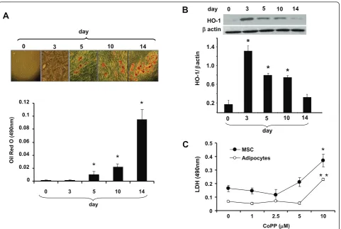
The temporal sequence of HO-1 expression was deter-mined over a 14-day period in MSC-derived adipocytes. The basal levels of HO-1 expression increased during MSC adipogenic culture growth and peaked at day 3 (Figure 1B). This was followed by gradual decrease in HO-1 expression, while lipid droplet area gradually increased over a period of 14 days (Figure 1A).
The effect of CoPP on cell viability during adipogenesis by LDH assay
Detection of cell membrane integrity is a rapid and simple approach to determine cell viability by measuring cellular LDH leakage in damaging cells. Our results showed that CoPP treatment in MSCs and adipocytes at day 14 have no cytotoxic effects at concentrations up to 5μM (Figure 1C). However, MSCs and adipocytes showed significant toxicity at a 10μM concentration of CoPP (P< 0.05).
The effect of CoPP on HO-1 expression, HO activity and cytokine levels during adipogenesis
We examined the effect of CoPP on lipid accumulation after 14 days, using standard culture conditions by
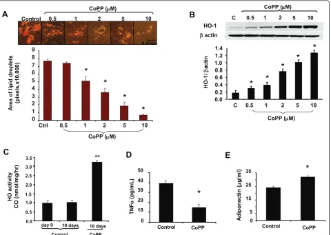
measuring Oil Red O-stained lipid droplet area (Figure 2A). The level of cells with Oil Red O-stained lipid dro-plets decreased as CoPP concentration increased. CoPP decreased lipid accumulation in a concentration-depen-dent manner. Oil Red O staining was barely detectable at the highest concentration of CoPP (10 μM), although untreated MSC-derived adipocyte progenitor cells were loaded with lipid droplets (control). Quantification of Oil Red O-stained cells showed an increase in the number of adipocytes in the absence of CoPP (77,093 ± 943 pixels) compared with the presence of CoPP (54,376 ± 5,366 and 3,526 ± 368 pixels) at 1 and 10 μM, respectively. The expression of HO-1 in the presence of CoPP increased in a dose-dependent manner (Figure 2B), which was consis-tent with our results showing decreased lipogenesis with increased concentrations of CoPP (Figure 2A). HO activ-ity, as measured by CO release, was increased in the presence of CoPP compared to the control (Figure 2C;
P< 0.05). HO activity increased reaching a maximum (P< 0.05) at day 10 and remained elevated. This was in contrast to the control where HO activity remained at the same level as day 0. One possible explanation of this discrepancy between HO-1 expression and HO activity could be differential effects of progressing adipogenesis on HO-1 expression and activity. Other investigators [42,43] have reported redox imbalances during the process of adipogenesis. We have previously shown that oxidative stress can induce HO expression while suppres-sing HO activity [44] (Figure 2C).
Adipose cell enlargement is associated with increased secretion of cytokines, which impairs the differentiation of pre-adipocytes and reduces adiponectin secretion. We examined the levels of TNFain the conditioned media of CoPP-treated MSC-derived adipocytes, and found that TNFalevels were significantly decreased (Figure 2D;P< 0.001) at day 14. In contrast, CoPP increased adiponectin levels were increased subsequent to CoPP treatment when compared to controls at day 14 (P< 0.05; Figure 2E).
Effect of CoPP and SnMP on adipogenesis and distribution of lipid droplet size, stained by BODIPY
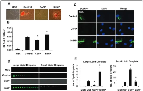
and decreased the number of small lipid droplets, com-pared with either culture treated with CoPP or control, indicating that this effect is mediated by increased HO activity (Figure 3D). The results were quantified and are shown in Figure 3E.
Effect of CoPP and SnMP on CD36 expression
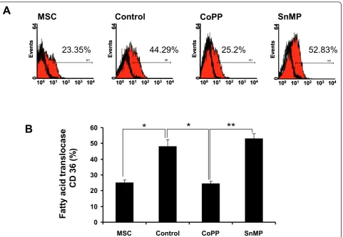
CD36, a fatty acid translocase, which mediates the trans-fer of fatty acids (FAs) into the cell [45], was measured in MSCs and MSC-derived adipocytes treated with either CoPP (5μM) or SnMP (5 μM). Cells were treated with adipogenic media and collected after 14 days. As seen in Figure 4A, during lipogenesis CoPP decreased CD36 expression (25.21 ± 1.67%) compared to control (44.29 ± 4.33%), while SnMP increased CD36 expression (52.83 ± 3.12%). The results are quantified in Figure 4B and show that CoPP decreased CD36 levels (P< 0.05) compared to control indicating decreased fatty acid uptake in adipo-cytes by upregulation of HO-1. As expected, SnMP restored CD36 levels to those seen in control, suggesting
that increased HO-1 expression and activity has anti-adi-pogenic response on adipocytes.
Effect of siRNA HO-1 on adipogenesis
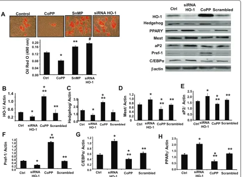
Upregulation of HO-1 by CoPP decreased lipid accumula-tion, which was measured as the relative absorbance of Oil Red O. To selectively assess the role of HO-1 on adiposity, HO-1 siRNA was added to the cell culture. Lipid accumu-lation was increased after HO-1 siRNA treatment as com-pared to MSCs treated with CoPP (Figure 5A;P< 0.05). Densitometry analysis showed that CoPP treatment increased HO-1 expression in MSC-derived adipocytes compared to the control, and this effect was reversed by siRNA HO-1 (Figure 5B;P< 0.01). The protein expression of Shh increased with CoPP treatment and was reversed by siRNA HO-1 (Figure 5C). Pref-1, an excellent marker for pre-adipocytes, which is extinguished during adipocyte differentiation [19], was increased by CoPP treatment and was decreased by siRNA HO-1 (Figure 5G). Upregulation of HO-1 by CoPP treatment decreased levels of the
0 3 5 10 14 day
HO-1 EEactin
A
C
B
0.2 0.6 1.0 1.4
0 3 5 10 14
HO-1
/
E
acti
n
day
*
*
*
0 3 5 10 14
day
0 0.02 0.04 0.06 0.08 0.1 0.12
0 3 5 10 14
Oil R
ed
O
(490nm)
day
*
*
*
0 0.1 0.2 0.3 0.4 0.5
0 1 2.5 5 10
LD
H
(
490nm)
MSC Adipocytes
CoPP (PM)
*
* *
Figure 1Expression of heme oxygenase-1 (HO-1), with time, on adipogenesis. (A) Pictures of lipid droplets of a representative experiment at days 3, 5, 10, and 14. Adipogenesis was measured as the relative absorbance of Oil Red O as described in Materials and methods (mean ± SD, *P< 0.05 vs control). (B) Western blot of HO-1 and actin in mesenchymal stem cell (MSC)-derived adipocytes from day 0 to day 14. Representative immunoblots and densitometry analysis are shown. Data are expressed as means ± SD;n= 4; *P< 0.05. (C) Lactic dehydrogenate (LDH) assay done to study the cytotoxicity at increasing concentrations of cobalt-protoporphyrin IX (CoPP). Data are expressed as means ± SD;
adipogenic markers Peg-1/Mest, aP2, C/EBPaand PPARg in MSC-derived adipocytes, and this effect was reversed by transduction with HO-1 siRNA (Figure 5D, E, E, G and 5H respectively;P< 0.02).
Effect of siRNA HO-1 on canonical Wnt signaling and Wnt-responsive genes
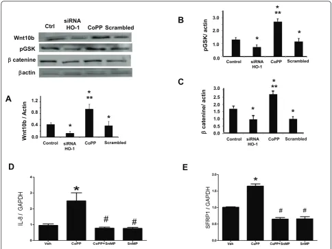
Upregulation of HO-1 by CoPP treatment increased protein expression of the canonical Wnt signaling cas-cade, Wnt10b, pGSK3band b-catenin in MSC-derived adipocytes as compared to the control. To selectively assess the role of HO-1 on adiposity, HO-1 siRNA was added to cell culture with a resultant decrease in Wnt10b, pGSK3b and b-catenin levels (Figure 6A, B and 6C respectively; P< 0.05). Canonical Wnt signaling cascade is known to activate IL-8 and SFRP1 [46]. The
levels of these Wnt responsive genes, IL-8 and SFRP1, were increased by CoPP treatment and these effects were reversed by concurrent treatment with SnMP (Figure 6D and E respectively;P< 0.05).
Effect of CoPP on canonical Wnt signaling cascade during adipogenesis
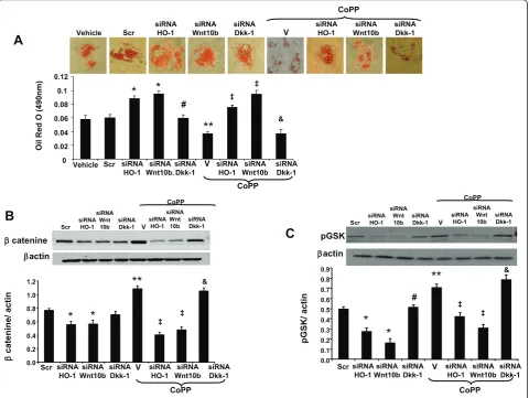
Our results show Oil Red O staining was increased after siRNA HO-1 treatment compared to MSCs treated with CoPP (Figure 7A). To elucidate the role of Wnt10b in the regulation of adipogenesis in MSC-derived adipocytes, we measured the effect of suppression of Wnt10b on adipo-genesis, using siRNA. The addition of siRNA Wnt10b increased lipid formation compared to the vehicle (P< 0.05), and this effect was not significantly altered by con-current administration of CoPP, suggesting a possible
A
C
B
*
0 1 2 3 4 5 6 7 8 9
Ctrl 0.5
CoPP (PM)
1 2 5 10
A
rea of
lipid droplet
s
(p
ixels,x10
,00
0)
*
*
*
Control 0.5 2 1 5 10
CoPP (PM)
HO-1
Eactin
C 0.5 1 2 5 10
CoPP (PM)
0.0 0.2 0.4 0.6 0.8 1.0 1.2 1.4
C 0.5
CoPP (PM)
1 2 5 10
*
*
*
*
*
HO-1
/
E
acti
n
D
E
*
0 10 20 30 40 50
Control CoPP
TNF
D
(pg/mL)
A
d
iponect
in (
P
g/ml)
5 15 25 35
Control CoPP
*
0
HO act
iv
it
y
CO (
n
mol/mg/hr)
0.5 1.5 2.5 3.5
day 0 10 days 10 days
Control CoPP
**
0.0 3.0
2.0
1.0
interaction between HO-1 and the Wnt10b pathway. Wnt antagonist, Dickkopf 1 (Dkk1), is secreted by human pre-adipocytes and promotes adipogenesis [47]. Further our results show that knockdown of Wnt antagonist Dkk1 by siRNA showed significant reduction in lipid accumulation compared to siRNA Wnt10b, and more importantly, administration of CoPP further significantly deceased lipo-genesis (P< 0.05; Figure 7A).
The protein expression ofb-catenin and phosphory-lated glycogen synthase kinase (GSK3)bwas measured to study the effect of Wnt10b inhibition by using siRNA (Figure 7B and 7C respectively). Our results showed decreased expression of b-catenin and pGSK3b when MSCs were treated with Wnt10b siRNA compared to the control (P < 0.05) and this effect was not reversed by concurrent treatment with CoPP. We next deter-mined whether inhibition of Dkk1 using siRNA affected these adipogenic markers. Our results further showed thatb-catenin and pGSK3blevels were increased com-pared to the MSCs treated with siRNA Wnt10b. More importantly, concurrent administration of CoPP signifi-cantly increased the gene expression ofb-catenin and
pGSK3b(P < 0.05) compared to the cells treated with siRNA Dkk1.
Discussion
This study demonstrates that the effects of HO-1 induc-tion in a cell-based model of adipogenesis are dependent upon activation of the Wnt canonical signaling pathway. HO-1 induction has reduced body weight and adiposity [25,48] and improved the metabolic profile in animal mod-els of obesity [24,49]. It has also been shown that upregu-lation of HO-1 reduces adipogenesis in cell cultures [34]. We show here in a cell-based model of adipogenesis (MSCs) that HO-1 induction mediates the recruitment of the Wnt canonical cascade, and entails reduced lipid accu-mulation comprised of smaller healthier adipocytes, reduced inflammation and improved adipokine secretion.
Differentiation of pre-adipocytes into adipocytes is regulated by a balance of transcriptional factors that can both positively and negatively influence differentiation. This is reflected by the appearance of various early, inter-mediate and late mRNA/protein markers and triglyceride accumulation. Several reports describe an association
MSC Control CoPP SnMP
B
A
Control CoPP SnMP MSC
Control
CoPP
SnMP
DAPI
BODIPY Merge
C
D
Large Lipid Droplets Small Lipid Droplets
Control
CoPP
SnMP MSC
0 1 2 3 4 5 6 7
MSCCtrl CoPPSnMP
No. of
lipid
droplet
s
Large Lipid Droplets
*
*
15 25 35
MSCCtrlCoPPSnMP
No. of
lipid
droplet
s
Small Lipid Droplets
*
E
Oil R
ed
O
(490nm)
0.00 0.05 0.10 0.15 0.20 0.25
*
*
5
*
between adipogenesis and Wnt signaling in the regula-tion of adult tissue homeostasis and remodeling [50,51]. Wnt10b is an endogenous regulator of adipogenesis that maintains pre-adipocytes in an undifferentiated state and functions as an adipogenic switch. The activation of Wnt10b contributes to the inactivation/phosphorylation of GSK3band consequently, elevated level ofb-catenin; it is the molecular node of the canonical Wnt signaling pathway. When Wnt signaling is active, GSK3 is inhib-ited. Conversely, when Wnt signaling is suppressed, GSK3 phosphorylatesb-catenin and targets it for ubiqui-tin-mediated degradation [11]. Our results describe a cri-tical link between the anti-adipogenic effects of HO-1 and stimulation of the Wnt canonical pathway. Two lines of evidence characterize this relationship: First, HO-1 induction in CoPP-treated cells was accompanied by increased levels of Wnt10b and its associated signaling mediators, namely, phosphorylated GSK3bandb-catenin. The concurrent reduction in adipocyte size and lipid accumulation establishes a link between this phenotype, that is, HO-1 induction and Wnt signaling. Second,
siRNA-mediated downregulation/inhibition of Wnt10b prevented HO-1 from reducing adipocyte hypertrophy in MSCs. Further our results show that inhibition of Dkk1 by siRNA decreased lipogenesis, and this effect was further enhanced by concurrent administration of CoPP. These observations indicate that activation of the Wnt canonical pathway plays a role in the prevention of adi-pocyte hypertrophy and the promotion of smaller heal-thier adipocytes in MSCs undergoing HO-1 induction. The precise molecular mechanism linking HO-1 to acti-vation of Wnt signaling is unclear. However, the restora-tion of the redox environment as a result of HO-1 induction [37] could contribute to activation of Wnt sig-naling. In this regard, it should be noted that chronic oxi-dative stress has been shown to suppress the Wnt canonical pathway [52,53] while enhancing lipid accumu-lation and hypertrophy in MSC-derived adipocytes. Furthermore, our studies show that upregulation of HO-1and increased HO activity lead to increased levels of the Wnt-responsive genes, IL-8 and SFRP1, which was reversed by the HO-1 inhibitor, SnMP. Thus, these
M1
0 1 2 3
M1 M1 M1
100 101 102 103 104
0
64
Ev
ent
s
100 101 102 103 104
0
64
Ev
ent
s
100 101 102 103 104
0
64
Ev
ent
s
100 101 102 103 104
0
64
Ev
ent
s
100 101 102 103 104
0
64
Ev
ent
s
100 101 102 103 104
0
64
Ev
ent
s
100 101 102 103 104
0
64
Ev
ent
s
100 101 102 103 104
0
64
Ev
ent
s
23.35% 44.29%
25.2% 52.83%
MSC Control CoPP
SnMP
A
B
*
* **
0 10 20 30 40 50 60
MSC Control CoPP SnMP
Fatty
acid
translocas
e
C
D
3
6
(%
)
results substantiate our hypothesis that Wnt10b may be considered an HO-1 gene target that when increased, ulti-mately results in a reduction in adipocyte hypertrophy.
Activation of Wnt/b-catenin signaling maintains pre-adipocytes in an undifferentiated state through inhibition of the adipogenic transcription factors, C/EBPa and PPARg[15-17]. C/EBPaand PPARghave been shown to activate adipocyte-specific genes and are involved in the growth arrest that is required for adipocyte differentiation. Our results show that the increased expression of HO-1 resulted in either maintaining pre-adipocytes in the undif-ferentiated state or slowed down this process, presumably through activation of Wnt/b-catenin and inhibition of C/ EBPaand PPARglevels. Pref-1 has also been shown to
participate in maintaining the pre-adipose phenotype. A decrease in Pref-1 expression is observed during adipo-cyte differentiation. In adipose tissue, Pref-1 is specifically expressed in pre-adipocytes but not in adipocytes and thus, is used as a pre-adipocyte marker [54]. In concor-dance with these observations, our results showed that upregulation of HO-1 increased Pref-1 expression, sug-gesting that HO-1 decreased adipocyte differentiation. Pref-1 prevents lipid accumulation and expression of adi-pocyte transcription factors such as PPARgand C/EBPa, as well as other late adipocyte markers, including FA synthase and FABP4/aP2 [54].CD36andaP2are PPARg target genes and their mRNA levels were significantly increased during adipocyte differentiation, leading to
Control CoPP SnMP siRNA HO-1
A
0.00 0.04 0.08 0.12 0.16 0.20CoPP SnMP siRNA HO-1 Ctrl
Oil Red O (
490
nm)
*
**
#Ctrl siRNA HO-1 CoPP Scrambled
HO-1 Eactin aP2 PPARࢢ Mest
B
HedgehogC D
F
E
HO -1 / A c ti n 0.2 0.6 1.0 1.4Ctrl siRNA CoPP Scrambled HO-1
**
*
*
Hedgehog/ A c tin 0.0 1.0 2.0 3.0Ctrl siRNA
HO-1 - CoPP Scrambled
*
*
**
0 0.2 0.4 0.6 0.8 1.0 1.2Ctrl CoPP Scrambled
M est Ac ti n
*
siRNA HO-1**
siRNA HO-1 0.0 0.5 1.0 1.5 2.0 2.5Ctrl CoPP Scrambled
aP2
Ac
ti
n
*
*
**
Scrambled Ctrl siRNA CoPP
HO-1 PP A R J Ac ti n
G
P re f-1 Ac ti nCtrl siRNA CoPP Scrambled HO-1 Pref-1
H
0.0 0.2 0.4 0.6 0.8 1.0 1.2Ctrl siRNA CoPP Scrambled HO-1
*
C/EBPĮ C/E B P Į Ac ti n**
**
**
**
*
*
**
0.0 0.5 1.0 1.5 2.02.5
*
*
**
**
0.0 0.2 0.4 0.6 0.8 1.0 1.2 1.4 1.6 1.8*
*
**
**
Figure 5Effect of heme oxygenase-1(HO-1) induction and suppression on adipogenic markers. (A) Effect of cobalt-protoporphyrin IX (CoPP), tin (stannic)-mesophorphyrin IX (SnMP) and siRNA HO-1 on lipid accumulation. Lipogenesis was measured as the relative absorbance of Oil Red O at day 14 after inducing adipogenesis as described in Materials and methods (mean ± SD, *P< 0.05 vs control, **P< 0.05 vs control,
#
increased lipid storage and lipogenesis [55]. Lipid accumu-lation of avian adipocytes is mainly dependent upon the FA transmembrane uptake process mediated by mem-brane proteins, such as fatty acid translocase (FAT/CD36) [45]. Our studies show that increased HO-1 gene expres-sion decreased aP2 and CD36 levels, suggesting that HO-1 mediated increase of Wnt10b could inhibit fatty acid accu-mulation and lipogenesis. Our study also showed that upregulation of HO-1 is associated with increased adipo-nectin levels and decreased inflammatory cytokine, TNFa. Increased pro-inflammatory and reduced anti-inflamma-tory cytokines reflect the functional consequences of upre-gulation of HO-1 in MSC-derived adipocytes [34]. Smaller adipocytes are considered to be healthy, insulin-sensitive adipocytes that are capable of producing adiponectin [48]. In light of this evidence, elevation of adiponectin along
with suppression of TNFasynthesis by adipocytes cul-tured in the presence of HO-1 induction complements the effect of the latter on adipocyte size. Together, these find-ings implicate the role of HO-1-Wnt signaling in bringing about reduced lipid accumulation and improved adipocyte function in MSC-derived adipocytes. Wnts,b-catenin and Shh, are essential to regulate the conversion of pre-adipo-cytes to adipopre-adipo-cytes [16,47]. In this regard, we also exam-ined Shh, which potentially works upstream of these known differentiation factors to reduce adipogenesis [8,27]. Upregulation of HO-1 increased Shh protein expression, which was reversed by siRNA of HO-1, confirming its role in decreasing adipocyte hypertrophy.
Our results show that the increase in Wnt10b in parallel with the increase in HO-1 gene expression by CoPP was associated with a significant reduction in levels of
Wnt10b
EEactin
D
Wnt
10b /
Ac
ti
n
Scrambled
0.0 0.4 0.8 1.2
Control siRNA CoPP HO-1
A
E
Ctrl siRNA HO-1 CoPP ScrambledpGSK
Ecatenine 0.0
pGSK/ actin 1.0
2.0 3.0
*
Control siRNA HO-1
- CoPP Scrambled
C
E
cat
enine/
act
in
0.0 0.5 1.0 1.5 2.0 2.5 3.0
*
Control siRNA HO-1
- CoPP Scrambled
B
*
**
*
**
*
**
*
*
#
#
*
*
Figure 6Effect of heme oxygenase-1 (HO-1) induction and suppression on adipogenic marker expression in mesenchymal stem cell (MSC)-derived adipocytes. (A-C) Representative immunoblots and densitometry analysis of Wnt10b, p-Glycogen synthase kinase (GSK),
Peg-1/Mest. A decrease in Peg-1/Mest is beneficial in the control of obesity, since upregulation of Peg-1/Mest occurs in obese adipose tissue in several models of obesity [21,56]. Our data demonstrate that the induction of HO-1 was effective in suppressing adipocyte differentiation, as evidenced by an increase in the canonical Wnt cascade and a decrease in Peg-1/Mest. These effects were reversed by blocking HO-1gene expression by siRNA, further demonstrating that HO-1 mediated-increase in Wnt10b, and decrease in Peg-1/Mest resulted in the maintenance of pre-adipocytes in their undifferentiated state with the slowing of the differentiation process. Taken together, these observations provide compelling evidence that HO-1-mediated increase in Wnt signaling and its associated genes modulate adipogenesis.
Conclusions
In conclusion, as represented in the schematic (Figure 8), our novel study demonstrates that upregulation ofHO-1 gene expression and increased HO activity decreased adi-pocyte differentiation, with an associated increase in the number of small healthy lipid droplets via interplay of the Wnt signaling cascade. This is evident by the fact that increased HO-1 expression and HO activity decreased adi-pocyte hypertrophy, decreased TNFalevels, increased adi-ponectin level and increased expression of the genes central to the canonical Wnt signaling cascade. Thus, crosstalk between HO-1 and Wnt10b could be employed therapeutically to suppress adipogenesis, and therefore constitutes an attractive drug development target to com-bat obesity-associated metabolic complications.
A
CoPP Vehicle Scr siRNA HO-1 siRNA Wnt10b siRNA Dkk-1 CoPP 0 0.02 0.04 0.06 0.08 0.1 0.12Vehicle Scr siRNA HO-1
siRNA Wnt10b.
siRNA Dkk-1
V siRNA HO-1 siRNA Wnt10b siRNA Dkk-1
*
# ‡ Ecatenine Eactin 0.0 0.2 0.4 0.6 0.8 1.0 1.2Scr siRNA HO-1 siRNA Wnt10b siRNA Dkk-1 siRNA HO-1 siRNA Wnt10b siRNA Dkk-1 V CoPP E cat enine / act in
*
& pGSK Eactin 0.0 0.1 0.2 0.3 0.4 0.5 0.6 0.7 0.8 0.9Scr siRNA HO-1 siRNA Wnt10b siRNA Dkk-1 siRNA HO-1 siRNA Wnt10b siRNA Dkk-1 V pGSK/ act in CoPP
*
*
# &B
C
siRNA HO-1 siRNA Wnt10b siRNA Dkk-1 VScr siRNA HO-1
siRNA Wnt
10b siRNA Dkk-1
CoPP V siRNA HO-1 siRNA Wnt
10b siRNA Dkk-1 Scr
siRNA HO-1
siRNA Wnt
10b siRNA Dkk-1
CoPP V siRNA HO-1 siRNA Wnt
10b siRNA Dkk-1
&
*
*
**
**
**
‡ ‡ ‡ ‡ ‡ Oil R ed O (490nm)Abbreviations
aP2: fatty-acid-binding protein 4 (FABP4); BODIPY: boron-dipyrromethene; C/ EBP: adipogenic transcription factors CCAAT/enhancer binding protein a; CoPP: cobalt-protoporphyrin IX; DAPI: 4’,6-diamidino-2-phenylindole; Dkk-1: Dickkopf 1; EDTA: ethylenediaminetetraacetic acid; EET: epoxyeicosatrienoic acids; FA: fatty acid; FAT/CD36: fatty acid translocase; FACS: fluorescence-activated cell sorter; FBS: fetal bovine serum; GC/MS: gas chromatography/ mass spectrometry; GSK3β: glycogen synthase kinase 3β; HMW: high molecular weight; HO-1: heme oxygenase-1; IL: interleukin; LDH: lactic dehydrogenate; Mest: Mesoderm-specific transcript; MSC: mesenchymal stem cells; NADPH: nicotinamide adenine dinucleotide phosphate-oxidase; OD: optical density; PBS: phosphate-buffered saline; PCR: polymerase chain reaction; Peg 1: Paternally expressed 1; PMSF: phenylmethylsulfonyl fluoride; PPAR: peroxisome proliferator activator receptor; Pref-1; Pre-adipocyte factor-1; SE: standard error; SFRP1: Secreted frizzled-related protein factor-1; Shh: Sonic
hedgehog; SnMP: tin (stannic)-mesophorphyrin IX; TNF: tumor necrosis factor; Wnts: Wingless-type.
Authors’contributions
LV contributed equally to this work performing cell culture and HO levels and contributed in conception and design. KS contributed equally to this work, drafted the manuscript and repeated all work done by LV to confirm the results. D-HK performed Oil red stain and measurements. NP designed the siRNA, treatments, and harvested stem cells from culture. MM performed western blots for signaling. LB performed the cytotoxic assay. SJP was involved in design and input for the significance for clinical application. NGA, PI, designed, accumulated data, compared figures and examined various experiments. JIS revised the discussion and input to redraw several figures and scheme. All authors read and approved the final manuscript for publication.
Wnt10b
EE-catenin
p-GSK3
E
HO-1
TNFD
IL-6
Large
Inflammed
Adipocytes
TNF
Peg-1/Mest
arg
PPARJ
aP2
CD36
C/EBPD
Adiponectin
hedgehog
Small
Adipocytes
Adipogenesis
Competing interests
The authors declare that they have no competing interests.
Acknowledgements
All authors had full access to the data and take responsibility for its integrity. All authors have read and agreed with the manuscript as written. This work was supported by National Institutes of Health grants DK56601, HL-34300 and BrickStreet Foundation Inc. We thank Jennifer Brown for her outstanding editorial assistance in the preparation of the manuscript.
Author details
1
Joan C. Edwards School of Medicine, Marshall University, 1600 Medical Center Drive, Huntington, WV, 25701-3655, USA.2Department of Drug
Science, Section of Biochemistry, University of Catania, Viale Andrea Doria, 6, 95125 Catania, Italy.3Department of Physiology and Pharmacology,
University of Toledo, 3000 Arlington Avenue, Toledo, OH, 43614-2598, USA.
4
Department of Medicine, New York Medical College, Munger Pavilion, Valhalla, NY, 10595, USA.5Brooklyn Fertility Center, Columbia University, 55
Central Park West, New York, NY 10023, USA.
Received: 10 August 2012 Revised: 7 December 2012 Accepted: 21 January 2013 Published: 12 March 2013
References
1. Mandrup S, Lane MD:Regulating adipogenesis.J Biol Chem1997,
272:5367-5370.
2. Billon N, Monteiro MC, Dani C:Developmental origin of adipocytes: new
insights into a pending question.Biol Cell2008,100:563-575.
3. Wislet-Gendebien S, Wautier F, Leprince P, Rogister B:Astrocytic and
neuronal fate of mesenchymal stem cells expressing nestin.Brain Res Bull
2005,68:95-102.
4. Ailhaud G:Adipose tissue as a secretory organ: from adipogenesis to the
metabolic syndrome.C R Biol2006,329:570-577.
5. Gesta S, Tseng YH, Kahn CR:Developmental origin of fat: tracking obesity
to its source.Cell2007,131:242-256.
6. Puri N, Sodhi K, Haarstad M, Kim DH, Bohinc S, Foglio E, Favero G, Abraham NG:Heme induced oxidative stress attenuates sirtuin1 and enhances adipogenesis in mesenchymal stem cells and mouse
pre-adipocytes.J Cell Biochem2012,113:1926-1935.
7. Vestergaard P:Bone metabolism in type 2 diabetes and role of
thiazolidinediones.Curr Opin Endocrinol Diabetes Obes2009,16:125-131.
8. James AW, Leucht P, Levi B, Carre AL, Xu Y, Helms JA, Longaker MT:Sonic Hedgehog influences the balance of osteogenesis and adipogenesis in
mouse adipose-derived stromal cells.Tissue Eng Part A2010,16:2605-2616.
9. Clevers H:Wnt/beta-catenin signaling in development and disease.Cell
2006,127:469-480.
10. Cadigan KM, Liu YI:Wnt signaling: complexity at the surface.J Cell Sci
2006,119:395-402.
11. Bennett CN, Ross SE, Longo KA, Bajnok L, Hemati N, Johnson KW, Harrison SD, MacDougald OA:Regulation of Wnt signaling during
adipogenesis.J Biol Chem2002,277:30998-31004.
12. MacDonald BT, Tamai K, He X:Wnt/beta-catenin signaling: components,
mechanisms, and diseases.Dev Cell2009,17:9-26.
13. Longo KA, Wright WS, Kang S, Gerin I, Chiang SH, Lucas PC, Opp MR, MacDougald OA:Wnt10b inhibits development of white and brown
adipose tissues.J Biol Chem2004,279:35503-35509.
14. Prestwich TC, MacDougald OA:Wnt/beta-catenin signaling in
adipogenesis and metabolism.Curr Opin Cell Biol2007,19:612-617.
15. Kawai M, Mushiake S, Bessho K, Murakami M, Namba N, Kokubu C, Michigami T, Ozono K:Wnt/Lrp/beta-catenin signaling suppresses adipogenesis by inhibiting mutual activation of PPARgamma and C/
EBPalpha.Biochem Biophys Res Commun2007,363:276-282.
16. Ross SE, Hemati N, Longo KA, Bennett CN, Lucas PC, Erickson RL, MacDougald OA:Inhibition of adipogenesis by Wnt signaling.Science
2000,289:950-953.
17. Okamura M, Kudo H, Wakabayashi K, Tanaka T, Nonaka A, Uchida A, Tsutsumi S, Sakakibara I, Naito M, Osborne TF, Hamakubo T, Ito S, Aburatani H, Yanagisawa M, Kodama T, Sakai J:COUP-TFII acts downstream of Wnt/beta-catenin signal to silence PPARgamma gene
expression and repress adipogenesis.Proc Natl Acad Sci USA2009,
106:5819-5824.
18. Rosen ED, MacDougald OA:Adipocyte differentiation from the inside out.
Nat Rev Mol Cell Biol2006,7:885-896.
19. Wang Y, Kim KA, Kim JH, Sul HS:Pref-1, a preadipocyte secreted factor
that inhibits adipogenesis.J Nutr2006,136:2953-2956.
20. Smas CM, Sul HS:Pref-1, a protein containing EGF-like repeats, inhibits
adipocyte differentiation.Cell1993,73:725-734.
21. Kamei Y, Suganami T, Kohda T, Ishino F, Yasuda K, Miura S, Ezaki O, Ogawa Y:Peg1/Mest in obese adipose tissue is expressed from the
paternal allele in an isoform-specific manner.FEBS Lett2007,581:91-96.
22. Takahashi M, Kamei Y, Ezaki O:Mest/Peg1 imprinted gene enlarges
adipocytes and is a marker of adipocyte size.Am J Physiol Endocrinol
Metab2005,288:E117-E124.
23. Peterson SJ, Drummond G, Kim DH, Li M, Kruger AL, Ikehara S, Abraham NG:L-4F treatment reduces adiposity, increases adiponectin
levels and improves insulin sensitivity in obese mice.J Lipid Res2008,
49:1658-1669.
24. Peterson SJ, Kim DH, Li M, Positano V, Vanella L, Rodella LF, Piccolomini F, Puri N, Gastaldelli A, Kusmic C, L’Abbate A, Abraham NG:The L-4F mimetic peptide prevents insulin resistance through increased levels
of HO-1, pAMPK, and pAKT in obese mice.J Lipid Res2009,
50:1293-1304.
25. Li M, Kim DH, Tsenovoy PL, Peterson SJ, Rezzani R, Rodella LF, Aronow WS, Ikehara S, Abraham NG:Treatment of obese diabetic mice with a heme oxygenase inducer reduces visceral and subcutaneous adiposity, increases adiponectin levels, and improves insulin sensitivity and
glucose tolerance.Diabetes2008,57:1526-1535.
26. Vanella L, Kim DH, Asprinio D, Peterson SJ, Barbagallo I, Vanella A, Goldstein D, Ikehara S, Kappas A, Abraham NG:HO-1 expression increases mesenchymal stem cell-derived osteoblasts but decreases adipocyte
lineage.Bone2010,46:236-243.
27. Neumann CJ:Hedgehogs as negative regulators of the cell cycle.Cell
Cycle2005,4:1139-1140.
28. Fontaine C, Cousin W, Plaisant M, Dani C, Peraldi P:Hedgehog signaling
alters adipocyte maturation of human mesenchymal stem cells.Stem
Cells2008,26:1037-1046.
29. Chhikara M, Wang S, Kern SJ, Ferreyra GA, Barb JJ, Munson PJ, Danner RL:
Carbon monoxide blocks lipopolysaccharide-induced gene expression by interfering with proximal TLR4 to NF-kappaB signal transduction in
human monocytes.PLoS One2009,4:e8139.
30. Abraham NG:Molecular regulation–biological role of heme in
hematopoiesis.Blood Rev1991,5:19-28.
31. Abraham NG, Kappas A:Pharmacological and clinical aspects of heme
oxygenase.Pharmacol Rev2008,60:79-127.
32. Li M, Peterson S, Husney D, Inaba M, Guo K, Kappas A, Ikehara S, Abraham NG:Long-lasting expression of HO-1 delays progression of type
I diabetes in NOD mice.Cell Cycle2007,6:567-571.
33. Berg AH, Scherer PE:Adipose tissue, inflammation, and cardiovascular
disease.Circ Res2005,96:939-949.
34. Kim DH, Burgess AP, Li M, Tsenovoy PL, Addabbo F, McClung JA, Puri N, Abraham NG:Heme oxygenase-mediated increases in adiponectin decrease fat content and inflammatory cytokines, tumor necrosis factor-alpha and interleukin-6 in Zucker rats and reduce adipogenesis in
human mesenchymal stem cells.J Pharmacol Exp Ther2008,325:833-840.
35. Vanella L, Kim DH, Sodhi K, Barbagallo I, Burgess AP, Falck JR, Schwartzman ML, Abraham NG:Crosstalk between EET and HO-1 downregulates Bach1 and adipogenic marker expression in
mesenchymal stem cell derived adipocytes.Prostaglandins Other Lipid
Mediat2011,96:54-62.
36. Kim DH, Vanella L, Inoue K, Burgess A, Gotlinger K, Manthati VL, Koduru SR, Zeldin DC, Falck JR, Schwartzman ML, Abraham NG:Epoxyeicosatrienoic acid agonist regulates human mesenchymal stem cell-derived adipocytes through activation of HO-1-pAKT signaling and a decrease in
PPARgamma.Stem Cells Dev2010,19:1863-1873.
37. Kim DH, Vanella L, Inoue K, Burgess A, Gotlinger K, Manthati VL, Koduru SR, Zeldin DC, Falck JR, Schwartzman ML, Abraham NG:EET-Agonist Regulates Human Mesenchymal Stem Cells-Derived Adipocytes Through Activation
of HO-1-pAKT Signaling and a decrease in PPARgamma.Stem Cells Dev
2010,19:1863-1873.
38. Chertkov JL, Jiang S, Lutton JD, Levere RD, Abraham NG:Hemin stimulation of hemopoiesis in murine long-term bone marrow culture.
39. Tavian D, Colombo R:Improved cytochemical method for detecting
Jordans’bodies in neutral lipid storage diseases.J Clin Pathol2007,
60:956-958.
40. Sambuceti G, Morbelli S, Vanella L, Kusmic C, Marini C, Massollo M, Augeri C, Corselli M, Ghersi C, Chiavarina B, Rodella LF, L’Abbate A, Drummond G, Abraham NG, Frassoni F:Diabetes impairs the vascular recruitment of normal stem cells by oxidant damage; reversed by
increases in pAMPK, heme oxygenase-1 and adiponectin.Stem Cells
2009,27:399-407.
41. Sodhi K, Inoue K, Gotlinger K, Canestraro M, Vanella L, Kim DH, Manthati VL, Koduru SR, Falck JR, Schwartzman ML, Abraham NG:Epoxyeicosatrienoic acid agonist rescues the metabolic syndrome phenotype of HO-2-null
mice.J Pharmacol Exp Ther2009,331:906-916.
42. Lowe CE, O’Rahilly S, Rochford JJ:Adipogenesis at a glance.J Cell Sci2011,
124:2681-2686.
43. Gummersbach C, Hemmrich K, Kroncke KD, Suschek CV, Fehsel K, Pallua N:
New aspects of adipogenesis: radicals and oxidative stress.Differentiation
2009,77:115-120.
44. Puri N, Zhang F, Monu SR, Sodhi K, Bellner L, Lamon BD, Zhang Y, Abraham NG, Nasjletti A:Antioxidants Condition pleiotropic vascular responses to exogenous H(2)O(2): role of modulation of vascular TP
Receptors and the heme oxygenase system.Antioxid Redox Signal2012,
18:471-480.
45. Pohl J, Ring A, Korkmaz U, Ehehalt R, Stremmel W:FAT/CD36-mediated long-chain fatty acid uptake in adipocytes requires plasma membrane
rafts.Mol Biol Cell2005,16:24-31.
46. Modder UI, Oursler MJ, Khosla S, Monroe DG:Wnt10b activates the Wnt,
notch, and NFkappaB pathways in U2OS osteosarcoma cells.J Cell
Biochem2011,112:1392-1402.
47. Christodoulides C, Laudes M, Cawthorn WP, Schinner S, Soos M, O’Rahilly S, Sethi JK, Vidal-Puig A:The Wnt antagonist Dickkopf-1 and its receptors
are coordinately regulated during early human adipogenesis.J Cell Sci
2006,119:2613-2620.
48. Nicolai A, Li M, Kim DH, Peterson SJ, Vanella L, Positano V, Gastaldelli A, Rezzani R, Rodella LF, Drummond G, Kusmic C, L’Abbate A, Kappas A, Abraham NG:Heme Oxygenase-1 Induction Remodels Adipose Tissue and Improves Insulin Sensitivity in Obesity-Induced Diabetic Rats.
Hypertension2009,53:508-515.
49. Sodhi K, Puri N, Inoue K, Falck JR, Schwartzman ML, Abraham NG:EET agonist prevents adiposity and vascular dysfunction in rats fed a high fat diet via a decrease in Bach 1 and an increase in HO-1 levels.
Prostaglandins Other Lipid Mediat2011,98:133-142.
50. Logan CY, Nusse R:The Wnt signaling pathway in development and
disease.Annu Rev Cell Dev Biol2004,20:781-810.
51. Taipale J, Beachy PA:The Hedgehog and Wnt signalling pathways in
cancer.Nature2001,411:349-354.
52. Vertino AM, Taylor-Jones JM, Longo KA, Bearden ED, Lane TF, McGehee RE Jr, MacDougald OA, Peterson CA:Wnt10b deficiency promotes coexpression of myogenic and adipogenic programs in myoblasts.
Mol Biol Cell2005,16:2039-2048.
53. Zhang Y, Sun Y, Wang F, Wang Z, Peng Y, Li R:Downregulating the canonical Wnt/beta-catenin signaling pathway attenuates the susceptibility to autism-like phenotypes by decreasing oxidative stress.
Neurochem Res2012,37:1409-1419.
54. Sul HS:Minireview: Pref-1: role in adipogenesis and mesenchymal cell
fate.Mol Endocrinol2009,23:1717-1725.
55. Lee J, Jung E, Lee J, Kim S, Huh S, Kim Y, Kim Y, Byun SY, Kim YS, Park D:
Isorhamnetin represses adipogenesis in 3T3-L1 cells.Obesity (Silver Spring)
2009,17:226-232.
56. Koza RA, Nikonova L, Hogan J, Rim JS, Mendoza T, Faulk C, Skaf J, Kozak LP:
Changes in gene expression foreshadow diet-induced obesity in
genetically identical mice.PLoS Genet2006,2:e81.
doi:10.1186/scrt176
Cite this article as:Vanellaet al.:Increased heme-oxygenase 1 expression in mesenchymal stem cell-derived adipocytes decreases differentiation and lipid accumulation via upregulation of the canonical
Wnt signaling cascade.Stem Cell Research & Therapy20134:28.
Submit your next manuscript to BioMed Central and take full advantage of:
• Convenient online submission
• Thorough peer review
• No space constraints or color figure charges
• Immediate publication on acceptance
• Inclusion in PubMed, CAS, Scopus and Google Scholar
• Research which is freely available for redistribution