Effect of 2% Chlorhexidine on the Bond Strength of Direct Composite Restoration to Dentin: An in vitro Study
IJPCDR
Effect of 2% Chlorhexidine on the Bond Strength of Direct
Composite Restoration to Dentin: An in vitro Study
1Sudheer Kumar Prabhu, 2VG Sam Joseph, 3Mini K John, 4Anulekh Babu, 5CU Vivek Chand, 6A Ajas
IJPCDR
ORIgInal aRtICle
10.5005/jp-journals-10052-0041
1Senior Resident, 2Professor and Head, 3,4Assistant Professor 5,6Postgraduate
1Government Dental College, Alapuzha, Kerala, India
2-6Department of Conservative dentistry and Endodontics
Government Dental College, Thiruvananthapuram, Kerala, India
Corresponding Author: CU Vivek Chand, Postgraduate Department of Conservative Dentistry and Endodontics Government Dental College, Thiruvananthapuram, Kerala, India e-mail: vivekchandcu@gmail.com
ABSTRACT
Context: Etching of dentin results in demineralization and activation of the dormant matrix metalloproteinases (MMPs). If the dentin bonding agent subsequently applied fails to penetrate the full depth of this demineralized zone, their collagenolytic effect results in deterioration of the resin–dentin bond over time. Anti-MMP agents like chlorhexidine (CHX) have a potential role in preventing this degradation.
Aim: To evaluate the effect of 2% CHX as a rewetting agent on the bond strength of direct composite restoration to dentin. Settings and design:In vitro study.
Materials and methods: Superficial dentin of the samples was exposed, etched, and divided into two groups. Group I specimens were rewet with water while CHX was used in group II. Composite cylinders of uniform dimension were built up and the specimens were subjected to shear bond strength testing immediately (at 24 hours) and after 6 months.
Statistical analysis: Independent t-test to determine whether differences if present were significant.
Results: Although the immediate shear bond strength values were higher for the CHX group, the difference was not statistically significant. After 6 months, the higher shear bond strength values were obtained for the CHX group and the difference was statistically significant.
Conclusion: Rewetting with 2% CHX has a beneficial effect on the resin–dentin bond over a 6-month storage period.
Keywords: Adhesion, Chlorhexidine, Composite resin, Matrix metalloproteinase, Shear bond strength.
Key messages: Reduction in bond strength subsequent to the time bound deterioration of the resin–dentin interface has been well documented. This study validates the role of CHX as a rewetting agent in addressing this problem. Modifications to the composite bonding protocol by integrating this philosophy have a promising role in future developments in adhesive dentistry. How to cite this article: Prabhu SK, Joseph VGS, John MK, Babu A, Chand CUV, Ajas A. Effect of 2% Chlorhexidine on the Bond Strength of Direct Composite Restoration to Dentin: An in vitro Study. Int J Prev Clin Dent Res 2016;3(3):187-191.
Source of support: Nil Conflict of interest: None
INTRODUCTION
Modern dental practice has shown a perceptible shift toward adhesive dentistry, with the advent of newer dental materials and a better understanding of the under-lying mechanisms. The introduction of composites and the development of reliable dental adhesives have indeed marked the beginning of a new era with a reduced need for cavity extension, facilitating better conservation of healthy tooth structure. However, one of the main prob-lems in that plague adhesive restorations is the decrease in the resin–dentin bond strength over time.1,2 This results in a progressive increase in microleakage, marginal staining, and weakening of the restoration, eventually leading to failure.
Matrix metalloproteinases (MMPs) are members of a large subfamily of zinc and calcium-dependent proteolytic enzymes (proteinases) responsible for remodeling and degradation of ECM components. The acid-etching procedure activates endogenous MMPs present within dentin.3,4 A decreasing gradient of resin monomer diffusion within acid-etched dentin5 results in incompletely infiltrated zones along the bottom of the hybrid layer. These zones contain denuded collagen fibrils6 and are sites susceptible to degradation by the activated MMPs. It is well known that chlorhexidine (CHX) is an inhibitor of MMP activity in vitro.7,8 Hence, the concept of applying a layer of CHX to the dentin surface emerged during the course of tooth preparation for a direct composite restoration.
AIM
To evaluate the effect of 2% CHX as a rewetting agent on the bond strength of direct composite restoration to dentin immediately and after 6 months of storage in artificial saliva.
MATERIALS AND METHODS
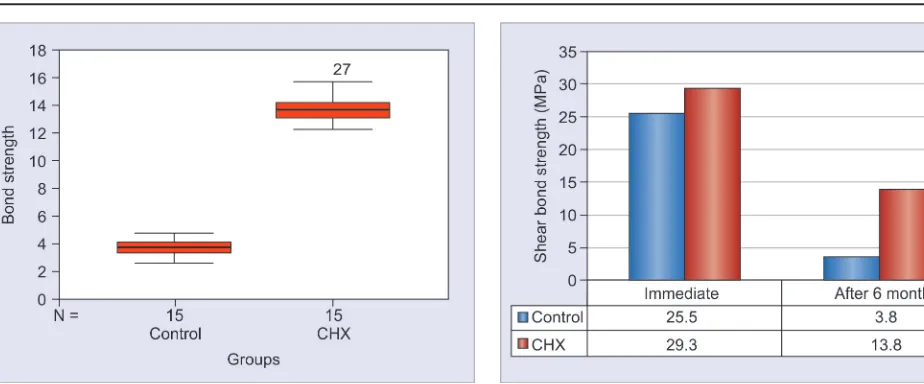
Graph 1: Box plot of the immediate shear bond strength
Institutional ethical committee clearance was obtained before proceeding with the study.
Sixty sound, caries-free human premolars, devoid of developmental anomalies, fluorosis, noncarious lesions, and other surface defects extracted for orthodontic purpose were collected from Department of Oral and Maxillofacial Surgery, Government Dental College, Thiruvananthapuram, Kerala. The samples were collected from patients in the 15 to 30 years age group.
Teeth Selection and Preparation
All the tooth samples were collected, stored, sterilized, and handled as per Occupational Safety and Health Admin-istration (OSHA) and the Center for Disease Control and Prevention (CDC) recommendations and guidelines. Teeth samples were cleansed of saliva, blood, and visible debris and soaked in hydrogen peroxide to remove the connec-tive tissue. They were then washed thoroughly with deion-ized water and used within 6 months of extraction. A flat and superficial dentin surface was exposed on each tooth after wet grinding the buccal enamel with a diamond disk. The enamel-free, exposed dentin surfaces were further polished with wet #600-grit silicon carbide paper for 60 seconds to standardize the smear layer.
Restorative Procedure
Sixty samples were divided into two groups of 30 samples each and prepared as follows:
Group I was taken as the control. The exposed dentin surface was acid-etched with 37% phosphoric acid for 15 seconds. After rinsing with water for 10 seconds and drying the etched surface, rewetting with water for 60 seconds was done, followed by gently blotting away any excess moisture present using a cotton pellet, resulting in a glistening surface. Adper Single Bond 2 (3M ESPE, St. Paul, MN, USA) was applied using an applicator tip. Two coats of dentin bonding agent were applied and gently air thinned for 5 seconds to evaporate the solvent. The adhesive was light cured with a halogen light set at 600 mW/cm2 for 10 seconds.
Group II was taken as the experimental group. The same protocol as above was followed, but for the difference in rewetting agent 2% CHX gluconate [Asep-RC, Anabond Stedman Pharma Research (P) Ltd.] was employed during the rewetting procedure.
After tooth sample preparation, composite resin (Filtek™ Z350 XT Universal Restorative, 3M ESPE) build-up was done on the bonded surfaces. Two increments of 1.5 mm each were placed into a brass ring of uniform dimensions (3 mm radius and 3 mm height)
bonding procedures were carried out by a single operator in a standardized environment.
From each group, half of the specimens were immediately subjected to a shear bond strength test after storage for 24 hours at 37°C and the other half after long-term storage of 6 months in artificial saliva.
Shear Bond Strength Testing
Shear bond strength was determined using a universal testing machine (INSTRON 3365, UK) at a cross-head speed of 1.00 mm/minute. The tooth sample was placed such that the tooth substrate–adhesive interface lay paral-lel to the direction of application of force. The force was applied by means of pulling a wire looped around the composite cylinder. Bond strength was calculated from the load applied and the area of the restoration as shear bond strength = load/area and expressed in megapascals (MPa).
Statistical Analysis
The data was tabulated using Microsoft Excel 2007 and analyzed using Statistical Package for the Social Sciences (SPSS) version 11.0 software. The mean and standard deviations were calculated for each group. The results were analyzed using independent t-test. The difference was considered statistically significant if the p-value obtained was < 0.05. The intergroup comparisons were made using independent t-test.
RESULTS
Effect of 2% Chlorhexidine on the Bond Strength of Direct Composite Restoration to Dentin: An in vitro Study
IJPCDR
Graph 2: Box plot of shear bond strength (after 6 months)
Graph 2 is a box plot showing the comparison between mean shear bond strength values of the two groups after the 6-month storage period. The mean shear bond strength values were 3.8 MPa (control) and 13.8 MPa CHX. Considerable reduction in the bond strength values was observed in both groups over the 6-month period. Independent t-test was used to compare the shear bond strengths of the two groups. The difference between values of both groups was observed to be statistically significant.
Comparison of Shear Bond Strength within the Groups
Mean shear bond strength values obtained in each group immediately and after 6 months were found to be different.
Table 1 provides a comparison of mean shear bond strength values within the control group between the immediate and after 6-month storage period.
Table 2 shows a comparison of mean shear bond strength values within the CHX group between the immediate and after 6-month storage period.
Graph 3 both the groups showed a reduction in shear bond strength following the 6-month storage period, with the decrease more apparent in the control group at the 6-month storage period. Paired t-test was used to compare these values and the difference was found to be highly statistically significant in the control and CHX groups.
DISCUSSION
Adhesion or bonding is the process of binding two adjoining materials and providing a resistance to their separation. Acid-etching brought about a substantial improvement in bonding to enamel and the bond was predictable. Durable bonding to dentin, however, has remained an elusive goal probably because of the mor-phological, histological, and compositional differences. Nakabayashi and Pashley (1998) described the nature of the resin–dentin interface as a “hybrid layer” comprising resin and dentin. This layer was found to disintegrate over time,3 primarily by two mechanisms: Hydrolytic and collagenolytic. After etching, the denuded collagen fibrils not enclosed by a protective resin covering stand exposed to the lytic action of endogenous enzymes called matrix metalloproteinases or MMPs. These are a multigene family within the metalloproteinase class of calcium-dependent zinc-containing endopeptidases capable of degrading all extracellular matrix (ECM) and basement membrane components including different collagens in both native and denatured.9,10 Mazzoni et al reported that endogenous MMPs are uncovered and/or activated by many, if not all dentin bonding procedures.
Developing new strategies to prevent the degradation of resin–dentin bonds is crucial to increase the longevity of bonded restorations. Due to the role of MMPs in the time-dependent degradation of this interface, it has been suggested that MMP inhibitors be employed to preserve it. Chlorhexidine, a widely used antimicrobial
Table 1: Comparison of shear bond strength between control groups
Groups Mean SD n Mean % decrease Paired t p-value
Immediately 25.5 6.3 15 85.3 12.87* 0.00 After 3.8 0.6 15
*Significant at 0.01 level
Table 2: Comparison of shear bond strength within the CHX group after 6 months
Groups Mean SD n Mean % decrease Paired t p-value
Immediately 29.3 5.1 15 52.8 11.02* 0.00 After 13.8 1.1 15
*Significant at 0.01 level
mechanism of anti-MMP action of CHX is still unclear. It was observed that adding calcium chloride to assay mixtures containing CHX almost completely prevented the inhibition of MMP. This observation implied that CHX may act via a cation-chelating mechanism. Also, CHX may affect essential sulfhydryl groups and/or cysteine present in the active site of MMPs. At high concentrations above 2%, the inhibitory action of CHX might be related to protein denaturation rather than by chelation of cations.11 Another advantage of using CHX is its substantivity, since it is able to bind to mineralized dentin for at least 12 weeks.12 Additionally, it has been shown that demineralized dentin can bind more CHX than mineralized dentin and that it may remain attached to demineralized dentin even after the application of bonding agent.13 Given that degenerative changes at the resin–dentin interface occur over a period of time, substantivity of CHX is undeniably an asset. Since the binding mechanism of CHX to demineralized dentin is electrostatic in nature and hence reversible,14 CHX molecules could be eventually displaced by competing cations derived from dentinal fluid or saliva and leach out of the denuded collagen matrix. For this reason, there is a prevailing notion that CHX binding to demineralized dentin merely postpones rather than permanently arrest bond degradation. Then again, for how long can CHX extend its protective effect is still a question that requires further research. Chlorhexidine proved to be beneficial only for etch-and-rinse adhesives, as it could not bind to the collagen matrix in the presence of an acidic environment as present in self-etch adhesives.15
In the present study, immediate shear bond strength testing provided values that served as a baseline for comparison with values obtained after the 6-month storage period. Comparison of immediate shear bond strength showed no statistically significant difference between the two groups (p > 0.05). Hence, CHX has no effect – favorable or otherwise, on the resin–dentin bond in the immediate time period. This has been a topic of contention since some studies reported a decrease in the bond strength due to CHX application.16,17 Ricci et al18 in their study found that CHX solution application did lead to high bond strength immediately.
The samples were stored in artificial saliva for a period of 6 months. The values obtained after the storage period are the result of both hydrolytic and collagenolytic disintegration. Since the setting was a static one, the changes that occur in dynamic conditions simulating a clinical setting could not be replicated. Since the samples were not subjected to cyclic loading, the marginal gaps and leakage produced thereof would also be minimal.
interface. The hydrolytic degradation in this scenario would possibly be minimal and the effect is uniform in all the samples.
After the 6-month storage period, the shear bond strength values showed a statistically significant difference (p < 0.01) between the two groups, with higher values for the CHX group. Hence, CHX has a beneficial effect in maintaining the resin–dentin bond strength over a period of 6-month. Intragroup analysis showed the bond strength values to diminish considerably in both the experimental and the control groups and the differences were statistically significant. Since CHX primarily prevents the collagenolytic degeneration, whatever drop in values present must be due to the hydrolytic changes in the interface, in part contributed by the water from the CHX solution itself. Since a 2% aqueous solution of CHX is used in this study, water could not be eliminated completely from the interface. The persistence of lytic changes at the resin–dentin interface could thus be attributed to the compromised moisture control rather than any ineffectiveness of CHX.
Another issue is the longevity of this protection CHX offers against collagenolysis. From the results of this study, it is seen that CHX maintained its protective action over 6 months. However, since the bonding between CHX and collagen is reversible, it is unlikely that this effect continues indefinitely. Further research and long-term clinical studies are required in this regard.
Effect of 2% Chlorhexidine on the Bond Strength of Direct Composite Restoration to Dentin: An in vitro Study
IJPCDR
of 1 to 2, showed no significant effect for CHX on the bond strength values. Furthermore, it can also be rightly assumed that CHX application must be done only onto etched dentin from which the acid has been thoroughly removed, for the desired effect.
CONCLUSION
Within the limitations of the present study, it can be concluded that
• 2% chlorhexidine gluconate did not have any
immediate effect – favorable or otherwise on the bond strength of resin to dentin.
• Rewetting with 2% CHX gluconate had a definite
positive effect on the resin–dentin bond strength over a 6-month storage period.
• There was a definite degradation of resin–dentin bond
strength with time as evidenced by the change in bond strength. This held true in both rewetting with water and 2% CHX gluconate.
ACKNOWLEDGMENTS
Authors would like to thank Dr. V Kalliyana Krishnan, BMT wing, SCTIMST, Poojapura, Thiruvananthapuram, Kerala, India for extending laboratory facilities to conduct this study. The authors are also indebted to Ms. Vibha C for helping with the testing procedures.
REFERENCES
1. De Munck J, Van Landyut K, Peumans M, Poitevin A, Lambrechts P, Braem M, Van Meerbeek B. A critical review of the durability of adhesion to tooth tissue: methods and results. J Dent Res 2005 Feb;84(2):118-132.
2. Breschi L, Mazzoni A, Ruggeri A, Cadenaro M, Di Lenarda R, De Stefano Dorgio E. Dental adhesion review: aging and stability of the bonded interface. Dent Mater 2008 Jan;24(1): 90-101.
3. Hashimoto M, Ohno H, Sano H, Kaga M, Oguchi H. In vitro degradation of resin-dentin bonds analyzed by microtensile bond test, scanning and transmission electron microscopy. Biomaterials 2003 Sep;24(21):3795-3803.
4. Wang Y, Spencer P. Hybridization efficiency of the adhesive- dentin interface with wet bonding. J Dent Res 2003 Feb;82(2): 141-145.
5. Spencer P, Wang Y. Adhesive phase separation at the dentin interface under wet bonding conditions. J Biomed Mater Res 2002 Dec 5;62(3):447-456.
6. Hashimoto M, Ohno H, Kaga M, Sano H, Endo K, Oguchi H. The extent to which resin can infiltrate dentin by acetone-based adhesives. J Dent Res 2002 Jan;81(1):74-78.
7. Pashley DH, Tay FR, Yiu C, Hashimoto M, Breschi L, Carvalho RM, Ito S. Collagen degradation by hostderived enzymes during aging. J Dent Res 2004 Mar;83(3):216-221. 8. Gendron R, Grenier D, Sorsa T, Mayrand D. Inhibition of the
activities of matrix metalloproteinases 2, 8, and 9 by chlorhexi-dine. Clin Diagn Lab Immunol 1999 May;6(3):437-439. 9. Perdigão J. Dentin bonding-variables related to the
clini-cal situation and the substrate treatment. Dent Mater 2010 Feb;26(2):e24-e37.
10. Sulkala M, Wahlgren J, Larmas M, Sorsa T, Teronen O, Salo T, Tjäderhane L. The effects of MMP inhibitors on human sali-vary MMP activity and caries progression in rats. J Dent Res 2001 Jun;80(6):1545-1549.
11. Komori PC, Pashley DH, Tjäderhane L, Breschi L, Mazzoni A, de Goes MF, Wang L, Carrilho MR. Effect of 2% chlorhexidine digluconate on the bond strength to normal versus caries-affected dentin. Oper Dent 2009 Apr;34(2):157-165.
12. Mohammadi Z, Abbott PV. Antimicrobial substantivity of root canal irrigants and medicaments: a review. Aust Endod J 2009 Dec;35(3):131-139.
13. Kim J, Gu L, Breschi L, Tjäderhane L, Choi KK, Pashley DH, Tay FR. Implication of ethanol wet-bonding in hybrid layer remineralization. J Dent Res 2010 Jun;89(6):575-580.
14. Blackburn RS, Harvey A, Kettle LL, Manian AP, Payne JD, Russell SJ. Sorption of chlorhexidine on cellulose: mechanism of binding and molecular recognition. J Phys Chem B 2007 Aug 2;111(30):8775-8784.
15. Curtis RV, Watson TF. Dental biomaterials: imaging, testing and modelling. Ed. by R. Curtis and T. Watson, Woodhead Publishing, Elsevier; 2014. 529p.
16. Gürgan S, Bolay S, Kiremitçi A. Effect of disinfectant applica-tion methods on the bond strength of composite to dentin. J Oral Rehabil 1999 Oct;26(10):836-840.
17. Sharma V, Rampal P, Kumar S. Shear bond strength of com-posite resin to dentin after application of cavity disinfectants – SEM study. Contemp Clin Dent 2011 Jul;2(3):155-159. 18. Ricci HA, Sanabe ME, Costa CA, Hebling J. Effect of
chlorhexi-dine on bond strength of two-step etch-and-rinse adhesive systems to dentin of primary and permanent teeth. Am J Dent 2010 Jun;23(3):128-132.
19. Turkun M, Cal E, Toman M, Toksavul S. Effects of dentin disinfectants on the shear bond strength of all-ceramics to dentin. Oper Dent 2005 Aug;30(4):453-460.
20. Say EC, Koray F, Tarim B, Soyman M, Gülmez T. In vitro effect of cavity disinfectants on the bond strength of dentin bonding systems. Quintessence Int 2004 Jan;35(1):56-60.