CC S.. = BY NC ND
http://doi.org/10.5114/bta.2019.90236
Antioxidant property and GCMS profile of oil extracted
from
Cocos nucifera
using a fermentation method
ALABI A.ADENIKE, PETER ADEGBOLA*, OLUMIDE S.FADAHUNSI Ladoke Akintola University of Technology, Ogbomoso, Nigeria
Abstract
Cocos nucifera L. is known as a tree of life because of its economic, domestic, and nutritional usefulness. Coconut oil (CO), which is derived from Cocos nucifera L., has received considerable attention because of its reported folkloric, nutritional, biological, and pharmacological properties. We previously reported the outstanding physico-chemical properties of CO; therefore, we analyzed its antioxidant activity and physico-chemical composition in this study. CO was extracted using a fermentation method with and without applying heat. Its antioxidant activity was investi-gated using the DPPH free radical scavenging method, the metal chelation capacity, the reduction of antioxidant power, and the nitric oxide scavenging capacity index. The compounds were identified using GC-MS. The data were expressed as a mean ± SEM and analyzed by one-way analysis of variance (ANOVA) using SPSS 21.0 with
data being considered significant at P < 0.05. The results showed that CO demonstrated a
concentration-de-pendent DPPH radical scavenging activity and nitric oxide scavenging capacity index, with the highest activity in the heat-extracted virgin CO (HEVCO). The ferric-reducing antioxidant power and metal chelation capacity
significantly varied (P < 0.05) between the HEVCO and the cold extracted virgin CO (CEVCO). The GCMS
ana-lysis of virgin CO identified important active compounds. The results revealed a higher content of phenolic com-pounds in HEVCO compared to CEVCO. In conclusion, applying heat favored incorporating phenolic comcom-pounds into CO and consequently improved the antioxidant potential of HEVCO compared to CEVCO.
Key words: Coconut oil; antioxidant; heat; phenolics; extraction; fermentation
Introduction
Coconut (Cocos nucifera L.) is a member of the Ara-caceae (palm) family and is extensively grown in the world’s tropical countries (Assa and Konan, 2010). In some parts, coconut tree is referred to as a “tree of life” or a “tree of thousand uses”, thus alluding to its eco-nomic, domestic, and nutritional usefulness, as well as the significance and importance of all the morphological parts of the tree (Bruce, 2004; Gervajio, 2007). Among the plant’s edible products, coconut oil (CO) has gained considerable attention because of its reported folkloric, nutritional, biological, and pharmacological properties (Prades et al., 2012; Shankar et al., 2013). CO is a color-less to pale brownish-yellow oil having a melting point of
23–26EC (Bezard et al., 1971; Thampan, 1988). CO is
derived by drying and pressing the dry pulp (copra) of coconut at a low temperature; however, there are other
methods for extracting CO that have been previously de-scribed in the literature (Aliwalas, 1970; Bernardini, 1970; Cancel et al., 1976; Leonard, 1983). CO is com-posed of fatty acids of some 12 or fewer carbons, which are known and classified as medium chain fatty acids (MCFA) (Babayan, 1988; Heydnger and Nakhasi, 1996; Conrado, 2003). In addition, there is a significant pre-sence of lauric acid in CO, which prevents the deposition of fats in blood vessels and consequently, prevents the risk of atherosclerosis (Conrado, 2003; Popova, 2011; Moigradean et al., 2013; Orsavova et al., 2015). Further-more, CO is reported to be highly resistant to peroxida-tion, a considerably low cholesterol content, as well as to peroxide and iodine value compared to other oils (ground-nut, melon, sunflower, and soybean) (Adegbola et al., 2018). CO has been reported to demonstrate significant beneficial and therapeutic activities such as anti-tumor
(Kopeć et al., 2011), hypocholesterolemic and anti-aging (Adam et al., 2007 ), anti-stress (Nevin and Rajamohan, 2006; Arunima and Rajamohan, 2013; Yeap et al., 2015), hepato-protective (Hanaa et al., 2013; Otuechere et al., 2014), anti-inflammatory and anti-pyretic (Intophuak et al., 2010); wound healing (Nevin and Rajamohan, 2010), blood sugar control (Garfinkel et al. 1992; Kasai et al., 2003) and sight-enhancing (Haliza et al., 2015) activities. These biological potentials are primarily in-fluenced by and attributed to presence of polyphenols, vitamin E, and lauric acid (Adam, 2007; Marina et al., 2009; Arlee et al., 2013 and Yousefi et al., 2013). CO’s physicochemical properties were established in our pre-vious study (Adegbola et al., 2018); however, this study evaluated the antioxidant activity and chemical compo-sition of CO, which was extracted by different methods.
Materials and methods
Materials
Coconut fruits were purchased from a local market in Esa-Oke, Osun, and all other chemicals that were used were of analytical grade.
Extraction of coconut oil
Matured coconuts were first crushed to obtain the coconut fruits. Then, the back of the fruits was removed and washed, and the fruits were ground and sieved such that we could obtain some coconut milk. Virgin CO (VCO) was then extracted from the coconut milk using a fermentation method. Subsequently, coconut milk was extracted from freshly harvested coconuts and fermen-ted by airborne lactic acid bacteria for 72 h (Srivastava et al., 2016), after which the oil phase was separated from the aqueous phase using a syringe. The resultant wet oil was heated at a low temperature for a short time period to remove the moisture and then finally filtered. For extracting the oil without heat, after separating into the oil and aqueous phase, the resultant oil in the upper phase was slowly dispensed using a syringe and then finally filtered (Marina et al., 2009).
DPPH radical scavenging activity of CO
The DPPH scavenging activity was determined using Brand-Williams et al.’s (1995) with slight modification. For preparing the stock solution, 40 mg was dissolved in methanol (100 ml) missing 3.5 ml of the stock solution
with methanol, the absorbance was obtained using a UV spectrophotometer at a wavelength of 517 nm.
Approxi-mately 100 μl of the oil sample with 1 ml methanol
DPPH solution was prepared and maintained in the dark for 2 h to allow the scavenging reaction to occur. The percentage of DPPH scavenging activity was calculated as follows:
DPPH scavenging activity (%) = [(A blank !A sample) / A blank] × 100
where A is the absorbance.
Ferric Reducing Antioxidant Power of CO
The ferric reducing antioxidant power (FRAP)of CO
was determined using Benzie and Strain’s (1996) me-thod as described by Ishtiaq et al. (2014). The FRAP reagent was prepared fresh using a 300 mM acetate buffer at pH 3.6 (3.1 g sodium acetatetrihydrate, 16 ml glacial acid made up to 1 : 1 with distilled water), 10 mM (2,4,6-tris (2-pyridyl)-s-triazine) in 40 mM HCl, and 20 mM FeCl3@6H2O in the ratio of 10 : 1 : 1 to afford the working reagent. Approximately 100 μl of the extracted sample was added to 1 ml FRAP reagent and the absor-bance was measured at 595 nm after 30 min. Trolox’s calibration curve was set up to estimate the activity ca-pacity of the samples. The result was expressed as milli-gram of Trolox equivalents per 100 milli-gram of the sample (mgTE/100 g of FW).
Determination of metal chelating activity of CO Chelation of Metal Ions – Cu2+
A violet-colored pyrocatechol reagent was used for determining Cu2+ chelating activity, as described by Saiga et al. (2003). A mixture of 1.0 ml of sodium acetate buf-fer (100 mM, pH 4.9), 100 ml of Cu (II) standard solu-tion (1.0 mg/ml), and 100 ml of the sample (200 μg) was prepared in a test tube and allowed to react for 5 min at room temperature. Then, to the mixture, 25 ml of a vio-let-colored pyrocatechol solution (4.0 mM) was added and the absorbance was obtained at 632 nm. The che-lating activity was extrapolated using the following for-mula:
Chelating activity (%) = (1 ! sample A632/control A632) × 100
Chelation of Metal Ions – Fe2+
of sodium acetate buffer (100 mM, pH 4.9), 100 ml of Fe(II) standard solution (1.0 mg/ml), and 100 ml of the sample (200 μg) was prepared in a test tube and allowed to react for 5 min at room temperature. Then, 50 ml of
a ferrozine solution (40 mM) was added and the Fe2+
chelating activity was determined by measuring the
for-mation of the Fe2+-ferrozine complex at 562 nm. The
following formula was used to extrapolate the
Fe2+ chelating activity (%) = (1 ! sample A562/control A562) × 100
where control A562 is the absorbance of control reaction (without sample) and sample A562 is the absorbance in the presence of a sample.
Nitric oxide radical scavenging activity of CO
To determine the nitric oxide radical scavenging acti-vity, reaction mixtures containing 2.0 ml of 10 mM NaN3 in a phosphate-buffered saline (pH = 7.4) and 1.0 ml of various concentrations (20–80 μg/ml) of the oil were in-cubated at 25EC for 150 min. Next, 1.0 ml of 0.33% sul-fanilic acid in 20% glacial CH3COOH was added to 0.3 ml of the incubated solution and allowed to stand for 5 min. Then, 0.5 ml of 0.1% (w/v) napthylethylenediamine di-hydrochloride was added to the mixture and incubated at 25EC for 30 min (Kuate et al., 2010; Kumar et al., 2010). The absorbance was measured at λmax = 540 nm using quercetin as a blank. Then, the nitric oxide sca-venging capacity index (NOSCI) of the oil sample was calculated as follows:
NOSCI % = (1 ! absorbance of test) / absorbance of blank) × 100
The NOSCI % was expressed as scavenging capacity index (SCI50), which is defined as the concentration (μg/ml) of the extract required to scavenge 50% of NO!
.
GCMS analysis of CO
The detection of compounds in VCO was performed on a GC-MS system (Agilent Technologies 789OA coupled with MSD VL5975C) with HP5MS column (30 × 0.0320 mm × 0.25 μm) we used He as the carrier gas at a constant flow rate of 2 ml/min. The sample in-jection volume was 1 ml, and the oven temperature was programmed to 80EC for 2 min at a rate of 10EC/min to 240EC with a holding time of 6 min. The samples were then run at a mass spectral scan range of 35!550 (m/z). The compounds were then identified by comparing the spectrum of the separated components with that of MS
library 2014, the National Institute of Standards and Technology (NIST), Maryland, USA.
Statistical analysis
All data were expressed as mean ± SEM. The values were analyzed by one-way analysis of variance (ANOVA) using Statistical Package for Social Sciences (SPSS) 21.0. The difference between the means was analyzed by Duncan’s multiple range test (DMRT) at P < 0.05.
Results and discussion
DPPH radical scavenging activity
The DPPH radical reaction has an advantage that the reaction process is not interrupted by certain side re-actions such as metal-ion chelation and enzyme inhibi-tion because of various additives. Moreover, the DPPH test is important because it recognizes free-radical sca-venging effects and not pro-oxidant activity (Ruiz et al., 2015). The method is based on the ability of antioxidants to donate protons and thus neutralize the free radical character of the DPPH and produce the non-radical DPPH (Saiqa et al., 2014). Figure 1 shows the DPPH ra-dical scavenging activity of VCO extracted with and without heat. A significant (P < 0.05) increase was obser-ved in the activity of VCO in a concentration-dependent manner. Furthermore, the activity in the HEVCO signi-ficantly increased (P < 0.05) compared with CEVCO. The highest activity of 10.6% and 8% was observed at
80 μg/ml for HEVCO and CEVCO, respectively. The
ob-servation made in this study supported the antioxidant activity previously described by Arlee et al. (2013) for VCO extracted using the fermentation method. The oil extracted by the fermentation process goes through va-rious processing steps such as heating during fermenta-tion and drying off water from the oil. Because phenolics are polar, they are easily dissolved in the aqueous phase of coconut milk; thus, the removal of water by heat might have incorporated more phenolic compounds into the HEVCO. Therefore, the removal of water might be responsible for the higher anti-oxidant activity observed in HEVCO.
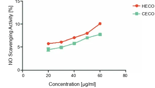
Nitric oxide radical scavenging activity
Concentration [ g/ml]:
D
P
P
H
s
cav
en
gi
n
g a
ct
ivi
ty
[
%
] 15
10
5
0
0 20 40 60 80 100
HECO CECO
HECO CECO 15
10
5
0
0 20 40 60 80
Concentration [ g/ml]:
N
O
Sc
av
e
ng
in
g
Ac
tiv
ity
[
%
]
Fig. 1. DPPH radical Scavenging activity of CO extracted by the cold and hot method; the values are expressed as mean ± SEM and considered significant at P < 0.05; CEVCO – cold
extracted virgin CO, HEVCO – hot extracted virgin CO
a significant role in various inflammatory processes. A persistent increase in the concentration of NO radical in the vascular endothelium causes direct toxicity to tis-sues and contributes to vascular collapse, which is as-sociated with septic shock (Tylor et al., 1997). Further-more, the chronic expression of NO radical is associated with various carcinoma and inflammatory conditions such as juvenile diabetes, multiple sclerosis, arthritis, and ulcerative colitis (Tylor et al., 1997). The peroxy-nitrite anion (ONOO.) was formed when NO reacted with superoxide radical and had a greater toxicity. More-over, its formation has been connected with reduced NO bioavailability in the vascular endothelium (Adegbola et al., 2017). The NO radical scavenging activity of VCO was measured as the nitric oxide scavenging capacity index increased in a concentration-dependent manner. Figure 2 shows a significant (P < 0.5) difference in the NO scavenging activity of the oil: it was higher in HEVCO than in CEVCO. The observations showed that VCO might possess some ability to prevent NO radical formation and be able to counteract its accompanying role in cardiovascular diseases.
Antioxidant and metal ion chelating activity of CO
The FRAP assay measures the total antioxidant ca-pacity in a sample by considering the oxidation-reduction potential (Ishtiaq et al., 2014). The interaction of anti-oxidants with the ferric tripyridyltriazine complex favors the reduction of the complex to produce ferrous tri-pyridyltriazine with an intense blue color (Gülçin, 2012). In this study, the ferric-reducing antioxidant power was
Fig. 2. Nitric oxide radical scavenging activity of CO extracted by the cold and hot method; the values are expressed as mean ± SEM and considered significant at P < 0.05; CEVCO – cold
extracted virgin CO, HEVCO – hot extracted virgin CO
measured as the equivalent of Trolox. As listed in Table 1, HEVCO showed the highest ferric-reducing potential of 93.15 ± 1.20 mg TE / 100 g. This ferric-reducing pro-perty is by breaking a free radical chain by donating a hydrogen atom (Duh et al., 1999). The metal ion chela-ting capacity is an important mechanism of antioxidant activity. Transition metals, especially ferrous ions, are potent catalysts that are capable of initiating lipid per-oxidation via a Fenton reaction in the cellular membrane (Moguel-Ordóñez et al., 2015). Moreover, ferrous ions can accelerate the peroxidation by the decomposition of lipid hydroperoxides into both alkoxyl and peroxyl radi-cals that are capable of abstracting hydrogen and ini-tiating the chain reaction of lipid peroxidation (Halliwell, 1991; Chang et al., 2002). The chelation of metal ions is a key strategy to avoid the generation of free radicals that are associated with redox-active metal catalysis (Mo-guel-Ordóñez et al., 2015). The significant metal ion che-lation observed with VCO, the highest in the HEVCO, is an indication of its potential to break the chain of lipid peroxidation (Table 1). Among the various transition me-tals, iron is known as the most important lipid oxidation pro-oxidant because of its high reactivity (Halliwell and Gutterridge, 1984; Kuate et al., 2010). As observed in
this study, VCO is a better Fe2+ chelator with 36.3
± 0.14% and 15.65 ± 0.49% activity than Cu2+ with 31.9
± 0.42% and 14.55 ± 0.21% activity for HEVCO and
Table 1. Antioxidant activity of CO extracted by the hot and cold method
Sample [mgTE/100 g]FRAP Cu2+ chelating activity[%] Fe2+ chelating activity[%]
HEVCO 93.15 ± 1.20a 15.65 ± 0.49a 36.3 ± 0.14a
CEVCO 90.49 ± 0.81b 14.55 ± 0.21b 31.9 ± 0.42b
The values are expressed as mean ± SEM and considered significant at P < 0.05; the value with different superscripts varied significantly; CEVCO – cold extracted virgin CO; HEVCO – hot extracted virgin CO
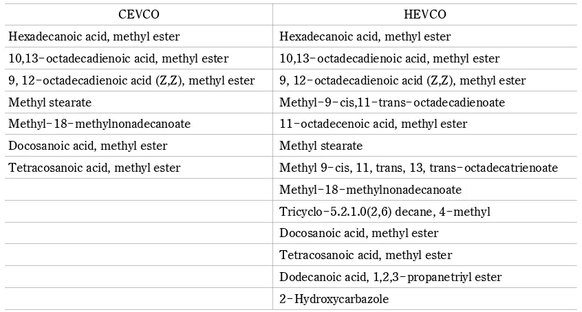
Table 2. Compounds identified in VCO extracted with cold and heat
CEVCO HEVCO
Hexadecanoic acid, methyl ester Hexadecanoic acid, methyl ester
10,13!octadecadienoic acid, methyl ester 10,13!octadecadienoic acid, methyl ester
9, 12!octadecadienoic acid (Z,Z), methyl ester 9, 12!octadecadienoic acid (Z,Z), methyl ester
Methyl stearate Methyl!9!cis,11!trans!octadecadienoate
Methyl!18!methylnonadecanoate 11!octadecenoic acid, methyl ester
Docosanoic acid, methyl ester Methyl stearate
Tetracosanoic acid, methyl ester Methyl 9!cis, 11, trans, 13, trans!octadecatrienoate
Methyl!18!methylnonadecanoate Tricyclo!5.2.1.0(2,6) decane, 4!methyl Docosanoic acid, methyl ester
Tetracosanoic acid, methyl ester
Dodecanoic acid, 1,2,3!propanetriyl ester 2!Hydroxycarbazole
CEVCO – cold extracted virgin CO; HEVCO – hot extracted virgin CO
study on HEVCO. As previously reported, heat favored the incorporation of additional phenolics into the VCO sample extracted with heat. Therefore, a higher phenolic content of HEVCO could contribute to the observed anti-oxidant activity.
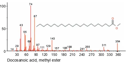
GC-MS profiles of compounds in CO
The constituents in VCO were identified using GCMS. The identified compounds in the HEVCO and CEVCO included certain fatty acids, as shown in Table 2. The fatty acid content included palmitic acid (C16 : 0), stearic acid (C18 : 0), oleic acid (C18 : 1), and PUFAs such as linoleic acid (C18 : 2) and linolenic acid (C18 : 3). Figure 3 and Figure 4 show the spectra and structure of the identified compounds. Some of these identified com-pounds have been associated with several biological functions such as antioxidant, inflammatory, anti-microbial, anticancer, and hypocholesterolemic activities
(Yu et al., 2005; Intahphuak et al., 2010; Chandrasekaran et al., 2011; Jegadeeswari et al., 2012; Upgade and Anusha, 2013; Parthipan et al., 2015).
(rume-Hexadecanoic acid, methyl ester
100
50
0
20 40 60 80 100 120 140 160 180 200 220 240 260 280
15 29
43 55 74
83 87
97 115129
143
157171 185 199213 227
239 270
O
O
100
50
0
20 40 60 80 100 120 140 160 180 200 220 240 260 280 300
15 29
41 55
59 67
81
95
109
123 135 150
184 178
191203 220233245 263
279 294
9, 12 octadecadienoic acid (Z,Z) , methyl ester ! !
O
O
20 40 60 80 100 120 140 160 180 200 220 240 260 280 300 100
50
0 100
50
0
O
O
O
O
30 60 90 120 150 180 210 240 270 300 330 360 390
1829 43
57
69 87
97 111 129
143
157 199 227242 283293 339 382 74
29 41
55
59 67
81
95
109
123 136 150
164 178
192204 220234246 263
279 294
Tetracosanoic acid, methyl ester 10, 13 octadecadienoic acid, methyl ester!
100
50
0
20 40 60 80 100 120 140 160 180 200 220 240 260 280 300
15 29 4355
83 87
97 111129
143
157 171185199213 227 241 255
287 298 74
1529 43
55
83 97 111129 87 74
143
157 171185199213227241255269 283
297 311 328
O
O
O O 100
50
0
20 40 60 80 100 120 140 160 180 200 220 240 260 280 300 320 340
Methyl 18 methylnonadecanoate! Methyl stearate
100
50
0
30 60 90 120 150 180 210 240 270 300 330 360 O
O
14 29
43
55
69 74
87
97 111 129
143
157 185199 241255 311
354
Docosanoic acid, methyl ester
Methyl 9 cis, 11 trans, 13 trans octadecadienoate! ! ! ! !
100
50
0
60 80 100 120 140 160 180 200 220 240 260 280 300
55
87
O
O
67 79
93
107 121
135 150
163
175 189 203 217 235 249261 292
Methyl 9 cis, 11 trans octadecadienoate! ! ! !
100
50
0
60 80 100 120 140 160 180 200 220 240 260 280 300
81
55
59 67
95
109
121 135 150 164 178
191201 220234245 262 294
O O
Tricyclo[5,2,1,0(2,6)]decane, 4 methyl! !
100
50
0
57 27
29
31 41
39
51 53
55
63 73 77
79 81 67
93 109
115 121
135
150
20 30 40 50 60 70 80 90 100 110 120 130 140 150 160
11-octadecanoic acid, methyl ester
20 40 60 80 100 120 140 160 180 200 220 240 260 280 300 100
50
0
O
O
29 41
55
59 69
87 97
111
123 137 152 166180192 204 222 246 264
296 74
100
50
0 15
29 43
55
83 87
97 111129
143
157171185199213227241255 297 326 74
O
O
Methyl 18 methylnonadecanoate!
283 269 311
29 43
57
183
98
311
129
71 257
227 298
O O 100
50
0
50 100 150 200 250 300 350 400 450 500 550 600 650
Dodecanoic acid, 1, 2, 3 propanetriyl ester!
200 325
367 425 480
550 620 508 395
439
O O O O
2 hydroxycarbazole!
100
50
0
NH
OH
27 384451 63 68 7782 87 91
101 110 127137 154
167 183
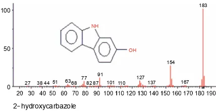
nic acid) is an isomer of conjugated linoleic acid (CLA) found in natural products (Kramer et al., 1998) that was reported to possess anti-inflammatory activity (O’Shea et al., 2004). 2!hydroxycarbazole is a phenolic alkaloid belonging to a wide group of compounds called carba-zole, which have diverse biological activities such as anti-oxidative, anti-inflammatory, antibacterial, and cytotoxic activities (Zhang et al., 2010; Bandgar et al., 2012; Bia-monte et al., 2013; Peng et al., 2013). The presence of
2!hydroxycarbazole in the HEVCO might have
influen-ced its antioxidant activity via the carbazole N!H and hydroxyl moiety. Lauric acid (dodecanoic acid) is one of the most abundant and important fatty acids found in CO (Ghani et al., 2018). Note that it was not detected by GC-MS analysis. Moreover, studies have reported that lauric content of CO varies with location, the variety of crop (Laureles et al., 2000), age of nuts (Balleza and Sierra, 1976), and time of harvest (Carandang, 2008).
Depending on the extraction method, compounds identified in the VCO varied among the samples, and they were present in higher amounts in HEVCO than in CEVCO. Previously, studies have reported that a higher temperature favors the incorporation of more phenolic compounds into oil (Marina et al., 2009; Srivastava et al., 2016). This might explain the reason for the number of compounds identified in the HEVCO. Because phenolics are polar and are easily dissolved in the aqueous layer of coconut milk, compared to the oil layer during fermenta-tion, some phenolic in CEVCO might have been lost during collection (Arlee et al., 2013).
Conclusions
This study confirms that CO has some antioxidant activity, which is responsible for its medicinal and nutri-tional benefits. In this study, the extraction methods influenced the activity and constituents of CO. More-over, it is evident that applying heat favored the incorpo-ration of phenolics into CO and consequently improved the antioxidant activity of HEVCO compared to CEVCO.
References
Adam S.K., Sulaiman N.A., Mat A.G., Jaarin K. (2007) Heating
reduces vitamin E content in palm and soy oils. MJBMB 15: 76–79.
Adegbola P., Aderibigbe I., Hammed W., Omotayo T. (2017)
Antioxidant and anti-inflammatory medicinal plants have
potential role in the treatment of cardiovascular disease: a review. Amer. J. Cardiovasc. Dis. 7(2): 19–32.
Adegbola P., Fadahunsi O.S., Alabi A.A. (2018) Comparative
antioxidant study of ripe and unripe plantain and the qualitative assessment of some food oil extracts. Annals. Food Sci. Technol. 19(4): 758–765.
Aliwalas A.R., Buccat C.P. (1970) Filtration extraction of gra-nulated coconut on a bench scale. Phillip. J. Sci. 96(3): 215–285.
Arlee R., Suanphairoch S., Pakdeechanuan P. (2013)
Differen-ces in chemical components and antioxidant-related sub-stances in virgin coconut oil from coconut hybrids and their parents. Food Res. Int. 20(5): 2103–2109.
Arunima S., Rajamohan T. (2013) Effect of virgin coconut oil
enriched diet on the antioxidant status and paraoxonase 1 activity in ameliorating the oxidative stress in rats – a com-parative study. Food Funct. 4(9): 1402–1409.
Assa R.R., Konan J.L. (2010) Physicochemical characteristics
of kernel during fruit maturation of four coconut cultivars (Cocos nucifera L.). Afr. J. Biotechnol. 9(14): 2136–2144. Babayan V.K. (1988) Medium chain triglycerides. In dietary fat
requirements in health and development. Ed. C.J. Beare-Rogers. AOCS press, Champain, Illinois (USA): 73–86.
Balleza C.F., Sierra Z.N. (1976) Proximate analysis of the
coconut endosperm in progressive stages of development. Philippines J. Crop Sci. 1: 37–44.
Bandgar B.P., Adsul L.K., Chavan H.V., Jalde S.S., Shringare S.N., Shaikh R., Meshram R.J., Gacche R.N., Masand V. (2012) Synthesis, biological evaluation, and docking stu-dies of 3-(substituted)-aryl-5-(9-methyl-3-carbazole)-1H-2-pyrazolines as potent anti-inflammatory and antioxidant agents. Bioorg. Med. Chem. Lett. 22: 5839–5844. Benzie I.F.F., Strain J.J. (1996) The ferric reducing ability of
plasma (FRAP) as a measure of ‘‘antioxidant power”: The FRAP assay. Anal. Biochem. 239: 70–76.
Bernardini E. (1970) Direct extraction of oil from oilseeds
without pressing. Riv. Ital. Sost. Gras. 47(8): 385–391. Bezard J., Bugaut M., Clement G. (1971) Triglyceride
composi-tion of coconut oil. J. Am. Oil Chem. Soc. 48(3): 134–139.
Biamonte M.A., Wanner J., Le Roch K.G. (2013) Recent
ad-vances in malaria drug discovery. Bioorg. Med. Chem. Lett. 23: 2829–2843.
Brand-Williams W., Cuvelier M.E., Berset C. (1995) Use of
a free radical method to evaluate antioxidant activity. Le-bensmitt.Wissensch. Tech. 28: 25–30.
Bruce F. (2004) The coconut oil miracle. Avery, USA: 1–7.
Cancel L.E., Rosario J.A., Hernandez E.R. (1976) Coconut oil
extraction from coconut milk presscake. J. Agric. Uni. Puerto Rico. 60(3): 281–293.
Carandang E.V. (2008) Health benefits of virgin coconut oil. www.pcrdf.org/artimages%5CVCO.doc.
Carter P. (1971) Spectrophotometric determination of serum
iron at the submicrogram level with a new reagent (ferro-zine). Anal. Biochem. 40: 450–458.
Chandrasekaran M., Senthilkumar A., Venkatesalu V. (2011)
esters from leaves of Sesuvium portulacastrum L. Eur. Rev. Med. Pharmcol. Sci. 15: 775–780.
Chang L.W., Ye W.J., Huang S.C., Duh P.D. (2002) Antioxidant
activity of sesame coat. Food Chem. 78: 347–354.
Conrado S.D. (2003) Coconut oil: atherogenic or not? (What
therefore causes atherosclerosis?). Philipp J. Cardiol. 31(3): 97–104.
Duh P.D., Tu Y.Y., Yen G.C. (1999) Antioxidant activity of the aqueous extracts of harng Jyur (Chrysanthemum mori-folium Ramat). LWT – Food Sci. Technol. 32: 269–277. Garfinkel M., Lee S., Opara E.C., Akwari O.E. (1992)
Insulino-tropic potency of lauric acid: a metabolic rationale for medium chain fatty acids (MCF) in TPN formulation. J. Surg. Res. 52(4): 328–333.
Gervajio G.C. (2005) Fatty acids and derivatives from coconut oil. John Wiley & Sons, USA: 1–56.
Ghani N.A.A., ChanniP A.A., Hwa P.C.H., Ja’afar F., Yasin
H.M., Usman A. (2018) Physicochemical properties,
anti-oxidant capacities, and metal contents of virgin coconut oil produced by wet and dry processes. Food Sci. Nutr. 6: 1298–1306.
Gülçin ¤. (2012) Antioxidant activity of food constituents: an overview. Arch. Toxicol. 86(3): 345–391.
Haliza A.M., Sharanjeet K., Ahmad R.G., Ng Chinn H., Nor H.A.S. (2015) Pilot study: the efficacy of virgin coconut oil as ocular rewetting agent on rabbit. Evid. Based Compl. Alt Med. 2015: 135987.
Halliwell B. (1991) Reactive oxygen species in living systems. Source biochemistry and role in human disease. Amer. J. Med. 91: 14–21.
Halliwell B., Gutteridge J.M. (1984) Oxygen toxicology,
oxy-gen radicals, tran-sition metals and disease. Biochem. J. 219: 1–4.
Hanaa M., Abd E.F., Lamiaa A.A.B. (2013) Hepatoprotective
effect of olive and coconut oils against oxidative stress-induced by 2, 4 dichlorophenoxyacetic acid. Indian J. Appl. Res. 3(12): 42–46.
Harada H., Yamashita U., Kurihara H., Fukushi E., Kawabata
J., Kamei Y. (2002) Antitumor activity of palmitic acid
found as a selective cytotoxic substance in a marine red alga. Antican. Res. 22: 2587–2590.
Heydnger J.A., Nakhasi D.K. (1996) Medium chain
triacyl-glycerols. J. Food Lipids.: 251–257.
Intahphuak S., Khonsung P., Panthong A. (2010)
Anti-inflam-matory, analgesic, and antipyretic activities of virgin coco-nut oil. Pharm Biol. 48(2): 151–157.
Ishtiaq S., Ahmad M., Hanif U., Akbar S., Mehjabeen S.H.K. (2014) Phytochemical and in-vitroantioxidant evaluation of different fractions of Amaranthus graecizans subsp. Silves-tris (Vill.) Brenan. Asian Pac. J. Trop. Biomed. 4(12): 965–997.
Jegadeeswari P., Nishanthini A., Muthukumaraswamy S.,
Mo-han V.R. (2012) GC-MS analysis of bioactive components
of Aristolochia krysagathra (Aristolochiaceae). J. Curr. Chem. Pharm. Sci. 2: 226–236.
Kasai M., Nosaka N., Maki H., Negishi S., Aoyama T., Naka-mura M., Suzuki Y., Tsuji H., Uto H., Okazaki M., Kondo
K. (2003) Effects of dietary medium and long-chain triacyl-glycerols (MLCT) on accumulation of body fat in healthy humans. Asia Pac. J. Clin. Nutr. 12(2): 151–160.
Kopeć A., Nowacka E., Piątkowska E., Leszczyńska T. (2011)
Charakterystyka i prozdrowotne właściwości steroli roś
lin-nych. Zywn. Nauk Technol. 3(76): 5–14.
Kramer J.K.G., Parodi P.W., Jensen R.G., Mossobad M.M.,
Yuraweczd M.P., Adlof R.O. (1998) Rumenic acid: a
pro-posed common name for the major conjugated linoleic acid isomer found in natural products. Lipids. 33(8): 835. Kuate D., Etoundi B.C.O., Soukontoua Y.B., Ngondi J.L., Oben
J.E. (2010) Comparative study of the antioxidant, free
radical scavenging activity and human LDL oxidation in-hibition of three extracts from seeds of a cameroonian spice, Xylopia parviflora (A. Rich.) Benth. (Annonaceae).
Int. J. Biomed. Pharm. Sci. 5(1): 18–30.
Kumar B.S.A., Lakshman K., Jayaveera K.N., Shekar D.S.,
Kumar A.A., Manoj B. (2010) Antioxidant and antipyretic
properties of methanolic extract of Amaranthus spinosus leaves. Asian Pacific J. Tropical Med. 3(9): 702–706. Laureles L.R., Rodriguez M.A.A., Caraos C.E., Reano G.A.,
Santos L.A.C., Mendoza E.M.T. (2000) Storage lipid varia-bility in promising coconut cultivars and hybrids: fatty acids and triacylglycerol composition. Philippines J. Crop Sci. 25: 42–54.
Leonard E. (1983) Sri Lankan inventor who makes life easier
for his countrymen. Cocomunity, APCC/QS/45/83,33–35. Mangunwidjaja D.S., Kardono S.R., Iswantini L.B.S.D. (2006)
Gas chromatography and gas chromatography – mass spectrometry analysis of Indonesian Croton tigliumseeds. J. Appl. Sci. 6: 1576–1580.
Maria J.R.P., Kannan P.S.M., Kumaravel S. (2011) GC-MS
Analysis of Lantana camara L. leaves. JPRD 2(11): 63–66. Marina A.M., CheMan Y.B., Nazimah S.A., Amin I. (2009)
Antioxidant capacity and phenolic acids of virgin coconut oil. Int. J. Food Sci. Nutr. 60(S2): 114–123.
Moguel-Ordóñez Y.B., Cabrera-Amaro D.L., Segura-Campos
M.R., Ruiz-Ruiz J.C. (2015) Studies on drying
characte-ristic, nutritional composition, and antioxidant properties of Stevia rebaudiana (Bertoni) leaves. Int. Agrophys. 29: 323–331.
Moigradean D., Poiana M.A., Alda L.M., Gogoasa I. (2013)
Quantitative identification of fatty acids from walnut and coconut oils using GC-MS method. J. Agroaliment Proc. Technol. 19(4): 459–463.
Nevin K.G., Rajamohan T. (2006) Virgin coconut oil
supple-mented diet increases the antioxidant status in rats. Food Chem. 99: 260–266.
Nevin K.G., Rajamohan T. (2010) Effect of topical application of virgin coconut oil on skin components and antioxidant status during dermal wound healing in young rats. Skin
Pharmacol. Physiol.23(6): 290–297.
O’Shea M., Bassaganya-Riera J., Mohede I.C.M. (2004)
Im-munomodulatory properties of conjugated linoleic acid. Am. J. Clin. Nutr. 79: S1199–1206.
Orsavova J., Misurcova L., Ambrozova J.V., Vicha R., Mlcek J.
contribution to dietary energy intake and dependence of cardiovascular mortality on dietary intake of fatty acids. Int. J. Mol. Sci. 16: 12871–12890.
Otuechere C.A., Madarikan G., Simisola T., Bankole O., Osho A. (2014) Virgin coconut oil protects against liver damage in albino rats challenged with the anti-folate combination, trimethoprim-sulfamethoxazole. J. Basic Clin. Physiol. Pharmacol. 25(2): 249–253.
Parthipan B., Suky M.G.T., Mohan V.R. (2015) GC-MS
Ana-lysis of Phytocomponents in Pleiospermium alatum (Wall. ex Wight & Arn.) Swingle, (Rutaceae). J. Pharmacogn. Phytochem. 4(1): 216–222.
Peng W.W., Zeng G.Z., Song W.W., Tan N.H. (2013) A new
cytotoxic carbazole alkaloid and two new other alkaloids from Clausena excavata. Chem. Biodivers. 10: 1317–1321.
Popova T. (2011) Fatty acid composition and oxidative
sta-bility of muscles in lambs fed coconut oil supplemented diet. Bulg. J. Agric. Sci. 17(3): 402–409.
Prades A., Dornier M., Diop N., Pain J.P. (2012) Coconut
water uses, composition and properties: a review. Fruits. 67: 87–107.
Ravi L., Krishnan K. (2017) Cytotoxic potential of
N-hexa-decanoic acid extracted from Kigelia pinnata leaves. Asian J. Cell Biol. 12: 20–27.
Ruiz J.C.R., Ordoñez Y.B.M., Basto A.M., Campos M.R.S.
(2015) Antioxidant capacity of leaf extracts from two
Stevia rebaudiana Bertoni varieties adapted to cultivation in Mexico. Nutr. Hosp. Aria. 31(3): 1163–1170.
Saiga A., Tanabe S., Nishimura T. (2003) Antioxidant activity of peptides obtained from porcine myofibrillar proteins by protease treatment. J. Agric. Food Chem. 51: 3661–3667. Saiqa I., Mansoor A., Uzma H., Shehla A., Mehjabeen Sairah H.K. (2014) Phytochemical and in-vitro antioxidant evalua-tion of different fracevalua-tions of Amaranthus graecizan ssubsp.
Silvestris (Vill.) Brenan. Asian Pac. J. Trop. Biomed. 4(12): 965–971.
Shankar P., Ahuja S., Tracchio A. (2013) Coconut oil: a
re-view. Agro Food Ind. Hi-Tec. 24(5): 62–64.
Srivastava Y., Semwal A.D., Majumdar A. (2016) Quantitative
and qualitative analysis of bioactive components present in virgin coconut oil. Cogent Food Agricult. 2: 1164929.
Thampan P.K. (1988) Glimpses of coconut industry in India.
Coconut Development Board, Cochin 1988.
Tylor B.S., Kion Y.M., Wang Q.I., Sharpio R.A., Billiar T.R., Geller D.A. (1997) Nitric oxide down regulates hepatocyte-induible nitric oxide synthase gene expression. Arch. Surg. 132: 1177–1183.
Upgade A., Anusha B. (2013) Characterization and medicinal
importance of phytoconstiuents of Carica papayafrom down south Indian region using gas chromatography and mass spectroscopy. Asian J. Pharm. Clinical Res. 6(4): 101–106.
Yeap S.K., Beh B.K., Ali N.M., Yusof H.M., Ho W.Y., Koh S.P. (2015) Antistress and antioxidant effects of virgin coconut oil in vivo. Exp. Ther. Med. 9: 1–39.
Yousefi M., Nateghi L., Rezaee K. (2013) Investigation of
physicochemical properties, fatty acids profile and sterol content in Malaysian coconut and palm oil. Ann. Biol. Res. 4(4): 214–219.
Yu F.R., Lian X.Z., Guo H.Y., McGuire P.M., Li R.D., Wang R.,
Yu F.H. (2005) Isolation and characterization of methyl
esters and derivatives from Euphorbia kansui (Euphorbia-ceae) and their inhibitory effects on the humanSGC-7901 cells. J. Pharm. Pharm. Sci. 8: 528–535.
Zhang F.F., Gan L.L., Zhou C.H. (2010) Synthesis,