Sagar Thapa 1 Original Article
Computed Tomography Differential Features of Anterior
Mediastinal Thymoma and Lymphoma
Sagar Thapa 1, Wen Ming1, Xiaofeng Wu1, Christina Gyawali 2, Ruikun Liao1, Zhu Zhang1
1 The First Affiliated Hospital of Chongqing Medical University, Chongqing, China 2 College of Stomatology, Chongqing Medical University, Chongqing, China
Corresponding Author: 13883669699@163.com
Abstract
Background To identify the CT features of anterior mediastinal thymoma and lymphoma and evaluate CT findings that may help in suggesting specific diagnosis among these tumors. Method
A total of fifty-eight chest CT scan studies with pathological diagnosis of thymoma (n = 30) and lymphoma (n =28) were retrospectively reviewed by two radiologists who were blind to the pathological results. The CT features showing cyst and calcification within the lesion, contrast enhancement, relation with pleura and adjacent vessels, bone invasion and lymph node enlargement were evaluated. Results In lymphoma (67.9%), the presence of associated mediastinal lymphadenopathy was significantly compared to thymoma. (P < 0.05). The presence of calcification within the lesion was significantly found in thymoma than the lymphoma (p <0.05). The presence of adjacent vessels relationship within the lesion was higher in lymphoma than the thymoma. (p <0.05) The remaining CT findings showed no significant difference among these diseases (p > 0.05). Conclusion In conclusion age, lymph nodes involvement, calcification and relationship to adjacent vessels on CT scan will be helpful to give specific diagnosis on anterior mediastinum lymphoma and thymoma.
.
Keywords
Mediastinum, Lymphoma, Thymoma, Computed Tomography
Introduction
The mediastinum is the central portion of thoracic cavity which is bounded by thoracic inlet superiorly, pleural cavity laterally and the diaphragm inferiorly1.It is also known as Veritable Pandora box because numerous organs are housed inside and also within which various lesions may occur1. Anterior mediastinum tumor compromises diverse groups of neoplasms and accounts 50% of all mediastinal masses2. Anterior mediastinum mass can be diagnosed as thymoma, thymic cyst, thymic carcinoma, mature teratoma, malignant lymphoma and malignant germ cell tumor2.
Computed tomography (CT) has the ability to demonstrate the extent and character of the lesion as well as the relationship of
masses to adjacent structures3. So, CT can be a choice for evaluating mediastinal masses. It’s very hard to diagnose anterior mediastinal mass based alone on CT findings; so the main aim of present study is to characterize CT features of common anterior mediastinum tumors focusing mainly on thymoma and lymphoma.
Methodology
Subjects
Sagar Thapa 2 in this study. A total of 58 cases were
available namely lymphoma and thymoma were included in our study among which 28 patients are of lymphoma (17 were male and 11 were female: age range: 13-61; mean age 30) and 30 cases of thymoma. (Male were 18 and female 12; age reference 83-30; mean age 52).
CT imaging
The CT examination was performed using a 64- slice helical CT (GE Discovery CT750 HD, Milwaukee, USA). In all patients, the CT examinations used a slice thickness of 5.00mm, a slice increment of 5.00mm, a pitch of 1.08, 120 kVp and 150- 200 mA, and a 512x512 image matrix through the thorax prior to and after intravenous injection of a contrast medium (iohexol; 300 mg iodine/ml; 1.5 ml/kg) at a rate of 3.0 ml/s. All CT images were captured at mediastinal (window width, 400- 450 HU; window level, 30-50 HU) and lung (window width, 1,000-1,500 HU; window level, -650 to – 500 HU) windows.
Image interpretation
CT scan was retrospectively reviewed by two radiologists who had more than 5 years’ experience in a chest CT interpretation. Both radiologists were aware that all patients had anterior mediastinal masses but were blind to the pathological results. CT images were assessed for tumor shape, size, contours, presence of necrotic or cystic portions, calcification within the tumor and also the invasion of adjacent structures. The degree of enhancement was also evaluated. Enlarged mediastinal lymph nodes were assessed.
Statistical Analysis
The difference in CT patterns among lymphoma and thymoma was statistically analyzed by Fisher’s exact test. A p value of 0.05 was considered statistically significant which was evaluated by the SPSS statistical software.
Results
The CT findings of thymoma and lymphoma are summarized and the P value was calculated which is shown in Table I.
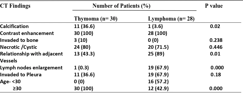
Table I Summary of the CT findings of thymoma and lymphoma.
CT Findings Number of Patients (%) P value
Thymoma (n= 30) Lymphoma (n= 28)
Calcification 11 (36.6) 1 (3.6) 0.02
Contrast enhancement 30 (100) 28 (100)
Invaded to bone 3 (10) 0 (0) 0.238
Necrotic /Cystic 24 (80) 20 (71.5) 0.446
Relationship with adjacent 13 (43.3) 25 (89) 0.01
Vessels
Lymph nodes enlargement 1 (0.3) 19 (67.9) 0.000
Invaded to Pleura 11 (36.6) 19 (67.9) 0.18
Age- <30 0 (0) 16 (57.2)
Sagar Thapa 3 Calcification was seen in 11 patients (36.6%) with thymoma and 1 patient (3.6%) with lymphoma (classical Hodgkin lymphoma misdiagnosed as thymoma). Calcification within the lesion was higher in thymoma than the lymphoma. (p <0.05) (Shown in Figure 1).
Figure 1 A- A 26-year-old female with Hodgkin’s lymphoma (Non- contrast CT) shows heterogeneous soft tissue mass at anterior mediastinum with inner calcification (arrow) B- A 66-year-old male with Thymoma (Non- contrast CT) showing heterogeneous anterior mediastinum mass with calcification (arrow).
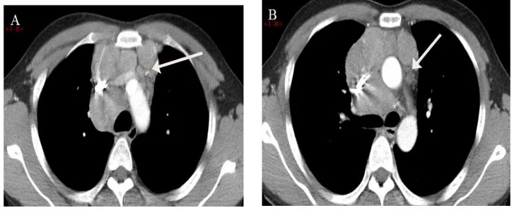
Relationship with adjacent vessels was seen in 13 patients (43.3%) with thymoma and 25 patients (89%) with lymphoma. In lymphoma, the presence of adjacent vessels relationship within the lesion was higher than the thymoma. (p <0.05) (Shown in Figure 2, 3).
Figure 2A- A 42-year-old male with lymphoma (Contrast CT) shows heterogeneous enhancing anterior mediastinum mass with pericardium effusion (arrow).
2B- A 16-year-old male with lymphoma (Contrast CT) shows heterogeneous mild enhancement mass in anterior mediastinum with compression of superior venacava, brachiocephalic trunk, common carotid artery, subclavian artery and
Sagar Thapa 4
Figure 3-A 34-year-old male with thymoma (Contrast CT) shows heterogeneous mild enhancement anterior mediastinum mass with
3A- Compression of superior venacava, brachiocephalic trunk, subclavian artery, common carotid artery (arrows).
3B- Compression of aorta (arrow).
Enlarged lymph nodes (mediastinal lymph nodes) was seen in 1patient (0.3%) with thymoma which was even misdiagnosed as lymphoma priorly and 19 patients (67.9%) with lymphoma (mediastinal lymph nodes-7, axillary lymph nodes-3, supraclavicular-4, and rest being multiple region lymph nodes enlargement). In lymphoma, the presence of lymph nodes within the lesion was higher than the thymoma. (p <0.05) (Figure 4).
Figure 4-A 29 year male with lymphoma shows mild homogenous enhancement with involvement of mediastinum lymph nodes (arrow) (A, B).
Sagar Thapa 5
Figure 5A- An 18-year-old male with lymphoma shows heterogeneous anterior mediastinal mass with huge inner cyst (arrow).
5B- A 38-year-old male with thymoma shows heterogeneous anterior mediastinal mass with inner calcification and cystic changes (arrow).
Pleura were invaded in 11 patients (36.6%) with thymoma and 19 patients (67.9%) with lymphoma. The presence of pleural invasion within the lesion was higher in lymphoma than the thymoma (p <0.05).
The contrast enhancement was seen in 30 patients (100%) with thymoma and 28 patients (100%) with lymphoma. Among 28 cases of lymphoma 23 cases have mild enhancement while 3 cases have moderate and 2 cases have obvious enhancement. Among 30 cases of thymoma 23 have mild enhancement 3 have moderate enhancement and 4 have obvious enhancement.
Adjacent bone was invaded in 3 patients (10%) with thymoma (2 cases were of breast bone destruction and 1 was adjacent rib) while no patients with lymphoma have adjacent bone invasion. The presence of adjacent bone invasion among these tumors showed no significant difference. (p > 0.05).
Age seems to be an important factor as all 30 cases of thymoma were above age of 30 and while for lymphoma 16(57.2%) were below 30 years old and 12(42.9%) were above the age of 30. Age seems to be significant as
lymphoma tends to be on younger patients than thymoma. (p<0.05)
Discussion
Primary anterior mediastinal masses are a heterogeneous group of neoplasm and non-neoplasm which includes congenital cyst, intrathoracic goiter and inflammatory disease4. It affects peoples of all age groups and remains an interesting diagnostic challenge. On CT imaging the commonest location of mediastinal mass is anterior mediastinum. Masses in this area are more likely to be malignant compared to those in other compartments5. Evaluation of mediastinal mass can be easily done by Chest CT studies and is capable of giving limited if not complete diagnosis1, 6.
Sagar Thapa 6 can be classified as Non- invasive (benign)
or invasive (malignant) thymoma. Only about one- third of thymoma is malignant which is characterized by invasion of tumor into mediastinal fat and mediastinal structures rather than the basis of histological9. On CT scan, Thymoma can be visualized as a well-defined homogenous or heterogeneous attenuation depending on the presence of hemorrhage, necrosis or cyst formation10. Focal calcification has been reported in 25% of patients, which is little less compared to our study i.e. 36% 11, 3. Calcification can be an important factor while differentiating thymoma but have to rule out other possible anterior mediastinal tumor such as germ cell tumor...
Lymphoma can be classified as Hodgkin
lymphoma (HL) and non- Hodgkin
lymphoma (NHL). Primary mediastinal lymphoma usually has HL, nodular sclerosing type mostly seen in adults while NHL is common as childhood mediastinal lymphoma. Most common lymphoma of the anterior mediastinum is Nodular sclerosing Hodgkin disease which occurs most commonly in woman12. Both HL and NHL are equally included in our present study. In our study diffuse large B cell, T-lymphoblastic and nodular sclerosing types were seen in almost equal numbers. On CT study, both HL and NHL with enlarged
mediastinal nodes demonstrates
homogenous attenuation with little or no enhancement. 21%-50% cases showed cystic appearance13 while calcification was seen rare prior to therapy but it does occur14. Whereas in our present study, one patient with classic HL (3.6%) had calcification.
Necrotic or cystic component was seen in 20-71.5% of cases. All 100% of lymphoma show contrast enhancement in our present study. HL and NHL most often involve
superior mediastinal lymph nodes (precarina, paratracheal and aortopulmonary nodes) with some different pattern of lymph node involving in these two groups. NHL is characterized by involvement of a single node group and posteriormediastinal nodes more commonly in patients rather than HL3. However, in present study these findings were not encountered. In the present study, the associated lymphadenopathy was seen in 71.5% of lymphoma cases and 25% of cases showed mediastinal invasion. The distribution of lymph node in our study is as: 19 patients (67.9%) have enlarged lymph nodes among which (mediastinal lymph nodes-7, axillary lymph nodes-3, supraclavicular-4, and rest being multiple region lymph nodes enlargement). Therefore, anterior mediastinal mass
associated with mediastinal
lymphadenopathy elsewhere is the strong indication of lymphoma.
Conclusion
In present study, many limitations were encountered which includes very less number of cases, variety of CT scanners used and variety of slice thickness on CT. This is a retrospective study which has a limited possibility of diagnosis (thymoma and lymphoma) which may affect imaging interpretation.
In conclusion age, lymph nodes
involvement, calcification and relationship to adjacent vessels on CT scan will be helpful to give specific diagnosis on anterior mediastinum lymphoma and thymoma.
Conflict of Interest None.
Sagar Thapa 6 References
1. Dasgupta, S., Bose, D., Bhattacharyya, N. K., Saha, M., Biswas, K., & Biswas, P. K. (2016). A clinicopathological study of mediastinal masses operated in a tertiary care hospital in Eastern India in 3 years with special reference to thymoma. Indian Journal of Pathology and Microbiology, 59(1), 20.
2. Tomiyama, N., Honda, O., Tsubamoto, M., Inoue, A., Sumikawa, H., Kuriyama, K., Nakamura, H. (2009). Anterior mediastinal tumors: diagnostic accuracy of CT and MRI. European journal of radiology, 69(2), 280-288.
3. Totanarungroj, K., Watcharaporn, C., & Muangman, N. (2010). Helpful CT findings for giving specific diagnosis of anterior mediastinal tumors. Medical journal of the Medical Association of Thailand, 93(4), 489.
4. Strollo, D. C., de Christenson, M. L. R., & Jett, J. R. (1997). Primary mediastinal tumors. Part 1*: tumors of the anterior mediastinum. Chest, 112(2), 511-522. 5. Aroor, A. R. A Study of Clinical
Characteristics of Mediastinal Mass 77-80.
6. Nenezić, D., & Stević, R. (2009).
Computerized tomography of
mediastinal tumors. Acta chirurgica Iugoslavica, 56(4), 69-75.
7. Wychulis, A. R., Payne, W. S., Clagett, O. T., & Woolner, L. (1971). Surgical treatment of mediastinal tumors: a 40 year experience. The Journal of thoracic and cardiovascular surgery, 62(3), 379. 8. Morgenthaler, T. I., Brown, L. R., Colby,
T. V., Harper, C. M., & Coles, D. T. (1993). Thymoma. Paper presented at the Mayo Clinic Proceedings.
9. Fon, G., Bein, M., Mancuso, A., Keesey, J., Lupetin, A., & Wong, W. (1982).Computed tomography of the anterior mediastinum in myasthenia
gravis. A Radiologic-pathologic correlative study. Radiology, 142(1), 135-141.
10. Rosado-de-Christenson, M., Galobardes, J., & Moran, C. (1992). Thymoma: radiologic-pathologic correlation. Radiographics, 12(1), 151-168.
11. Brown, L. R., & Aughenbaugh, G. L. (1991). Masses of the anterior mediastinum: CT and MR imaging. AJR. American journal of roentgenology, 157(6), 1171-1180.
12. Marchevsky, A. M., & Kaneko, M. (1992). Surgical pathology of the mediastinum: Raven Pr.
13. Salonen, O., Kivisaari, L., & Somer, K. (1984). Differential diagnosis of anterior upper mediastinal expansions by
contrast-enhanced computed
tomography. Computerized radiology, 8(4), 217-222.
14. Panicek, D. M., Harty, M., Scicutella, C., & Carsky, E. (1988). Calcification in untreated mediastinal lymphoma. Radiology, 166(3), 735-736.