Pyelonephritis
WILLIAM F. FALLS, JR., M.D.
Medical Service, Veterans Administration Hospital, and Department of Medicine, Medical College of Virginia, Health Sciences Division of Virginia Commonwealth University, Richmond, Virginia
The purpose of this paper is to review current thinking about pyelonephritis. Pyelonephritis may be defined as a "bacterial infection of the kidney which affects the parenchyma, the pelvis, and the calyces. It
occurs in two forms, acute and chronic."1
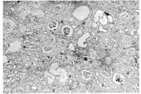
Clinically, acute pyelonephritis is characterized by the symptoms of dysuria, frequency, urgency, chills, fever, and flank pain. The urine sediment con-tains numerous white blood cells, bacteria, and white blood cell casts (Fig I). A quantitative culture of the first voided urine in the morning yields a growth of greater than I ()5 colonies per ml. Renal histologic examination reveals an acute interstitial inflamma-tory reaction with polymorphonuclear leukocytes and microabscess formation (Fig 2). If the infection remits either spontaneously or with antibiotic ther-apy, the involved areas may heal with formation of a contracted, fibrotic scar. In contrast to the acute in-fection, chronic pyelonephritis may be totally asymp-tomatic. Examination of the urine sediment usually shows changes similar to those in acute disease except that the quantities of cells and casts may be Jess remarkable. The classic pathologic picture of chronic pyelonephritis includes interstitial infiltration by mononuclear cells, scarring, tubular dilatation (thy-roidization) and periglomerular fibrosis (Fig 3). The typical chronic pyelonephritic kidney is shrunken, contains multiple scars, and is atrophic.
The pathologic picture of pyelonephritis is not entirely specific, particularly in the chronic state where other processes which cause an interstitial inflammatory reaction may give a similar appear-ance. An incomplete list of nonbacterial causes of
Correspondence and reprint requests to Dr. William F. Falls, Jr., Renal Section, VA Hospital, Richmond, VA 23249.
132
chronic interstitial nephritis includes: analgesic abuse, other drug toxicity, tuberculosis, sarcoidosis, gouty nephropathy, hypercalcemic nephropathy, and ischemic vascular disease.2 Initially, these disorders may be suspected because of the presence of sterile pyuria, but later, superimposed bacterial infection may cloud the picture.
Bacteria invade the kidney by two principal routes: hematogenous and retrograde.3
Hemato-genous spread occurs infrequently, but when it does, it is the means whereby most staphylococcal and streptococcal infections are initiated in the kidney. These infections may be severe and associated with multiple abscess formation. Most bacterial infections of the kidney are caused by gram-negative organisms that reach the kidney via retrograde spread from the lower urinary tract; the natural habitat of the major-ity of these organisms is the gastrointestinal tract from which they spread to the urethra and then into the bladder and up the ureters. Approximately one fourth of all bacterial infections involve the urinary tract, but only a portion of these ascend above the bladder to the renal parenchyma.
Several factors are now recognized as being of major importance in the pathogenesis of urinary tract infection, the first of which is sex. There is clearly a higher incidence in females at all ages but particularly in the childbearing years. Only in later life with the a:dvent of prostatic hypertrophy and associated ob-structive uropathy does the incidence tend to increase in the male. The increased incidence in females prob-ably relates to the shorter urethra, the absence of the antibacterial action of prostatic fluids, and trauma during intercourse. Recently it has also been recog-nized that disturbances in the bacterial resistance of the vaginal vestibule secretions may be the factor
FALLS: PYELONEPHRITIS 133
Fig I-Urine sediment from a patient with acute pyelonephritis, showing white blood cells, a white blood cell cast, a granular cast and numerous bacteria (X 1,000).
which initially allows bacterial colonization of the urethra in females.•
Instrumentation of the urinary tract is frequently associated with introduction of bacteria into the bladder. Unfortunately, the instrumentation is usu-ally undertaken for evaluation of an anatomicusu-ally abnormal tract. Bladder urine is normally sterile. When pathogenic organisms are introduced into the bladder of a normal, unobstructed animal or man, they are rapidly cleared because of the combined effects of the bacteriostatic properties of normal urine, the dilution of organisms by voiding, and the resistance of the bladder mucosa to bacterial coloni-zation.5 On the other hand, in the obstructed state there is a residual pool of relatively stagnant urine; the bacterial clearing mechanisms are no longer oper-ative and infection ensues. In this context neurogenic bladder dysfunction may have the same propensity to infection as overt obstructive uropathy.
In recent years numerous studies have indicated that ureteral reflux is probably the most important factor which allows for initiation and perpetuation of renal parenchymal infection. Congenital reflux be-comes a problem in early childhood and is usually caused by an abnormal placement of the ureter in the bladder wall.6 In the immature kidney it is likely that even sterile reflux may result in calyceal scarring and contracture of the parenchyma. This type of reflux carries an ominous prognosis and frequently requires surgical correction. Acquired reflux (Fig 4) may ap-pear in older individuals and may be related to blad-der infection, adjacent bladder diverticulae, or neuro-genic mechanisms.7 When reflux is severe, it may be associated with intrarenal reflux in the polar regions of the kidney. Some authorities think that this may be the cause of atrophic pyelonephritis.8
134 FALLS: PYELONEPHRITIS
\•
1111
•.
'
.
~
'
·
Fig 2-Light microscopic representation (H & Estain) of a renal biopsy section, showing the picture of acute pyelonephritis. Glomerular architecture is relatively well maintained, but there is a marked interstitial inflammatory reaction with both polymorphonuclear and mononuclear leukocytes. A white blood cell cast fills the collecting duct on the right. (X 80).
relaxing effects of high estrogen concentrations, may predispose to the development of pyelonephritis in pregnancy.a Fully 30% of pregnant patients with
asymptomatic bacilluria will have an episode of acute
pyelonephritis during a given pregnancy if left un-treated.9
Controversy continues to surround the issue of whether or not diabetes mellitus predisposes to the development of pyelonephritis, and recent evidence
suggests that its incidence is higher in diabetic
women.1
°
Clearly, when pyelonephritis develops in a diabetic patient, the infection itself is more likely to be severe.The cortex appears to be relatively resistant to bacterial colonization, and most parenchymal infec-tions begin in the medulla where increased suscepti-bility to infection probably relates to the lower blood
supply to the medulla, the hypertonicity of the
med-ullary interstitium which depresses phagocytosis, the
high level of ammonia which interferes with activa-tion of the complement system, and the tendency for granulocytes to emigrate from the medullary area.a
Eighty-five percent of new urinary tract infec-tions which develop outside the hospital are caused by Escherichia coli organisms and the remaining 15%
by other organisms, most of which are also
gram-negative.11 When infection develops in the hospital setting or occurs after instrumentation, the bacterial flora is likely to be different and is usually composed of gram-negative organisms with a greater degree of bacterial resistance than E coli.
FALLS: PYELONEPHRITIS
135
Fig 3-Light microscopic autopsy section (H & E stain) from a patient with chronic pyelonephritis. Marked interstitial inflammatory reaction in association with scarring, tubular dilatation, and periglomerular fibrosis is present. Secondary arteriolar thickening has also occurred ( X 20).
age if the reflux is not corrected. It has been suggested that the small shrunken kidney seen in later life may be the result of atrophic pyelonephritis in childhood.
Papillary necrosis may be one of the most devas-tating complications of urinary tract infection, partic-ularly if it occurs in diabetes mellitus with obstruc-tion. This constellation of disturbances was formerly associated with a high frequency of sepsis and death, but it is seen less frequently since the advent of mod-ern antibiotic therapy. At the present time it is more likely that papillary necrosis may present insidiously with slow deterioration in renal function, perhaps in conjunction with the passage of small pieces of papil-lary tissue in the urine. Contemporary studies12 sug-gest that this latter picture may be seen more fre-quently with another underlying disease such as analgesic abuse or sickle cell disease, rather than as a manifestation of pyelonephritis (Fig 5).
Struvite stone formation is another common complication of urinary tract infection and is caused by infection with urea-splitting organisms, particu-larly those of the proteus species. This type of infec-tion is seen all too frequently in patients with obstruc-tive uropathy or neurogenic bladder dysfunction. Under either of these circumstances a continuous cycle of stone formation-infection-stone formation may develop. The composition of most "staghorn" calculi which develop in an obstructed, infected uri-nary tract is struvite. Infection is virtually impossible to clear from the urine in this situation.
136
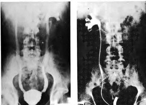
Fig 4-Voiding cystourethrogram in a patient with acute cystitis
and bilateral reflux. Moderate ureteral dilatation is present and the contrast medium refluxes into the calyceal system. The left calyceal
system is also clearly dilated.
as gallium scanning and CT scanning, may be of
great diagnostic aid. In addition, sonography of the
retroperitoneum and abdomen may be of diagnostic
help.
Gram-negative sepsis remains a frequent
compli-cation of urinary tract infection and may be
associ-ated with instrumentation or the presence of an
in-d wel I ing urethral catheter; therapy requires
aggressive treatment with measures to support the
circulation while administering a bactericidal
antibi-otic.
Whether or not chronic pyelonephritis
pre-disposes to the development of severe hypertension
continues to be a subject of debate. Some evidence in both children and adults would suggest that the chronically scarred kidney may be associated with
FALLS: PYELONEPHRITIS
Fig 5-Retrograde urogram in a patient with papillary necrosis
secondary to analgesic abuse. There is marked distortion of the
calyceal system on the right. Necrotic, papillary debris is present in
several calyces, displacing the contrast medium and giving rise to the characteristic "ring" sign.
activation of a pressor hypertensive mechanism;13
however, most patients with chronic infection remain
normotensive or, at worse, mildly hypertensive.
Per-haps a more important aspect of the association is the
fact that progression of pyelonephritis, like that of
any other type of renal disease may be accelerated by
the presence of superimposed hypertension; there-fore, elevated blood pressure in a patient with pyelo-nephritis should be treated vigorously.
A number of disturbances in relation to preg-nancy have been correlated statistically with the
pres-ence of urinary tract infection. The most notable of
these are anemia and decreased birth weight of the
fetus. After a period of skepticism about the relation-ship of these abnormalities to infection, recent studies
ear-FALLS: PYELONEPHRITIS
Cin
mi/min
140
CpAH
m
1
/min
700
137
F.F.
0/o
-.- p
<.025
p<
.005
N.S.f
-
-600
-~
30
D
INITIAL
+
--
••
120
500
....
D
>5 Yff:
400
20
rt
100
80
60
300
40
200
10
20
100
Fig 6-Comparison of mean ± SEM values for C,,,, CPAH, and FF between initial measurements early after injury and similar
deter-minations after more than five years in 18 patients with spinal cord injuries and persislent bacilluria.
lier, there is a high incidence of acute pyelonephritis
in pregnant females with bacill uria, and this condi-tion, clearly, should be treated.9
For years pyelonephritis was considered to be a major cause of end-stage renal disease. Careful eva
lu-ation of patients entering dialysis and transplant pro-grams, however, has indicated that this is not the
case.14 It is likely that many of the cases of interstitial
nephritis which were thought to be caused by
infec-tion were in actuality the end result of one of the other causes of interstitial disease mentioned earlier.
Present estimates would suggest that no more than
15% of the patients reaching end-stage disease have infection as the primary cause of renal failure and virtually all of these are complicated by the presence of stones, reflux, or other anatomic disturbance.
Information about the natural history of urinary
tract infection in man is still incomplete. As noted
above, end-stage renal disease in the absence of an
anatomic or neurologic defect in the urinary tract is rare despite the frequency of culture-proven
bacil-luria. We have recently attempted to define more
clearly the association of chronic bacill uria and
dete-rioration in renal function by a· prospective analysis of the changes in inulin clearance (C1n) and PAH
clearance (CPAH) in a group of patients with per-sistent bacilluria (spinal cord injury patients). C1n is an accurate measure of the glomerular filtration rate
and CPAH indicates the renal plasma flow.
Figure 6 demonstrates that at a mean period of five years after the initial determinations, a
statisti-cally significant reduction in mean CPAH from an initial value of643 ml/min shortly after injury to 556 ml/min at the time of study had occurred in 18 study patients. Over the same period, there was no s ignifi-cant change in Cn; there was also a significant reduc-tion in the filtration fraction (FF). This index reflects
the ratio of C1n/CPAH and is an indicator of the state of perfusion in the kidney. Diseases which produce
renal ischemia may be associated with a low FF. Each of the study patients had a neurogenic bladder and persistent bacilluria. A number had experienced one or more severe complications such as renal
stones, reflux, or sepsis. Yet, at the time of study, all
138
kidneys of these patients; however, barring superim-position of some other insult, renal failure is unlikely to be the cause of their death.
There will always be differences of opinion about the work-up and therapy of patients with urinary tract infection. Outlined below are some principles which are generally applicable to these issues:
1. A quantitative culture, colony count, and an-tibiotic sensitivity determination should be performed in all pregnant women and in all individuals suspected of having a urinary tract infection.
2. A patient with the typical clinical picture of acute pyelonephritis or gram-negative sepsis should be treated with a bactericidal antibi-otic to which the organism is sensitive for at least 10 days.
3. Radiographic studies including an intra-venous pyelogram and voiding cystourethro-gram should be done after the first episode of acute pyelonephritis in the adult male and in all children. Similar studies should be done in any female with recurrent infection or an ele-vated serum creatinine. Further urologic eval-uation should be done in all patients in whom these studies identify a structural abnormal-ity. Surgical correction of obstruction and se-vere reflux should be undertaken. Mild reflux in children may be observed during a period of antibiotic therapy and can be expected to disappear in 50% of cases by age 6 years and in 65% by age 14 years.6
4. The clinical picture of cystitis and asympto-matic bacilluria may be treated with bacte-riostatic agents. If recurrence occurs, ,the above-mentioned radiologic studies should be done. If these are negative, prophylactic or suppressive therapy may be tried. In the fu-ture, tests for antibody coating of bacteria may be helpful in differentiating upper tract infection with renal parenchymal in-volvement from lower tract infection.15
5. Reculture should be done after the com-pletion of therapy for a urinary tract infection and on several occasions over the next two years to determine the presence of recurrence or reinfection.
In summary, current thoughts about the patho-genesis, natural history, complications, and manage-ment of pyelonephritis have been reviewed. It is now clear that chronic urinary tract infection in the ab-sence of some anatomical disturbance or
complica-FALLS: PYELONEPHRITIS
tion seldom leads to renal failure. Efforts should therefore be directed toward discovering and correct-ing derangements in the anatomical integrity of the urinary tract at an early stage of infection.
Acknowledgment: This work was funded by the
Veterans Administration (MRIS 2737). The author is deeply indebted to Dr. Peter Schatzki and Dr. Bar-bara E. Kipreos of the Veterans Administration Hos-pital, Richmond, Virginia, for their assistance in the preparation of the pathologic material presented in the illustrations.
REFERENCES
I. HEPTINSTALL RH: Pathology of the Kidney. 2 ed. Boston, Little Brown and Company, 1974, pp 837-927.
2. SUKI WN, EKNOYAN G: Tubulo-interstitial disease, in Brenner
BM, Rector FC (eds): The Kidney. Philadelphia, WB Saunders
Company, 1976, pp 1113-1144.
3. MoNTGOMERIE JZ, GuzE L: The renal response to infection in Brenner BM, Rector FC (eds): The Kidney. Philadelphia, WB Saunders Company, 1976, pp 1079-1112.
4. FOWLER JE, STAMEY TA: Studies of introital colonization in women with recurrent urinary infections. VII. The role of bacterial adherence. J Urol 117:472-476, 1977.
5. NORDEN CW, GREEN GM, KASS EH: Antibacterial
mecha-nisms of the urinary bladder. J Clin Invest 47:2689-2700, 1968.
6. BELMAN AB: The clinical significance of vesicoureteral reflux. Ped Clin NA. 23:707-720, 1976.
7. AMAR AD, SINGER B, LEWIS R, ET AL: Vesicoureteral reflux in
adults. A twelve-year study of 122 patients. Urology. 3: 184-189, 1974.
8. HODSON J, MAUNG TM, MCMANAMON PJ, ET AL: Reflux
nephropathy. Kidney Int, suppl 4, 1975, pp 50-58.
9. BRUMFITT W: The effects of bacteriuria in pregnancy on mater-nal and fetal health. Kidney Int, suppl 4, 1975, 113-119.
10. FORLAND M, THOMAS v, SHELOKOV A, ET AL: Urinary tract infections in patients with diabetes mellitus. JAMA 238: I 924-1926, 1977.
11. KUNIN CM: Detection, Prevention and Management of Urinary
Tract Infections, 2 ed. Philadelphia, Lea and Febiger, 1974.
12. MAHER JF: Toxic nephropathy, in Brenner BM, Rector FC
(eds): The Kidney. Philadelphia, WB Saunders Company,
1976, pp 1355-1393.
13. KINCAID-SMITH P: Vascular obstruction in chronic
pyelone-phritis kidneys and its relation to hypertension. Lancet
FALLS: PYELONEPHRITIS
14. SCHECHTER H, LEONARD CD, SCRIBNER BH: Chronic pyelo-nephritis as a cause of renal failure in dialysis candidates.
JAMA 216:514-517, 1971.
139