An in vitro Analysis of Different Desensitizing Agents applied on Human Dentin IJPCDR
An in vitro Analysis of Different Desensitizing
Agents applied on Human Dentin
1Poorva Chouksey, 2Adarsha M Shankaregowda, 3Usha H Lingareddy, 4Ashwini Paramashivaiah 5Vijayalakshmi Lakshminarasimhaiah, 6Upasana Lingaiah
IJPCDR
ORIgInal aRtICle 10.5005/jp-journals-10052-0142
1Postgraduate Student, 2Professor, 3Principal, Professor and Head, 4Associate Professor, 5,6Reader and Associate Professor 1-5Department of Conservative Dentistry and Endodontics Vokkaligara Sangha Dental College & Hospital, Bengaluru Karnataka, India
6Department of Oral Medicine and Radiology, V S Dental College & Hospital, Bengaluru, Karnataka, India
Corresponding Author: Adarsha M Shankaregowda, Professor Department of Conservative Dentistry and Endodontics Vokkaligara Sangha Dental College & Hospital, Bengaluru Karnataka, India, e-mail: [email protected]
ABSTRACT
The aim of the study was to evaluate the ability of four topical desensitizing agents on dentinal tubule impediment utilizing confocal laser scanning microscopy (CLSM). Buccal cervical areas of 45 extracted human molars were smoothed and wet-cleaned with SiC paper, trialed by utilization of 17% ethylenedi-aminetetraacetic acid (EDTA) so as to mimic the clinical aspect of overly sensitive dentin of cervical surfaces.
The teeth were randomly divided into one control group (n = 5) and four experimental groups, as indicated by the dentin surface treatments: group I: GLU; group II: MSC; group III: NAN; group IV: TMD; group V: control. The samples were analyzed under the confocal laser examining magnifying lens. The extents of totally blocked, halfway impeded, and open tubules inside each group were calculated.
The ratios of totally and incompletely blocked tubules to the total tubules for all the groups were determined, and the data were statistically analyzed utilizing nonparametric tests and
statistical significance was calculated. The depth of penetration
was greatest for MS Coat (1.35), while it was least for Gluma (1.07). The difference among all the groups was not
statisti-cally significant for depth of penetration. Gluma desensitizer
demonstrated all the more totally impeded tubules (0.44) as well as partially blocked tubules (0.37). The distinctions among all
the groups were statistically significant for tubule impediment (p ≤ 0.05).
Keywords: Confocal laser microscopy, Dentinal hypersensitiv-ity, Topical desensitizing agents.
How to cite this article: Chouksey P, Shankaregowda AM, Lingareddy UH, Paramashivaiah A, Lakshminarasimhaiah V, Lingaiah U. An in vitro Analysis of Different Desensitizing Agents applied on Human Dentin. Int J Prev Clin Dent Res 2018;5(1):5-10.
Source of support: Nil
Conflict of interest: None
INTRODUCTION
Dentin hypersensitivity (DH) is neither an uncommon issue nor a recent one. However, it remains an ineffec-tively understood area and consequently, there appears to be no effective or everlasting treatment for this painful clinical condition. Albeit different hypotheses have been proposed to explain the mechanism of DH. It is as yet indistinct how stimuli applied to the external dentin surface may stimulate nerve fibers.1
Dentin hypersensitivity has been defined as a short, sharp pain arising from exposed dentin in response to stimuli—typically thermal, evaporative, tactile, osmotic, or chemical—and which cannot be ascribed to any other dental defect or disease.2 Dentin sensitivity
(DS) is observed frequently. Its predominance has been accounted for to be between 4 and 69% among adults.3
The existing literature on the prevalence of DH is highly varied as a result of the use of largely different methods of evaluation. The DH varies from 15 to 70 years of age and the peak incidence is between 20 and 40 years.4
The highest incidence of DH has been recorded on the buccal cervical zone of teeth.
The most commonly affected teeth are canines > premolars > incisors > molars. Chronic trauma from toothbrushing, acid erosion from the environment, gastric regurgitation or dietary substances, anatomical factors, the gingival recession caused by periodontitis, or periodontal surgery are some of the factors that have been implicated.5
Strikingly, an essentially higher extent of left vs right contralateral teeth was reported in right-handed patients with DH. Clinical trials have demonstrated that everyday utilization of desensitizing toothpaste twice daily requires 2 to 4 weeks to show any significant desensitization. If after using desensitizing toothpaste, the patient’s DS remains a concern, clinicians should reassess the differen-tial diagnosis and consider in-office treatments beginning with topically applied desensitizing agents.4
For the most part, as a predisposing factor to DH, the dentin needs to become exposed, as a result of the loss of enamel and/or gingival recession.6 Addy et al7
and argues in favor of toothpaste adding to DH, presum-ably because of their abrasiveness.7 The hydrodynamic
hypothesis is the most commonly accepted mechanism to explain DH. As indicated by this hypothesis, painful stimuli coming from the oral environment act on the surface of the uncovered dentin and cause a rapid fluid movement within the dentinal tubules. According to this speculation, proposed by Brännstrom et al,8 clinical
research has been stimulated by proposing two strategies for desensitizing dentin: (1) minimize the capacity of the intradental nerves to respond to fluid movements and (2) decrease stimuli-evoked fluid shifts in the dentinal tubules by reducing dentine permeability.9 The agents
that impede dentinal tubules may cause protein precipita-tion, crystal precipitation on or in dentinal tubules, or con-ventional restorative techniques may block the tubules.10
Dentinal tubules can be obliterated on the surface and/ or occluded with the tubule orifices.
However, superficial occlusion of the tubules can be removed by daily toothbrushing, dissolution of the precipitate facilitated by saliva, or consumption of acidic beverages, causing transient desensitizing effects. Suc-cessful treatment with durable outcome has been related to intratubular deposition, which reduced the fluid flow or totally seals the tubule lumen.11 The mechanism of
action of various chemical desensitizing agents is still not well understood.
Therefore, the purpose of this study was to evaluate the ability of four topical desensitizing agents on the dentinal tubule occlusion using CLSM. Although dif-ferent speculations have been proposed to explain the mechanism of DH, it is still unsure how stimuli applied to the outer dentin surface may stimulate nerve fibers.1
Dentin hypersensitivity has been defined as a short, sharp pain arising from exposed dentin in response to stimuli, typically thermal, evaporative, tactile, osmotic, or chemical, and which cannot be ascribed to any other dental defect or disease.2 Dentin sensitivity is observed
frequently.
Its prevalence has been accounted for to be between 4 and 69% among adults.3 The existing literature on
the prevalence of DH is highly varied as a result of the use of widely different methods of evaluation. The DH varies from 15 to 70 years of age and the peak incidence is between 20 and 40 years.4
The most astounding occurrence of DH has been accounted for on the buccal cervical region of teeth. The teeth most commonly influenced are canines > premolars > incisors > molars. Chronic trauma from toothbrushing, acid erosion from the environment, gastric regurgitation or dietary substances, anatomical factors, the gingival
However, brushing without dentifrice brings down DH scores, while brushing with toothpaste increases them, and contends for toothpaste adding to DH, probably on account of their abrasiveness.7 Therefore,
the purpose of this study was to assess the capacity of four topical desensitizing agents on the dentinal tubule impediment utilizing CLSM. The DH is related to the fluid flow inside the dentinal tubules, and as indicated by Poiseuille’s law, this movement of fluid is directly proportional to the fourth power of the radius.12
As a consequence, any diminution in the radius of the tubule opening would be required to lessen dentin perme-ability and as such should be effective in treating DH.13
The hypersensitive dentin surfaces uncover that they have more patent tubules per unit area than nonsensitive dentin.
Dentin will only be sensitive if the tubules are patent from the pulp to the oral environment, and this patency will alter with formation and elimination of the smear, hence, resulting in an episode condition.14 The present
investigation has demonstrated that the four desensitiz-ing agents tested all impeded dentinal tubules, though to various levels. Absi et al15 and Yoshiyama et al16 reported
that in naturally desensitized dentin, the majority of the tubules were blocked.
Based on transmission electron microscopy (TEM), Yoshiyama et al16 revealed that tubular impediments
could be because of the extension of the intratubular dentin layer or deposition of substances in the tubules. A portion of the impediments in their investigation were crystals of inorganic salts, however, some might be organic in origin. However, the nature of the impeding layer is imperative.
Pashley and Carvalho17 noticed that tubules
appar-ently occluded with a smear plug are porous to both solvent and solute. In this way, the surface appearance alone may not correlate with sensitivity or permeability.18
Research has demonstrated that the ideal DH treatment should imitate normal desensitizing procedures prompt-ing spontaneous impediment of open dentin tubules.19
Gluma desensitizer has the longest history of utilization as a desensitizing agent in clinical settings.
Since glutaraldehyde is a biologic fixative, it has been recommended that the dentinal tubules are impeded as an effect of reaction with plasma proteins from the dentinal fluid that diminishes the diameter of the dentinal tubules. Hydroxyethyl methacrylate (HEMA), a compound of dentin bonding agents, is a hydrophilic monomer with the capacity to penetrate into acid etched and moist dental hard tissue. Subsequently,20 this precipitation advances
An in vitro Analysis of Different Desensitizing Agents applied on Human Dentin IJPCDR
MATERIALS AND METHODS
Forty-five human premolar teeth with no decays or previ-ous restorations, extracted for periodontal reasons, were selected from a pool of freshly extracted teeth. Dental plaque, calculus, and external debris were removed using manual and ultrasonic scalers. The teeth were stored in 1% chloramine T solution at 37°C.
Teeth were decoronated and divided into four experi-mental groups containing 10 samples each and control group of five samples, namely group I: GLUMA power gel by Heraeus Kulzer; group II: MS Coat by Sun Medical; group III: Nanoseal by Nippon; group IV: Teethmate desensitizer by Kuraray Noritake; and group V: Control group. The teeth were sectioned below cementoenamel junction using a water-cooled diamond saw (Struers minitom).
From each buccal surface of the tooth crown, a sec-tioned sample (5 mm length × 3 mm width) was obtained including the cervical area. To remove enamel and expose the underlying dentin cervical area, each fragment was ground (600-grit) flat on a polishing machine. The exposed dentin surfaces were wet-polished with 1000- and 1200-grit silicon carbide abrasive paper using Struers Labapol-5, simulating hypersensitive dentin in cervical regions. The exposed dentinal surface was cleaned with EDTA (pH 7.4) for removal of the smear layer using 17% EDTA liquid for 4 minutes (every 30 seconds replaced). The products were applied according to manufacturer’s instructions.
Then the samples were examined by CLSM into two planes, x–y (treated surface) and x–z or y–z (optical sec-tions perpendicular to the treated surface) at 50× mag-nification to clearly visualize the dentinal tubules and possibly precipitated salts, crystals, protein coagulation. The total number of tubules was counted from the various images captured by the CLSM.
Out of the total tubules, those that were completely impeded, incompletely occluded, and open tubules were counted. The ratio of completely impeded tubules to the total tubules as well as the ratio of incompletely occluded tubules to the total tubules was calculated. The results were statistically compared using one-way analysis of variance (ANOVA) followed by Tukey’s post hoc analysis.
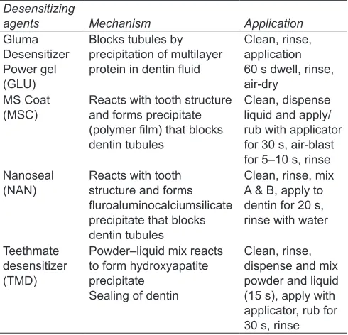
The desensitizing agents, composition, and manufac-turer are given in Table 1. Desensitizing agents, mecha-nism, and application are given in Table 2.
RESULTS
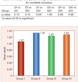
The depth of penetration was highest for MSC and lowest for Gluma. However, insignificant differences were detected regarding the depth of penetration of four desensitizing agents into the dentinal tubules.
The partial occlusion was greatest for Gluma and least for Toothmate. Analysis of variance revealed significant differences among partial occlusion for the desensitiz-ing agents between Gluma and Nanoseal; Gluma and Toothmate (Tables 3 to 10 and Graphs 1 to 3).
The complete occlusion was greatest for Gluma fol-lowed by MSC and NAN, and was found to be least for Toothmate. Moreover, ANOVA followed by Tukey’s
post hoc analysis revealed significant differences among complete occlusion for the desensitizing agents between Gluma and Toothmate.
DISCUSSION
Gluma desensitizer has the mechanism that purportedly depends on aggregate or incomplete occlusion of the
Table 1: Desensitizing agents, composition, and manufacturer
Desensitizing
agents Composition Manufacturer
Gluma desensitizer power gel (GLU)
Gluteraldehyde,
hydroxyethylmethacrylate, pyrogenic silica, water, dye
Heraeus Kulzer, Hanau, Germany MS Coat (MSC) Polymethylmethacrylate,
polystyrene sulfonic acid copolymer, oxalic acid, fluoride, water
Sun Medical Co., Shiga, Japan
Nanoseal (NAN)
Fluoride-calcium-aluminum-silicate glass in aqueous dispersion Phosphoric acid aqueous solution Nippon Shika Yakuhin Co., Ltd., Shimonoseki, Japan Teethmate desensitizer (TMD) Powder: tetra-calcium phosphate, dicalcium phosphate anhydrous Liquid : water and preservative
Kuraray Noritake Dental Inc., Okayama, Japan
Table 2: Desensitizing agents, mechanism, and application
Desensitizing
agents Mechanism Application
Gluma Desensitizer Power gel (GLU)
Blocks tubules by precipitation of multilayer protein in dentin fluid
Clean, rinse, application 60 s dwell, rinse, air-dry
MS Coat
(MSC) Reacts with tooth structure and forms precipitate (polymer film) that blocks dentin tubules
Clean, dispense liquid and apply/ rub with applicator for 30 s, air-blast for 5–10 s, rinse Nanoseal
(NAN) Reacts with tooth structure and forms fluroaluminocalciumsilicate precipitate that blocks dentin tubules
Clean, rinse, mix A & B, apply to dentin for 20 s, rinse with water
Teethmate desensitizer (TMD)
Powder–liquid mix reacts to form hydroxyapatite precipitate
Sealing of dentin
tubules by protein coagulation and precipitation upon response with glutaraldehyde and hydroxyethyl meth-acrylate.21 In a spectroscopic examination, the response
mechanism among glutaraldehyde and 2-HEMA was portrayed as a two-step reaction.
Glutaraldehyde reacts with serum albumin inciting precipitation that causes a second step polymerization
Table 5: Mean partial obliteration of tubules
Group I Group II Group III Group IV
0.58 0.36 0.46 0.32
0.32 0.4 0.44 0.2
0.44 0.36 0.3 0.32
0.5 0.28 0.24 0.36
0.5 0.34 0.22 0.3
0.36 0.44 0.32 0.3
0.34 0.32 0.36 0.48
0.48 0.42 0.44 0.44
0.38 0.3 0.46 0.32
0.48 0.38 0.32 0.38
Table 6: Comparison of mean partial occlusion between four study groups using one-way ANOVA test followed by Tukey’s post hoc
analysis
Groups n Mean SD Standard error Min Max f-value p-value
I 10 0.37 0.12 0.04 0.24 0.64 4.343 0.01* II 10 0.27 0.11 0.04 0.12 0.46
III 10 0.23 0.11 0.03 0.08 0.42 IV 10 0.21 0.08 0.02 0.08 0.36 SD: Standard deviation; *p-value ≤0.05 is significant
Table 7: Multiple comparison using Tukey’s post hoc analysis for partial occlusion
Group GI vs GII GI vs GIII GI vs GIV GII vs GIII GII vs GIV GIII vs GIV
p-value 0.19 0.03* 0.01* 0.83 0.60 0.98 *p-value ≤0.05 is significant
Table 9: Comparison of mean complete occlusion between four study groups using one-way ANOVA test followed by Tukey’s
post hoc analysis
Groups n Mean SD Standard error Min Max f-value p-value
I 10 0.44 0.08 0.03 0.32 0.58 3.114 0.04* II 10 0.36 0.05 0.02 0.28 0.44
III 10 0.36 0.09 0.03 0.22 0.46 IV 10 0.34 0.08 0.02 0.2 0.48 SD: Standard deviation; *p-value ≤0.05 is significant
0.64 0.22 0.22 0.24
0.26 0.18 0.34 0.26
0.44 0.34 0.16 0.2
0.34 0.46 0.34 0.08
0.42 0.2 0.42 0.24
0.38 0.16 0.16 0.18
0.34 0.34 0.08 0.14
0.28 0.42 0.24 0.36
0.32 0.12 0.16 0.24
Table 10: Multiple comparison using Tukey’s post hoc analysis for complete occlusion
Group GI vs GII GI vs GIII GI vs GIV GII vs GIII GII vs GIV GIII vs GIV
p-value 0.13 0.10 0.04* 1.00 0.95 0.98 *p-value ≤0.05 is significant
Graph 1: Comparison of mean depth of penetration between four study groups
Group I Group II Group III Group IV
0.24 0.26 0.18 0.18
0.64 0.22 0.22 0.24
0.26 0.18 0.34 0.26
0.44 0.34 0.16 0.2
0.34 0.46 0.34 0.08
0.42 0.2 0.42 0.24
0.38 0.16 0.16 0.18
0.34 0.34 0.08 0.14
0.28 0.42 0.24 0.36
0.32 0.12 0.16 0.24
Table 4: Comparison of mean depth of penetration between four study groups using one-way ANOVA test
Groups n Mean SD Standard error Min Max f-value p-value
I 10 1.07 0.38 0.12 0.47 1.56 0.666 0.58 II 10 1.35 0.61 0.19 0.73 2.85
An in vitro Analysis of Different Desensitizing Agents applied on Human Dentin IJPCDR
of HEMA.22 Specimens treated with Gluma desensitizer
demonstrated a resinous layer of thickness 1 to 2 µm blocking the surface of the tubules. This is as per the findings of Joshi and Gowda23 and Arrais et al.24 For its
water solvency, HEMA is outstanding, and the HEMA substance of Gluma may hence, advance profound infiltration of the GA component into the tubules.25 This
would explain the greater presence of precipitates in the tubules. MSC contains oxalic acid and a fluoride contain-ing acid polymer.
As indicated by the manufacturer, calcium oxalate upon application to dentin is precipitated and the acid polymer is claimed to give a surface sealing film. NanoSeal is a desensitizing compound introduced in the Japanese market recently. Regarding the composition, this product seems to be a spin-off from silicate cement.
It is hypothesized that, upon application of the acidic mix to the dentin surface CaF2, Ca3PO4 and phosphosili-cate are precipitated into dentinal tubule entrances and on intertubular dentin. Immediately after application and throughout the entire assessment time, visual analog scale rating was reduced by almost three scores. The slight regain in sensitivity recorded at 6 months recall might indicate that the precipitate is gradually removed by mechanical action and/or erosion in dietary acids. Teeth-mate Desensitizer is a calcium phosphate-based Teeth-material. Amid over two decades, there has been extensive enthusiasm to create calcium phosphate mixes for treat-ment of DH. Calcium phosphate compounds are trans-ferred to hydroxyapatite, the main mineral phase in teeth. This means that such products can be characterized as true biocompatible and biomimetic materials.
Teethmate Desensitizer is the first calcium phosphate-containing desensitizer biomimetic materials to be marketed. In order to remove smear layer, control group specimen were polished and later etched showed patent
tubules and similar tubule density as described for sensi-tive areas.
In our investigation, the vast majority of the tubules in the control group was observed to be open, with some of them blocked with a smear layer. On the contrary, a large portion of the tubules in the segments treated with desensitizing agents—GLU, MSC, NAN, TMD—were partially or completely occluded.
In the present investigation, we have demonstrated that professionally applied in-office products containing Gluma desensitizer, MSC, NAN, and TMD are equipped for blocking the dentin tubules to varying degrees and may have clinical potential to decrease DH. All desensi-tizers impeded the tubules, yet Gluma has demonstrated prevalent outcomes regarding tubule impediment.
The outcome of the present study is constrained to physical findings of the change in the dentinal tubules and do not present in vivo differences that may be produced because of the physiological impact of these desensitizing operators. Contrasts between our outcomes and those of other studies may be related to the dentin specimen used, etching process, time and application mode of desensitiz-ing agents, or a combination of these variables.
CONCLUSION
Overall, Gluma desensitizer was found to be most effec-tive. The newer materials MSC, NAN, and TMD appear to be promising in-office desensitizing agents. However, further research is required to access the dissolution resistance or solubility level of precipitates that occlude the dentinal tubules and also their ability to reduce fluid flow through dentin.
REFERENCES
1. Pereira JC, Segala AD, Gillam DG. Effect of desensitizing agents on the hydraulic conductance of human dentin
Graph 2: Comparison of mean partial occlusion between four
Consensus-based recommendations for the diagnosis and management of dentin hypersensitivity. J Can Dent Assoc 2003 Apr;69(4):221-226.
3. Guentsch A, Seidler K, Nietzsche S, Hefti AF, Preshaw PM, Watts DC, Jandt KD, Sigusch BW. Biomimetic mineralization: long-term observations in patients with dentin sensitivity. Dent Mater 2012 Apr;28(4):457-464.
4. Pashley DH, Tay FR. Dentin hypersensitivity: current state of the art and science. Dentin Hypersensitivity Consensus Monogr 2008 Jan;4(9 Special Issue):1-12.
5. von Troil B, Needleman I, Sanz M. A systematic review of the prevalence of root sensitivity following periodontal therapy. J Clin Periodontol 2002 Dec;29(Suppl 3):173-177.
6. Shiau HJ. Dentin hypersensitivity. J Evid Based Dent Pract 2012 Sep;12(3 Suppl):220-228.
7. Addy M, Absi EG, Adams D. Dentine hypersensitivity. The effects in vitro of acids and dietary substances on root-planed and burred dentine. J Clin Periodontol 1987 May;14(5): 274-279.
8. Brännstrom M, Astrom A. A study on the mechanism of pain elicited from the dentin. J Dent Res 1964 Jul-Aug;43: 619-625.
9. Wang Z, Sa Y, Sauro S, Chen H, Xing W, Ma X, Jiang T, Wang Y. Effect of desensitising toothpastes on dentinal tubule occlusion: a dentine permeability measurement and SEM
in vitro study. J Dent 2010 May;38(5):400-410.
10. Pereira JC, Martineli AC, Tung MS. Replica of human dentin treated with different desensitizing agents: a methodological SEM study in vitro. Braz Dent J 2002;13(2):75-85.
11. Pashley DH, Carvalho RM, Pereira JC, Villanueva R, Tay FR. The use of oxalate to reduce dentin permeability under adhesive restorations. Am J Dent 2001 Apr;14(2):89-94. 12. Pashley DH. Dentin permeability and its role in the
pathobio-logy of dentin sensitivity. Arch Oral Biol 1994 Feb;39:S73-S80. 13. Brauer DS, Karpukhina N, Law RV, Hill RG. Structure of
fluoride-containing bioactive glasses. J Mater Chem 2009 Jun;19(31):5629-5636.
15. Absi EG, Addy M, Adams D. Dentine hypersensitivity: a study of the patency of dentinal tubules in sensitive and non-sensitive cervical dentine. J Clin Periodontol 1987 May;14(5):280-284.
16. Yoshiyama M, Masada J, Uchida A, Ishida H. Scanning electron microscopic characterization of sensitive vs. insensitive human radicular dentin. J Dent Res 1989 Nov;68(11):1498-1502. 17. Pashley DH, Carvalho RM. Dentine permeability and dentine
adhesion. J Dent 1997 Sep;25(5):355-372.
18. Orchardson, R. Strategies for the management of dentine hypersensitivity. In: Addy M, Embery G, Edgar WM, Orchard-son R, editors. Tooth wear and sensitivity: clinical advances in restorative dentistry. London: Martin Dunitz; 2000. pp. 315-325. 19. Markowitz K, Pashley DH. Discovering new treatments for
sensitive teeth. The long path from biology to therapy. J Oral Rehabil 2008 Apr;35(4):300-315.
20. Swift EJ, Perdigao J, Heymann HO. Bonding to enamel and dentin: a brief history and state of the art. Quintessence Int 1995 Feb;26(2):95-110.
21. Morris MF, Davis RD, Richardson BW. Clinical efficacy of two dentin desensitizing agents. Am J Dent 1999 Apr;12(2): 72-76.
22. Qin C, Xu J, Zhang Y. Spectroscopic investigation of the function of aqueous 2-hydroxyethylmethacrylate/glutaral-dehyde solution as a dentin desensitizer. Eur J Oral Sci 2006 Aug;114(4):354-359.
23. Joshi S, Gowda AS, Joshi C. Comparative evaluation of NovaMin desensitizer and Gluma desensitizer on dentinal tubule occlusion: a scanning electron microscopic study. J Periodontal Implant Sci 2013 Dec;43(6):269-275.
24. Arrais CA, Chan DC, Giannini M. Effects of desensitizing agents on dentinal tubule occlusion. J Appl Oral Sci 2004 Jun;12(2):144-148.