stage-specific larval camouflage
pattern-associated genes in the swallowtail butterfly,
Papilio xuthus
Futahashi
et al.
R E S E A R C H A R T I C L E
Open Access
Comprehensive microarray-based analysis for
stage-specific larval camouflage
pattern-associated genes in the swallowtail butterfly,
Papilio xuthus
Ryo Futahashi
1,2, Hiroko Shirataki
1, Takanori Narita
3,4, Kazuei Mita
5and Haruhiko Fujiwara
1*Abstract
Background:Body coloration is an ecologically important trait that is often involved in prey-predator interactions through mimicry and crypsis. Although this subject has attracted the interest of biologists and the general public, our scientific knowledge on the subject remains fragmentary. In the caterpillar of the swallowtail butterflyPapilio xuthus, spectacular changes in the color pattern are observed; the insect mimics bird droppings (mimetic pattern) as a young larva, and switches to a green camouflage coloration (cryptic pattern) in the final instar. Despite the wide variety and significance of larval color patterns, few studies have been conducted at a molecular level compared with the number of studies on adult butterfly wing patterns.
Results:To obtain a catalog of genes involved in larval mimetic and cryptic pattern formation, we constructed expressed sequence tag (EST) libraries of larval epidermis forP. xuthus, andP. polytesthat contained 20,736 and 5,376 clones, respectively, representing one of the largest collections available in butterflies. A comparison with silkworm epidermal EST information revealed the high expression of putative blue and yellow pigment-binding proteins inPapilio species. We also designed a microarray from the EST dataset information, analyzed more than five stages each for six markings, and confirmed spatial expression patterns by whole-mountin situ hybridization. Hence, we succeeded in elucidating many novel marking-specific genes for mimetic and cryptic pattern formation, including pigment-binding protein genes, the melanin-associated geneyellow-h3, the ecdysteroid synthesis enzyme gene3-dehydroecdysone 3b-reductase, andPapilio-specific genes. We also found many cuticular protein genes with marking specificity that may be associated with the unique surface nanostructure of the markings. Furthermore, we identified two transcription factors,spaltand ecdysteroid signal-relatedE75, as genes expressed in larval eyespot markings. This finding suggests thatE75 is a strong candidate mediator of the hormone-dependent coordination of larval pattern formation.
Conclusions:This study is one of the most comprehensive molecular analyses of complicated morphological features, and it will serve as a new resource for studying insect mimetic and cryptic pattern formation in general. The wide variety of marking-associated genes (both regulatory and structural genes) identified by our screening indicates that a similar strategy will be effective for understanding other complex traits.
Keywords:Butterfly, color pattern evolution, expressed sequence tag (EST), larval body marking, microarray,Papilio polytes, Papilio xuthus
* Correspondence: [email protected]
1
Department of Integrated Biosciences, Graduate School of Frontier Sciences, University of Tokyo, Kashiwa, Chiba 277-8562, Japan
Full list of author information is available at the end of the article
Background
Body coloration is an ecologically important trait that is often involved in prey-predator interactions through mimicry and crypsis (camouflage) [1]. Because butter-flies and moths spend most of their lives as larvae, which have soft bodies, they have developed a wide range of mechanisms to protect themselves from preda-tors such as birds. The larval body markings of butter-flies differ completely between closely related species and between individuals of the same species in different life stages [2,3]. Despite the variety and significance of the larval color patterns, few studies have been con-ducted at a molecular level compared with the available research on adult butterfly wing patterns [4-9].
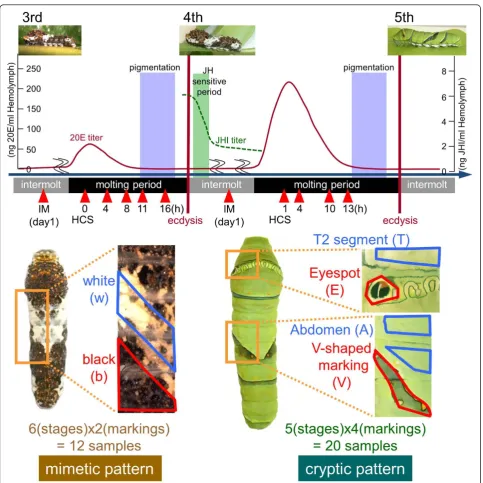
Spectacular changes in color pattern are observed in the caterpillar of the swallowtail butterflyPapilio xuthus. As a young larva, it mimics bird droppings (mimetic pat-tern), and during the final molting period, it switches to a green camouflage coloration (cryptic pattern). In addi-tion, larvae in the final instar stage have a large eyespot on their thoracic segment that is believed to be useful for avoiding predation. One of the main factors involved in larval pattern formation is insect hormones. Previously, we revealed that larval pattern switch is regulated by juvenile hormone (JH) [10]. The decline of JH titers on the first day of the fourth instar stage was the important factor controlling the formation of a green cryptic pattern in the fifth instar stage. The pattern transition occurs through ecdysis, and ecdysteroids appear to regulate the expression of several pigmentation genes. Topical appli-cation of 20-hydroxyecdysone alters the expression tim-ing of several pigmentation genes [11,12]. Compartim-ing gene expression between stages and markings represents a promising strategy for identifying the genes responsible for larval pattern formation.
Previously, we found several pigmentation genes using a cDNA subtraction and candidate gene approach [10,11,13-16]. Six melanin synthesis (or associated) genes,tyrosine hydroxylase (TH), dopa decarboxylase
(DDC),yellow,tan,laccase2andguanosine triphosphate cyclohydrolase I(GTP-CH I) are highly expressed in the presumptive black regions andebonyis highly expressed in the presumptive red region [16]. We also reported that the combination ofbilin-binding protein 1 (BBP1) and
yellow-related gene(YRG) correlated perfectly with larval blue, yellow, and green coloration in threePapiliospecies [12,17]. However, it was difficult to obtain novel mark-ing-specific genes expressed at particular stages or genes with relatively low expression. For butterfly adult wing patterns, expressed sequence tag (EST) construction [18-20] and microarray analysis [6,21] have been con-ducted to obtain marking-associated genes. Microarray analysis revealed that the expression of both a patterning
gene (transcription factor geneoptix) and effector gene (ommochrome synthesis genecinnabar) are associated with red wing patterns inHeliconiusspecies [6,21].
In this study, we constructed an EST library of larval epidermis with more than 20,000 clones, and designed a microarray with the aim of comprehensively revealing the molecular mechanisms of larval mimetic and cryptic pat-tern formation. We performed microarray-based screening for marking-specific genes using six markings at 11 differ-ent stages, and verified the marking specificity of candidate genes by whole-mountin situhybridization. We identified many novel marking-specific genes, including novel blue and yellow pigment-binding protein genes; a novelyellow
family gene, the expression of which prefigures black cuti-cular markings; cuticuti-cular protein genes associated with marking-specific cuticular nanostructures; marking-asso-ciated regulatory genes; marking-specific ecdysteroid synthesis pathway genes; andPapilio-specific marking-associated genes. The data presented in this study provide a new resource to understand insect mimetic and cryptic pattern formation.
Results and discussion
Construction of aPapilioexpressed sequence tag database
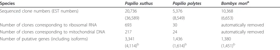
To comprehensively search the genes involved in larval body pattern formation, we constructed cDNA libraries derived from the whole epidermis during the third and the fourth molting periods, and sequenced 20,736 clones of
P. xuthusand 5,376 clones ofP. polytes(Table 1). We clas-sified 4,114 and 1,614 nonredundant EST clusters and sin-gletons fromP. xuthusandP. polytes, respectively (Table 1 and see Additional files 1, 2, 3 and 4). From these, 773 and 178 clusters were considered isoforms or premature forms of other clusters (Additional files 3 and 4). Excluding these clusters, we identified 3,341 and 1,436 putative gene clus-ters (Additional files 1 and 2) forP. xuthusandP. polytes, respectively [DDBJ: AK401027-AK405767]. We assigned a serial number to each cluster in descending order of clone numbers (Additional files 1 and 2). We also determined full-length cDNA sequences for the 150 most highly expressed genes ofP. xuthusthrough random amplifica-tion of cDNA ends technique. Among 3,341 gene clusters ofP. xuthus, 2,746 genes had ORFs encoding predicted proteins longer than 50 amino acids, of which 2,276 had significant sequence similarities to monarch butterfly
Comparison of highly expressed genes among two
Papiliospecies andBombyx
We previously reported a full-length EST database of lar-val epidermis in the fourth molting period of the silkworm
B. mori[25]. BecauseP. xuthusandP. polyteslarvae have a completely different appearance fromB. morilarvae, there is a possibility that the combination of highly expressed genes may be different betweenPapilio and
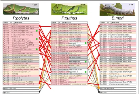
Bombyx. We color-coded the 30 most highly expressed genes in accordance with the similarity of the abundance of EST clones (Figure 1). The 30 most highly expressed genes ofP. xuthusandP. polytesincluded many cuticular protein family genes, similar toB. mori. While the details of the 30 most highly expressed genes ofP. xuthuswere very similar to those ofP. polytes(for example, 63% of the 30 most highly expressed genes were shared byP. xuthus
andP. polytes, as indicated by the red shading in Figure 1), the combination of highly expressed genes were different from those ofB. mori(Figure 1), which may reflect the dif-ferences of the larval appearance betweenPapilio and
Bombyx. For example, three genes,Px-0010(Pp-0008),
Px-0019(Pp-0013) andPx-0024(Pp-0004), were included among the 30 most highly expressed genes in both
P. xuthusandP. polytes (indicated by green circles in Figure 1), but homologs of these genes were not found in theB. moriepidermal EST database. Several cuticular pro-tein genes were only found inB. mori, and many cuticular protein genes ranking high inPapiliospecies were not among the 100 most highly expressed genes inB. mori
(Figure 1). Although the accuracy of expression levels was unclear based only on the clone numbers of the EST data-base, the relatively high expression of the 30 most highly
expressed genes ofP. xuthuswas subsequently confirmed by microarray analysis. Notably,Px-0010andPx-0024had sequence similarity with bilin-binding protein (BBP), and
Px-0019had sequence similarity with takeout/JH-binding protein (JHBP), which we will discuss in detail in subse-quent sections.
Microarray-based screening for marking-specific genes in
P. xuthus
One advantage of thePapiliolarva is its sufficiently large size for separating each marking by dissection, that is, these larvae are convenient for performing microarray analysis of the markings. In addition to the EST dataset, to construct thePapilio microarray we also indepen-dently cloned 77 genes known to be associated with wing-marking patterns or ecdysteroid signal cascade (includingDistal-less,cinnabar, andecdysone receptor) [21,26-30] by using degenerate primers (Additional file 6). We used 32 samples in the microarray (six stages and two markings for mimetic pattern and five stages and four markings for cryptic pattern, Figure 2).
To determine whether we could compare microarray data across all samples, we first compared the normalized sig-nal intensity of housekeeping genes (ribosomal protein genes). We found that these genes were ubiquitously expressed as expected, and their normalized signal inten-sity among the samples was within two-fold of their aver-age values in most cases (Additional files 7 and 8). We also checked several melanin synthesis genes, for which we have previously reported their stage-specific and mark-ing-specific expressions, and confirmed the expected spe-cific expression (see below). Thus, we considered that a comparison of the normalized signal intensity across all of the examined samples is reliable for screening of the novel marking- and stage-specific genes.
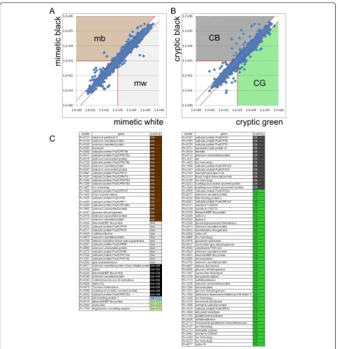
To screen for marking-specific genes, we compared the average normalized signal intensities between mimetic white (mw) and mimetic black (mb) samples, and between cryptic green (CG) (thoracic 2 segment and abdomen region with no markings) and cryptic black (CB) samples (eyespot and V-shaped markings). We categorized genes with an average signal intensity more than two-fold Table 1 Summary ofP. xuthus,P. polytesandB. moriexpressed sequence tag data sets
Species Papilio xuthus Papilio polytes Bombyx moria
Sequenced clone numbers (EST numbers) 20,736 5,376 10,368 (36,589) (8,549) (6,653)
Number of clones corresponding to ribosomal RNA 693 30 automatically removed Number of clones corresponding to mitochondrial DNA 217 24 automatically removed Number of putative genes (including isoforms) 3,341 1,436 1,380
(4,114)b (1,614)b (1,451)b
a
Reported in [25];b773 clusters ofP. xuthus, 178 clusters ofP. polytes, and 71 clusters ofB. moriwere considered to be isoforms/premature forms of other clusters. EST: expressed sequence tags.
Table 2 Summary ofP. xuthusgene set
Category Numbers
P. xuthusgenes (total) 3,341
P. xuthusgenes with coding sequences (> 50 amino acids) 2,746
with homology toDanaus plexippusproteins (P< 1e-10) 2,276 with homology toBombyx moriproteins (P< 1e-10) 2,189 with homology toDrosophila melanogasterproteins
(P< 1e-10)
change between mb and mw, and/or CG and CB, and an average signal intensity higher than 1,000 as marking-spe-cific genes. Using these criteria, we regarded 34 genes as mb-, 13 as mw-, 24 as CB- and 48 as CG-enriched genes (Figure 3). Among the marking-specific genes, nine genes (Px-0011,Px-0108,Px-0220,Px-0245,Px-0248,Px-0430,
Px-0470, Px-0559andPx-0652) were regarded as both mb- and CG-enriched genes. This group contained mela-nin synthesis and associated genes, the takeout/JHBP gene family, cuticular protein genes, the 3-dehydroecdysone 3b-reductase (3DE 3b-3b-reductase) gene and genes of unknown function. In contrast, three genes (Px-0019,Px-0364and
Px-1769) were regarded as both mw- and CG-enriched genes.Px-0019was aPapilio-specific highly expressed gene based on a comparison of the 30 most highly expressed genes (indicated by green circles in Figure 1). One gene, Px-0010, already reported as BBP1, was
regarded as mb- and CG-enriched gene. This is reasonable becauseBBP1expression was detected in blue spots within the black region of the mimetic pattern and the green region of the cryptic pattern [12,15].
To screen for instar- and stage-specific genes, expression profiles were grouped by self-organizing maps with Gene-Pattern software [31], which is often used to summarize microarray data. By self-organizing maps analysis, expres-sion profiles were grouped into 13 co-expresexpres-sion clusters (Figure 4). The largest cluster (cluster C) contained approximately one third of the genes with constant expression profiles (for example, housekeeping genes such as ribosomal protein genes). Other clusters contained intermolt-enriched genes (IM3, IM3-4 and IM4) and genes enriched in the early (EM3, EM3-4 and EM4), mid-dle (MM3, MM3-4 and MM4), and late stages of molting (LM3, LM3-4 and LM4). Genes belonging to clusters Figure 1Comparison of the 30 most highly expressed genes betweenP. xuthus,P. polytesandB. mori.B. moridata is from [25]. The 30 most highly expressed genes were color-coded in accordance with similarity of abundance of epidermal expressed sequence tag clones (red indicates genes which are among the 30 most highly expressed genes of bothP. xuthusandP. polytes, orP. xuthusandB. mori; orange indicates genes which are among the 30 most highly expressed genes in one library and the 100 most highly expressed genes in another library; gray indicates the genes which are among the 30 most highly expressed genes in one species and out of the 100 most highly expressed genes in other species; and white indicates genes among the 30 most highly expressed genes in one species but not found in another species). Green circles indicate thePapilio-specific highly expressed genes ranking in the 30 most highly expressed genes of bothP. xuthusandP. polytes
other than cluster C were expected to be regulated by ecdysteroid because their expression levels differed in the molting period when the ecdysteroid titer changes (see Figure 2). The third instar- (IM3, EM3, MM3 and LM3) and fourth instar-enriched genes (IM4, EM4, MM4 and LM4) were expected to be positively and negatively
regulated by JH, respectively. The genes with similar expression patterns between the third and fourth instars (IM3-4, EM3-4, MM3-4, LM3-4 and C) were assumed to be JH-independent genes. The marking specificity and instar and/or stage specificity for each gene is indicated in Additional file 1.
Identification of several candidates for blue and yellow pigment-binding proteins
One of the most obvious characteristics of thePapilio lar-val cryptic pattern is the overall green coloration. Lepidop-teran larval green coloration consists of blue bile pigment
(bilin) and yellow carotenoids in general [32]. These pig-ments usually exist as pigment-protein complexesin vivo
[33]. We previously reported that the combination of
BBP1andYRGcorrelated perfectly with larval blue, yellow and green coloration in three Papilio species [12]. Figure 3Screening for marking-specific genes.(A)Scatter plots of normalized signal intensity of mimetic white (xaxis, average of six stages) versus mimetic black (y axis, average of six stages). Genes with average intensity more than two-fold (red dashed lines) between mimetic black and mimetic white, and also with an average signal intensity higher than 1,000 (red solid lines) were categorized as marking-specific genes.(B)
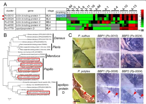
Although the high expression ofBBP1(Px-0010) in the CG region was indicated by the EST database, microarray analyses and whole-mountin situhybridization (Figure 5A, C), YRG (Px-0080) expression was relatively low (Additional file 7). This imbalance in the expression level of putative blue and yellow pigment-binding proteins sug-gested the presence of other pigment-binding proteins. No gene homologous to the carotenoid-binding protein reported from the silkwormB. mori[34] was found in the
PapilioEST datasets. Instead,Px-0019(related to the take-out/JHBP gene family) andPx-0024(related to the lipoca-lin family), both among the 30 most highly expressed genes in the EST library, appeared to be good candidates for carotenoid-binding protein and BBP genes (see Figure 1). BothPx-0019andPx-0024were CG-enriched in the
microarray-based gene expression analysis (Figure 3, Addi-tional file 9). The expression profile ofPx-0024was very similar to that ofBBP1(Px-0010) (Figure 5A). Molecular phylogeny indicated thatPx-0024and itsP. polytes ortho-logPp0004were members of the lipocalin family, a puta-tive binding protein for lipophilic substances, including the BBP ofPieris rapaeand insecticyanin (BBP) of Man-duca sexta(Figure 5B). Thus, we namedPx-0024(and its orthologPp0004),BBP2. Molecular phylogeny also indi-cated that these genes had lineage-specific gene duplica-tion, andP. xuthusandP. polyteshad at least four and five paralogous genes, respectively. Almost identical marking-specific expression betweenBBP1andBBP2in bothP. xuthusandP. polyteswas indicated by microarray analysis and whole-mount in situ hybridization (Figure 5C),
suggesting that these two genes may function coordinately as blue pigment-binding proteins. As it is supposed that BBPs of the saturniid silkwormRhodinia fugaxexist as a dimer in epidermis [35], it is possible thatBBP1andBBP2
form heterodimers. Notably, bombyrin ofB. moriis not associated with blue coloration, and molecular phylogeny indicated that the blue pigment-binding proteins ofPieris,
ManducaandPapiliodo not form a single cluster, sug-gesting that blue pigment-binding proteins evolved inde-pendently to serve a common physiological role in each organism.
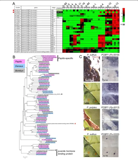
RegardingPx-0019, its spatial expression pattern asso-ciated with the cryptic pattern was similar to those of
BBP1andBBP2inP. xuthus(Figure 6C). Similar to the expression pattern ofYRG reported previously [12], the expression ofPp-0013 (the ortholog ofPx-0019) inP. polyteswas not detected in the blue spot region (Figure 6C) whereas BBP1 and BBP2 expression was clearly detected (red arrowheads in Figure 5C). Molecular phy-logeny indicated that Px-0019formed a large cluster consisting of the takeout/JHBP gene family and 23 other genes in theP. xuthusEST database (Figure 6B). Nota-bly, this cluster included the carotenoid-binding protein of the locust Schistocerca gregaria(red asterisk in Figure 6B) [36], suggesting thatPx-0019is one of theP. xuthus
carotenoid-binding proteins.Px-0019formed aPapilio -specific cluster with three other Papiliogenes (indicated by‘Papilio-specific’in Figure 6B). Among the 24Papilio
takeout/JHBP genes, Px-0019and its closest homolog gene Px-0220had the strongest marking specificity in the microarray expression profile (Figure 6A). In con-trast to that ofPx-0019, the expression level of Px-0220
was higher in eye spot regions and V-shaped markings (Figure 6A). Consistent with the microarray results, the spatial expression pattern ofPx-0220perfectly correlated with the yellow spot region of the V-shaped markings (Figure 6C). The expression patterns ofPx-0019and Px-0220strongly suggest that thesePapilio-specific family genes bind yellow carotenoid pigments with different spatial regulation. We therefore named these genes putative carotenoid-binding proteins (PCBP1 and PCBP2, respectively). Moderate PCBP1 (Px-0019) expression was also detected in the white region of the mimetic pattern by microarray analysis and in situ
hybridization, suggesting that this gene also colocalizes with white pteridine/uric acid pigments (Figure 6A, C).
Because JHBP has high ligand specificity [37,38] and it has been assumed to be monomeric in solution [39], PCBP1 and PCBP2 may bind to different types of caro-tenoids. Other takeout/JHBP genes (for example, Px-0150 and Px-0591) also had marking specificity and were enriched in the fourth instar (Figure 6A), suggest-ing that these genes also have a supportsuggest-ing role in caro-tenoid binding. Because the presence of several
carotenoids, including alpha-carotene, beta-carotene and lutein, have been reported forP. xuthus [40], these take-out/JHBP family genes ofP. xuthus may each recognize a different carotenoid.
Notably, the locust carotenoid-binding protein (red asterisk in Figure 6B) did not form a single cluster with
Px-0019orPx-0220, which is similar to the case of BBP, suggesting that both blue and yellow pigment-binding proteins evolved convergently among insect species. Green coloration among lepidopteran larvae appears to have emerged independently in the phylogenetic tree [2]. The independent occurrence of BBPs and carote-noid-binding proteins within the lipocalin family genes and takeout/JHBP family genes may reflect the conver-gent evolution of larval green coloration.
Identification of the novel marking-specific melanin synthesis gene,yellow-h3
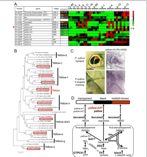
We previously reported that several melanin-related genes were associated with stage- and species-specific black larval cuticular markings in threePapilio species [10-14,16]. The microarray results were consistent with our previous findings. BothTH (Px-0470) andDDC( Px-1800) were strongly expressed in the black markings during the latter half of the molting period; yellow( Px-0108) andlaccase2(Px-0292) were strongly expressed in the black markings during the middle of the molting period; tan (Px-1031) was strongly expressed in the cryptic black markings at the latter half of the molting period; andGTP-CH I(Px-1001) was strongly expressed in the black markings only in specific stages (Figure 7A). In addition to these genes, Yellow-f-related genes have been reported as dopachrome-converting enzymes for melanin biosynthesis (for example,D. melanogaster yellow-f and yellow-f2 and the Aedes aegypti dopa-chrome-converting enzyme, Figure 7B) [41,42]. In B. mori and D. melanogaster, 10 and 14 yellow family genes (which have major royal jelly protein motifs) have been reported (Figure 7B) [40,42]. We obtained all yel-low family genes inP. xuthus(red boxes in Figure 7B) excludingyellow-b. In lepidopteran species, yellow-f3
and yellow-f4 are the closest homologs toDrosophila yellow-f/yellow-f2 (Figure 7B) [43].
Unexpectedly, yellow-f3(Px-4077) and yellow-f4( Px-0903) ofP. xuthuswere not upregulated in black mark-ings, but instead were CG-enriched (Figure 7A). Conver-sely, yellow-h3 (Px-0430) was upregulated in black markings, and it exhibited a very similar expression pat-tern to that ofyellow (Figure 7A). Using whole-mount
Figure 6Expression pattern of putative carotenoid-binding protein genes.(A)Heat map of 24takeout/juvenile hormone-binding protein
(JHBP) family genes inP. xuthus. Red indicates positive values and green indicates negative values (color spectrum bar is shown to the right; N. D., not detected). Red circles indicate the genes examined by whole-mountin situhybridization in (C). Stage and marking of each sample is shown above. The stage-specific co-expression cluster and marking specificity of each gene are also shown. See also Figures 2 to 4.(B)Neighbor joining tree oftakeout/JHBPfamily genes based on their amino acid sequences. The numbers at the tree nodes represent the bootstrap values. The scale bars indicate the evolutionary distance between the groups.P. xuthus,D. plexippusandB. morigenes are shaded in red, blue and gray, respectively. Red asterisk indicates carotenoid-binding protein of locustSchistocerca gregaria, and black asterisks indicate takeout protein of
Drosophila melanogasterand JHBP ofManduca sexta. Accession numbers are shown in parenthesis. Predicted genes ofD. plexippusandB. mori
Figure 7Spatial expression pattern of melanin synthesis genes.(A)Heat map of the relative expression level of melanin synthesis/related genes andyellowfamily genes inP. xuthus. Red indicates positive values and green indicates negative values (color spectrum bar is shown to the right; N.D., not detected). Red circle indicates the gene examined by whole-mountin situhybridization in (C). Stage and marking of each sample is shown above. The stage-specific co-expression cluster and marking specificity of each gene are also shown. See also Figures 2 to 4.(B)
Neighbor joining tree ofyellowfamily genes based on their amino acid sequences. The numbers at the tree nodes represent the bootstrap values. The scale bars indicate the evolutionary distance between the groups. Accession numbers are shown in parenthesis. Red boxes indicate
theP. xuthusgenes. Yellow-g gene has not been reported in lepidopteran species [43].(C)Spatial expression patterns ofyellow-h3mRNA inP.
xuthuslarva during the fourth molt. Numbers in each panel indicate molt stage-hours after head capsule slippage.(D)Biosynthetic pathway
underlying the formation of melanin. Dopamine, N-beta-alanyldopamine (NBAD) and N-acetyldopamine (NADA)are used in the production of Dopamine-melanin (black and brown), NBAD pigment (yellow or reddish brown), and NADA pigment (transparent) in insects [5,16,45,62]. Aa
DCE:Aedes aegyptidopachrome conversion enzyme; BH4: tetrahydrobiopterin; Bm:Bombyx mori; DDC: dopa decarboxylase; Dm:Drosophila
melanogaster;GTP: guanosine triphosphate; GTP-CH I: GTP cyclohydrolase I; Hm:Heliconius melpomene; NADA: N-acetyldopamine; NAT:
arylalkylalamine-N-acetyltransferase; NBAD: N-beta-alanyldopamine; NBH2: dihydroneopterin triphosphate; Px:Papilio xuthus. TH: tyrosine
regions inHeliconius wings by RT-PCR, but its spatial expression patterns have not been clarified [43]. Our data, combined with the Heliconiusresults, suggest that
yellow-h3functions in melanin biosynthesis in lepidop-teran species (Figure 7D) similarly to yellow-fand yel-low-f2inDrosophila.
Based on our microarray analysis, three yellow family genes, yellow-f3,yellow-h2and yellow-e, were regarded as CG-enriched genes (Figures 3 and 7A).yellow-f3was highly expressed during the late molting period similar toTHand DDC, andyellow-h2andyellow-ewere highly expressed during the middle of the molting period simi-lar toyellow(Figure 7A), implying that these genes may function in inhibiting melanin pigmentation. In the silk-worm B. mori, yellow-e disruption promoted melanin pigmentation in the larval head and tail, where strong
yellow-e expression was detected [44], which is consis-tent with our results. In melanin synthesis, it is assumed that arylalkylamine-N-acetyltransferase (NAT) activity is involved in synthesis of N-acetyldopamine, a precursor of colorless cuticle [5,45,46]. However, disruption of
NAT in B. mori resulted in an overall blackish pheno-type only in the adult stage, whereas it had little effect on larval pigmentation [46,47]. Our EST datasets did not contain NAT or NAT-like genes, suggesting that
NATgene is not the primary factor for colorless cuticu-lar production in cuticu-larval stages. Because green is the color of the epidermis seen through the colorless cuticle, the observation of CG-enrichedyellowfamily genes (CG in Figure 3) implies thatyellow family genes are major negative regulators of melanin pigmentation in the larval stage instead of theNATgene (Figure 7D). Taxa-specific gene duplication found betweenDrosophilaand lepidop-teran species (for example,yellow-f,yellow-h, Figure 7B), and the reverse function implicated foryellow-f family genes between Diptera and Lepidoptera suggest that the function of theyellowgene family has diversified among insect taxa.
Marking-specific cuticular protein genes
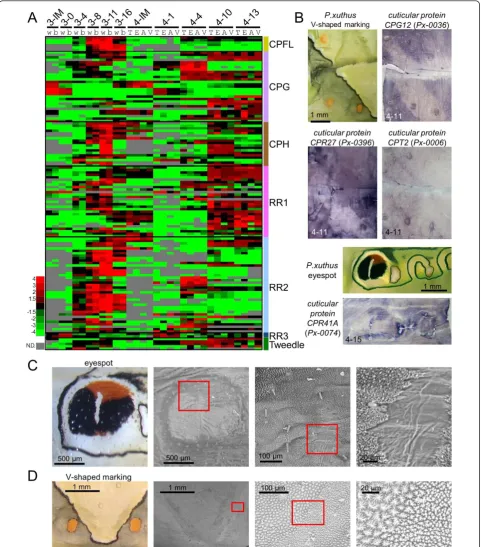
The 30 most highly expressed genes of the Papilio epi-dermal EST dataset included many cuticular protein genes similar to B. mori (Figure 1) [25]. In B. mori, more than 200 cuticular protein genes have been reported [48,49]. The heat map of 128 cuticular protein genes ofP. xuthus indicated the clear stage specificity of most of the cuticular protein genes (Figure 8A). Cuticu-lar protein genes can be divided into several groups based on amino acid sequence similarity [48,50]. Many genes with a CPFL or RR2 motif were highly expressed in the third instar (mimetic pattern) although stage spe-cificity was not completely associated with this motif, as we reported previously [15]. Interestingly, many cuticu-lar protein genes also displayed marking specificity
(Figure 3 and Additional file 7), which was confirmed by
in situhybridization (Figure 8B). The cuticular proteins
CPG12 (glycine-rich motif,Px-0036) and CPR27(RR-1 motif, Px-0396) were highly expressed in the CG region as well as the yellow spot region in V-shaped markings (Figure 8B), and CPT2 (Tweedle motif,Px-0006) was highly expressed in the yellow spot region as well as the boundary of the V-shaped region (Figure 8B). Strong expression of CPR41A (RR1 motif, Px-0074) was detected in the red regions of the eyespot (Figure 8B). To examine whether the different spatial expression of the cuticular protein genes affected exoskeletal struc-tures, we examined the cuticular surface by electron microscopy. We found that cuticular structures were different at each marking (Figure 8C, D), and we could easily recognize the eyespot region through electron microscopy (Figure 8C). The surface nanostructure was fine in the black region of the eyespot and coarse in the yellowish green region around the eyespot. The surface nanostructure of the red region was intermediate between the black and green regions. The white stripe center of the eyespot had a very smooth surface. We previously reported that muscle was attached to this white stripe region [14]. The surface nanostructure dra-matically changed at the boundary of the V-shaped markings (Figure 8D). The surface nanostructure of the black stripe region was fine and the green region was coarse similar to the eyespot. These results indicated that color pattern and surface structure are tightly related, which is similar to the adult wing scale in which clear correlations between color and structure have been reported [51,52]. Although the precise role of each cuti-cular protein remains unclear, specific cuticuti-cular proteins may have the function of transporting or maintaining the specific cuticular pigments.
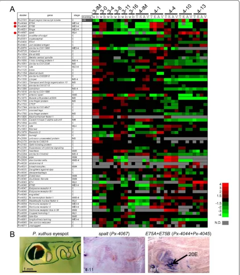
Candidates for marking-associated patterning genes Although several regulatory genes have been reported for butterfly adult wing pattern formation [6,7,26,29], it is still unclear which gene controls the larval body markings. We surveyed the marking specificity of 76 regulatory genes such as transcription factors and signal transducers. According to microarray analysis, nine genes (trithorax, polybromo, aristaless, cut, doublesex
(male type), optomotor blind, ultrabithorax, vein and
wingless) were expressed at lower levels than measure-able, and most genes had no clear marking specificity (Figure 9A). However, we found thatE(spl) region tran-script mbeta(Px-2191) had clear marking specificity (CB in Figure 3) and four other genes E75A, E75B, fringe
Figure 9Expression pattern of candidates for patterning genes.(A)Heat map of the relative expression level of 67 regulatory genes (transcription factors and signal transducers). Red indicates positive values and green indicates negative values (color spectrum bar is shown to the right; N.D., not detected). Red circles indicate the genes examined by whole-mountin situhybridization in (B). Stage and marking of each sample is shown above. The stage-specific co-expression cluster and marking specificity of each gene are also shown. See also Figures 2 to 4.(B)
we could not detect the positive signals of the four other genes, perhaps because of the low signal intensity in the examined stage (whole-mount in situhybridization is applicable after the middle stage, 11 hours after head capsule slippage (HCS), of the molting period when cuticular apolysis is complete). Notably,spaltexpression coincided with the black markings of several butterfly wing patterns [7,29]. Taken together, spaltappeared to be a positive regulator of melanin pigmentation in both larvae and adult butterflies.
E75AandE75Bare involved in the ecdysteroid signaling cascade, and their expression is induced by ecdysteroids [30]. We therefore examined the effect of ecdysteroids by the topical application method [11] and found that higher
E75expression is maintained in black eyespot regions (Figure 9B; probes were designed to target common regions ofE75AandE75B). The expression pattern ofE75
coincided with the eyespot pattern, which was similar to
yellow[11], suggesting thatE75had a marking specificity in the early molting stage. InManduca sexta, bothE75A
andE75Bare involved in the stage-specific gene expres-sion ofDDCboth directly and indirectly [30]. Our results suggest thatE75Aand/orE75Bregulate both the marking specificity and stage specificity of several black marking-associated genes. As we described previously, several cuti-cular protein genes also exhibited marking specificity and stage specificity (Figure 8). Although it is still not clear how color and nanostructure are determined, one possible explanation is that marking-specific transcription factors involved in the ecdysteroid signaling cascade regulate both pigmentation and cuticular protein genes. E75is one strong candidate mediator of the hormone-dependent coordination of larval pattern and nanostructure formation.
AlthoughspaltandE75were the only two transcription factor genes for which we were able to detect marking-associated expression by whole-mountin situ hybridiza-tion, microarray analysis suggested thatE(spl) region tran-script mbeta and fringe, both involved in the Notch signaling pathway, also have marking specificity. In the butterfly wing pattern, Notchis involved in intervein markings [53], andfringeis upregulated by 20-hydroxyec-dysone [54]. The Notch signaling pathway may also be involved in hormone-dependent pattern formation in but-terfly larvae.
The marking-specific ecdysteroid biosynthesis enzyme gene,3-dehydroecdysone 3b-reductase
Unexpectedly, we found that the3DE 3b-reductasegene (Px-0248), which converts inactivated 3-dehydroecdysone to ecdysone [55,56], had clear marking specificity during the late stage of the molting period. According to whole-mountin situhybridization findings,3DE 3b-reductase
was highly expressed in black markings in both mimetic
and cryptic patterns (Figure 10), which is very similar to theTHandDDCexpression patterns. The simultaneous expression of3DE 3b-reductaseand melanin synthesis enzyme genes suggested that localized regulation of ecdysteroid titers in the epidermis is also involved in marking-specific gene regulation. Relatively high concen-trations of ecdysone in the black markings may be impor-tant for regulating theTHandDDCexpressions during the pigmentation stages.
Papilio-specific marking-associated genes
Among 2,746P. xuthus genes (ORF > 50 amino acids), more than 400 genes had no sequence similarity with the genome sequences of other insects, including silkworm and monarch butterfly (Additional file 5). Through micro-array analysis and whole-mountin situhybridization, we found three Papilio-specific genes (that is, has no sequence similarity with known proteins including the monarch butterflyDanaus,Bombyxgenome sequences, and the Butterfly Base sequence database [57] with clear marking specificity (Px-0559,Px-3233andPx-3244), and one gene,Px-0011, with only low sequence similarity with the monarch butterfly (that is, butterfly specific).Px-0559,
Px-3233andPx-3244expression coincided with black, yel-low and eyespot markings, respectively (Figure 10). Px-0011expression was also strongly associated with black markings (Figure 10). These proteins have no known domains and display no similarity to any characterized proteins. Px-0011 and Px-3233 have signal peptide sequences, andPx-0011,Px-0559andPx-3233are threo-nine-, argithreo-nine-, and tyrosine-rich proteins, respectively (Additional file 1). These genes were expressed during the middle or late stages of the molting period, suggesting that these genes were structural proteins. Although the precise function of these genes is currently unknown, this is the first example of species-specific genes associated with pat-tern formation.
Conclusion
with the cuticular pigmentation of various insect orders [5,45,60-62], which suggests their conserved role in cuti-cular pigmentation across insect taxa. Conversely, our
genes and takeout/JHBP family genes (Figures 5, 6, 7). We also found that there were severalPapilio-specific genes with clear marking specificity, suggesting that spe-cies-specific genes contribute to marking formation. Notably, some of the regulatory genes involved in larval pattern appeared to be different from those of adults (for example,Distal-less involvement in eyespots was less obvious in larvae), whereas others appeared to participate in both larval and adult pattern formation (for example,
spalt expression associated with black markings). Furthermore, we found that both the transcription factor
E75and the ecdysteroid synthesis enzyme3DE 3b-reduc-tasehad clear marking specificity, suggesting their invol-vement in ecdysteroid-dependent coordinated gene regulation in larval pattern formation. Ecdysteroid is also involved in butterfly adult wing pattern [28], which sug-gests the possibility that these genes contribute to pattern formation in the adult wing. The wide variety of mark-ing-associated genes identified by our screening indicates that the strategy utilized in this study is effective for clari-fying pattern formation in species without genome infor-mation. The gene collection and the expression profile presented in this study will be invaluable for exploring not onlyPapiliobut also insects in general.
Methods
Experimental animals and developmental staging
P. xuthus was either purchased from Eiko-Kagaku (Osaka, Japan), kindly provided by Dr. Akira Yamanaka (Yamaguchi University, Japan), or collected from the field. Larvae were reared on leaves of Zanthoxylum ailanthoides(Rutaceae) at 25°C under long-day condi-tions (16 h light and 8 h dark). The staging of the molt-ing period was based on the time when HCS occurred, as well as spiracle and hair pigmentation [12].
Construction and sequencing of the cDNA library
Total epidermal RNAs ofP. xuthusandP. polyteswere isolated from 50 individuals per species during the molt-ing period (35 individuals at third molt and 15 indivi-duals at fourth molt) using TRI reagent (Sigma-Aldrich, St. Louis, MO, USA) according to the manufacturer’s instructions. After the attached muscle and fat body were removed, whole dorsal integuments from the thoracic 2 segment to the abdominal 7 segment were dissected from larvae. Total RNAs were mixed and subjected to cDNA library construction.
The cDNA library construction was carried out by the Dragon Genomics Center (Takara Bio Inc., Mie, Japan) by the following procedure. The mRNA was purified using an Oligotex-dT30 Super mRNA purification Kit (Takara, Bio Inc., Mie, Japan), and then cDNA libraries were constructed using a cDNA synthesis Kit (Strata-gene, La Jolla, CA, USA). First-strand cDNA synthesis
was carried out with oligo d(T)18 primers. Synthesized
cDNA were size-selected and ligated into theEcoRI and
XhoI sites of pBluescript II SK(+) vector (Stratagene). The ligated products were then transformed into com-petent DH10BEscherichia colicells by electroporation. The transformed cells were plated onto Luria Broth media and incubated overnight.
For sequencing of cDNA libraries, white colonies were randomly selected and sequenced from both ends using an ABI prism 3130 Genetic Analyzer (Applied Biosystems, Foster City, CA, USA), which was carried out at National Institute of Genetics (Shizuoka, Japan) with the support of the MEXT Genome Support Project for theP. xuthus
cDNA library, and at the National Institute of Agrobiolo-gical Sciences (Ibaraki, Japan) for the P. polytescDNA library. All EST sequences have been deposited [DDBJ: FY174038-FY210626, FY302525-FY358875]. Among these ESTs, sequences corresponding to ribosomal RNA [DDBJ: AB674749] and mitochondrial DNA [DDBJ: EF621724] were eliminated. The remaining ESTs were subjected to cluster analysis using a Phred/Phrap/Consed software package [63]. After automatic clustering, we checked every sequence manually for each alignment, and divided and reassembled the putative chimeric sequences. SignalP 3.0 [24] was used for signal peptide prediction.
Microarray experiments and data analysis
The Papilio 15 K oligo-microarray slide (60-mer oligonu-cleotides on 15,208 spots, Agilent Technologies, Palo Alto, CA, USA) was constructed using 8,000 Papilio
unique sequences (3,376 fromP. xuthus, 4,612 fromP. polytesand 12 fromP. machaon). We could not design an appropriate probe for 42 genes because of low sequence complexity (indicated by‘n.e.’in Additional file 1). Probe sequences were designed using Agilent’s web portal eArray [64], and one or two probes each for
P. xuthus, P. machaonandP. polytes gene were con-structed (probe sequences ofP. xuthusare shown in Additional file 7).
We performed two color microarray hybridizations. Mimetic white, cryptic thorax and cryptic abdomen were independently labeled with cyanine 3-CTP (Cy3), and mimetic black, cryptic eyespot, and cryptic V-shaped markings were independently labeled with cya-nine 5-CTP (Cy5) in all stages. Double-stranded cDNA and labeled cRNA were synthesized using the Low RNA Input Linear Amplification Kit according to the manu-facturer’s instructions (Agilent Technologies). Total RNA (400 ng each for Cy3 and Cy5) was converted to cDNAs with T7 promoter oligo-dT primer and Moloney Murine Leukemia Virus reverse transcriptase. Second-strand cDNA was transcribed to cRNA with T7 RNA polymerase and 10 mM Cy3 or Cy5. The labeled cRNA was purified using an RNeasy Mini Kit (Qiagen, Tokyo, Japan).
Hybridization was performed using an In situ Hybridi-zation Kit Plus (Agilent Technologies). One microgram each of Cy3-labeled cRNA and Cy5-labeled cRNA were mixed, fragmented and hybridized to each oligo-microar-ray at 65°C for 17 h at 4 rpm. The aroligo-microar-rays were washed in 6 × saline sodium citrate, 0.005% TritonX-102 for 10 min at room temperature and in 0.1 × saline sodium citrate, 0.005% TritonX-102 for 5 min at 4°C. Intensities of the hybridized probes were detected with an Agilent G2565BA Microarray Scanner with 10 mm scan resolu-tion, and the signals were extracted with G2565AA Fea-ture Extraction Software v.7.1 (Agilent Technologies). Normalization was performed‘per spot’and‘per chip’ using the GENESPRING program. The low expression level spots in all cells were removed (fluorescence inten-sity is approximately lower than 100). Microarray data were deposited in the Gene Expression Omnibus [GEO: GSE37920]. The normalized fluorescence intensity of all samples and each probe sequence are shown in Addi-tional file 7.
We used microarray-based screening to obtain the genes involved in the overall steps of pattern formation, with the following strategies: comparing gene expression between different markings to identify marking-specific genes; comparing gene expression between mimetic and cryptic patterns to identify instar-specific genes; and checking the gene expression profile during the molting periods to identify stage-specific genes. To do this, we dissected larval epidermis at 11 different stages (Figure 2, red arrowheads) for microarray screening. Because cryptic patterns are assumed to be determined in the JH-sensitive period (green box in Figure 2) [10], six different stages before the JH-sensitive period were expected to be associated with the mimetic pattern and five different stages after the JH-sensitive period were analyzed for genes associated with cryptic pattern. For the mimetic pattern, we separately dis-sected the white region (w) and the black region (b), whereas for the cryptic pattern, we separately dissected
the thoracic 2 segment (T), eyespot (E), abdomen region with no markings (A) and V-shaped markings (V) (Figure 2). Expression profiles of 11 developmental stages or six markings (mimetic white, mimetic black, cryptic thorax, cryptic eyespot, cryptic abdomen and cryptic V-shaped marking) were grouped by self-organizing maps with Gen-ePattern software [31]. This algorithm is often used to summarize microarray data. We obtained expression pro-files in terms of relative expression rather than absolute expression levels by dividing the average expression levels of each gene.
Phylogenetic analysis
Alignment was conducted based on translated protein sequences using Clustal W program implemented in MEGA5, and phylogenetic trees were constructed by the Neighbor joining method with the MEGA5 program [65]. The confidence of the various phylogenetic lineages was assessed by the bootstrap analysis.
Whole-mountin situhybridization
Whole dorsal integuments from the thoracic 2 segment to the abdominal 7 segment were dissected from larvae. After the body fat and muscle attached to the epidermis were carefully removed, the larval epidermis was fixed immediately in 4% paraformaldehyde in PBS (137 mM NaCl, 8.10 mM Na2HPO4, 2.68 mM KCl and 1.47 mM
KH2PO4, pH 7.4). Whole-mount in situ hybridization
was performed as described previously [13,15-17]. An RNA probe for each gene was prepared using the DIG RNA Labeling Kit (Roche Biochemicals, Mannheim, Germany). A color reaction for digoxigenin-labeled anti-sense RNA probes was performed at room temperature in 100 mM Tris-HCl, 100 mM NaCl and 50 mM MgCl2
(pH 9.5) containing 3.5 μL/mL 5-bromo-4-chloro-3-indolyl-phosphate, 4-toluidine salt and 4.5μL/mL nitro-blue tetrazolium chloride. Digoxigenin-labeled sense strand probes were used as negative controls.
Scanning electron microscopy analysis
Cuticular nanostructure of eyespot and V-shaped mark-ing region was observed by scannmark-ing electron micro-scopy using a Hitachi TM-1000 scanning electron microanalyzer (Hitachi High-Technologies Corp., Tokyo, Japan).
Additional material
BLASTP analysis toDrosophila melanogasterpeptides (Flybase v. 5.42); DmCG: CG number of best hit from BLASTP analysis toD. melanogaster
peptides; DmE-value: Evalue of best hit from BLASTP analysis toD.
melanogasterpeptides; BmGene: best hit from BLASTP analysis toBombyx
moripeptides; BmE-value: Evalue of best hit from BLASTP analysis toB. moripeptides; DpGene: best hit from BLASTP analysis toDanaus plexippuspeptides; DpE-value: Evalue of best hit from BLASTP analysis to
D. plexippuspeptides; Remarks: gene name of each cluster; accession number: GenBank accession number; co-expression cluster: co-expression cluster by microarray analysis (n.d.: not detected: n.e.: not examined); marking specificity: average intensity of mimetic black (mb) vs. mimetic white (mw) and/or cryptic green (CG) vs. cryptic black (CB) over two-fold is indicated.
Additional file 2: List of 1,436P. polytesnonredundant genes. Explanation of column contents are the same as Additional file 1.
Additional file 3: List of identified putative isoforms or premature transcripts ofP. xuthus.
Additional file 4: List of identified putative isoforms or premature transcripts ofP. polytes.
Additional file 5: The Venn diagram ofP. xuthusgenes. The numbers of homologous genes shared betweenP. xuthusepidermal expressed sequence tags and other insect genomes are shown (cutoff threshold E values:P< 1e-10by BLASTP search).
Additional file 6: List of 77 additionally clonedP. xuthusgenes. Explanation of column contents is the same as Additional file 1.
Additional file 7: Microarray result ofP. xuthusgenes. Probe sequences, normalized signal intensity of each sample, sum of signal intensity, and relative expression level of each sample are shown.
Additional file 8: Heat map of the relative expression level of 78 ribosomal protein genes inP. xuthus. Red indicates positive values and green indicates negative values (color spectrum bar is shown to the right; N.D., not detected). Stage and marking of each sample is shown above. The sage-specific co-expression cluster and marking specificity of each gene are also shown. See also Figures 2 to 4.
Additional file 9: Heat map of the relative expression level of the 30 most highly expressed genes inP. xuthus. Stage and marking of each sample is shown above. Red indicates positive values and green indicates negative values (color spectrum bar is shown to the right; N.D., not detected). The stage-specific co-expression cluster and marking specificity of each gene are also shown. See also Figures 2 to 4.
Abbreviations
3DE 3b-reductase: 3-dehydroecdysone 3b-reductase; BBP: bilin-binding protein; CB: cryptic black; CG: cryptic green; Cy3: cyanine 3-CTP; Cy5: cyanine 5-CTP;DDC:dopa decarboxylase; EST: expressed sequence tag;GTP-CH I:
guanosine triphosphate cyclohydrolase I; HCS: head capsule slippage; JH: juvenile hormone; JHBP: juvenile hormone-binding protein; mb: mimetic black; mw: mimetic white; NAT: arylalkylamine-N-acetyltransferase; ORF: open reading frame; PBS: phosphate-buffered saline; PCBP: putative carotenoid-binding protein; RT-PCR: reverse transcriptase polymerase chain reaction;TH:
tyrosine hydroxylase;YRG:yellow-related gene.
Acknowledgements
We are very grateful to Dr. Asao Fujiyama for arranging the EST project, to Dr. Akira Yamanaka for kindly providing many larvae ofP. xuthus, to Dr. Tetsuya Kojima for help with scanning electron microscopy analysis, to Dr. Yuki Nakamura for help with the microarray analysis, to Mrs. Kyoko Chagi for technical support ofin situhybridization, and to Dr. Mizuko Osanai-Futahashi for her critical reading of the manuscript. This work was supported by Grant-in-Aid for Scientific Research on Priority Areas“Comparative Genomics”from the Ministry of Education, Culture, Sports, Science and Technology of Japan 20017007 (to HF), Grants-in-Aid for Scientific Research“Genome Science” 221S0002 (to HF), Grants-in-Aid for Scientific Research 22128005 (to HF), and Grant-in-Aid for JSPS fellows 07J03511 (to RF).
Author details
1Department of Integrated Biosciences, Graduate School of Frontier Sciences,
University of Tokyo, Kashiwa, Chiba 277-8562, Japan.2Bioproduction Research Institute, National Institute of Advanced Industrial Science and Technology, Tsukuba, Ibaraki 305-8566, Japan.3National Institute of Genetics,
Yata 1111, Mishima, Shizuoka 411-8540, Japan.4Nihon University College of
Bioresource Sciences, Fujisawa, Kanagawa 252-0880, Japan.5National Institute
of Agrobiological Sciences, Owashi, Tsukuba, Ibaraki 305-8634, Japan.
Authors’contributions
RF and HF designed the research. RF performed the manual assembly and annotation of EST datasets. RF and HS analyzed the EST database and generatedin situhybridization data. TN sequenced the cDNA library ofP. xuthus, and KM sequenced the cDNA library ofP. polytes. RF performed and analyzed the microarray data. RF prepared figures and RF and HF wrote the manuscript. All authors read and approved the final manuscript.
Competing interests
The authors declare that they have no competing interests.
Received: 21 March 2012 Accepted: 31 May 2012 Published: 31 May 2012
References
1. Ruxton GD, Sherratt TN, Speed MP:Avoiding Attack: The Evolutionary Ecology of Crypsis, Warning Signals and MimicryOxford: Oxford University Press; 2005.
2. Scoble MJ:The LepidopteraNew York: Oxford University Press; 1992. 3. Prudic KL, Oliver JC, Sperling FA:The signal environment is more
important than diet or chemical specialization in the evolution of warning coloration.Proc Natl Acad Sci USA2007,104(49):19381-19386. 4. Monteiro A:Alternative models for the evolution of eyespots and of serial homology on lepidopteran wings.BioEssays2008,30(4):358-366. 5. Wittkopp PJ, Beldade P:Development and evolution of insect
pigmentation: genetic mechanisms and the potential consequences of pleiotropy.Semin Cell Dev Biol2009,20(1):65-71.
6. Reed RD, Papa R, Martin A, Hines HM, Counterman BA, Pardo-Diaz C, Jiggins CD, Chamberlain NL, Kronforst MR, Chen R, Halder G, Nijhout HF, McMillan WO:optixdrives the repeated convergent evolution of butterfly wing pattern mimicry.Science2011,333(6046):1137-1141. 7. Joron M, Frezal L, Jones RT, Chamberlain NL, Lee SF, Haag CR, Whibley A,
Becuwe M, Baxter SW, Ferguson L, Wilkinson PA, Salazar C, Davidson C, Clark R, Quail MA, Beasley H, Glithero R, Lloyd C, Sims S, Jones MC, Rogers J, Jiggins CD, ffrench-Constant RH:Chromosomal rearrangements maintain a polymorphic supergene controlling butterfly mimicry.Nature
2011,477(7363):203-206.
8. Monteiro A, Podlaha O:Wings, horns, and butterfly eyespots: how do complex traits evolve?PLoS Biol2009,7(2):e37.
9. Conceicao IC, Long AD, Gruber JD, Beldade P:Genomic sequence around butterfly wing development genes: annotation and comparative analysis.PloS One2011,6(8):e23778.
10. Futahashi R, Fujiwara H:Juvenile hormone regulates butterfly larval pattern switches.Science2008,319(5866):1061-1061.
11. Futahashi R, Fujiwara H:Regulation of 20-hydroxyecdysone on the larval pigmentation and the expression of melanin synthesis enzymes and
yellowgene of the swallowtail butterfly,Papilio xuthus.Insect Biochem Mol Biol2007,37(8):855-864.
12. Shirataki H, Futahashi R, Fujiwara H:Species-specific coordinated gene expression and trans-regulation of larval color pattern in three swallowtail butterflies.Evol Dev2010,12(3):305-314.
13. Futahashi R, Fujiwara H:Melanin-synthesis enzymes coregulate stage-specific larval cuticular markings in the swallowtail butterfly,Papilio xuthus.Dev Genes Evol2005,215(10):519-529.
14. Futahashi R, Fujiwara H:Expression of one isoform of GTP cyclohydrolase I coincides with the larval black markings of the swallowtail butterfly,
Papilio xuthus.Insect Biochem Mol Biol2006,36(1):63-70.
16. Futahashi R, Banno Y, Fujiwara H:Caterpillar color patterns are determined by a two-phase melanin gene prepatterning process: new evidence fromtanandlaccase2.Evol Dev2010,12(2):157-167. 17. Futahashi R:Whole-mountin situhybridization of sectioned tissues of
species hybrids to detectcis-regulatory changes in gene expression pattern.Methods Mol Biol2011,772:319-328.
18. Papanicolaou A, Joron M, McMillan WO, Blaxter ML, Jiggins CD:Genomic tools and cDNA derived markers for butterflies.Mol Ecol2005,
14(9):2883-2897.
19. Papanicolaou A, Gebauer-Jung S, Blaxter ML, Owen McMillan W, Jiggins CD:
ButterflyBase: a platform for lepidopteran genomics.Nucleic Acids Res
2008,36(Database):D582-587.
20. Beldade P, Rudd S, Gruber JD, Long AD:A wing expressed sequence tag resource forBicyclus anynanabutterflies, an evo-devo model.BMC Genomics2006,7:130.
21. Reed RD, McMillan WO, Nagy LM:Gene expression underlying adaptive variation inHeliconiuswing patterns: non-modular regulation of overlappingcinnabarandvermilionprepatterns.Proc Biol Sci2008,
275(1630):37-45.
22. Zhan S, Merlin C, Boore JL, Reppert SM:The monarch butterfly genome yields insights into long-distance migration.Cell2011,147(5):1171-1185. 23. The International Silkworm Genome Consortium:The genome of a
lepidopteran model insect, the silkwormBombyx mori.Insect Biochem Mol Biol2008,38(12):1036-1045.
24. SignalP 3.0 Server.[http://www.cbs.dtu.dk/services/SignalP-3.0/]. 25. Okamoto S, Futahashi R, Kojima T, Mita K, Fujiwara H:Catalogue of
epidermal genes: genes expressed in the epidermis during larval molt of the silkwormBombyx mori.BMC Genomics2008,9:396.
26. Carroll SB, Gates J, Keys DN, Paddock SW, Panganiban GE, Selegue JE, Williams JA:Pattern formation and eyespot determination in butterfly wings.Science1994,265(5168):109-114.
27. Brunetti CR, Selegue JE, Monteiro A, French V, Brakefield PM, Carroll SB:The generation and diversification of butterfly eyespot color patterns.Curr Biol2001,11(20):1578-1585.
28. Koch PB, Merk R, Reinhardt R, Weber P:Localization of ecdysone receptor protein during colour pattern formation in wings of the butterflyPrecis coenia(Lepidoptera: Nymphalidae) and co-expression with Distal-less protein.Dev Genes Evol2003,212(12):571-584.
29. Monteiro A, Glaser G, Stockslager S, Glansdorp N, Ramos D:Comparative insights into questions of lepidopteran wing pattern homology.BMC Dev Biol2006,6:52.
30. Hiruma K, Riddiford LM:The molecular mechanisms of cuticular melanization: the ecdysone cascade leading to dopa decarboxylase expression inManduca sexta.Insect Biochem Mol Biol2009,39(4):245-253. 31. Reich M, Liefeld T, Gould J, Lerner J, Tamayo P, Mesirov JP:GenePattern
2.0.Nat Genet2006,38(5):500-501.
32. Riley CT, Barbeau BK, Keim PS, Kezdy FJ, Heinrikson RL, Law JH:The covalent protein structure of insecticyanin, a blue biliprotein from the hemolymph of the tobacco hornworm,Manduca sextaL.J Biol Chem
1984,259(21):13159-13165.
33. Law JH, Wells MA:Insects as biochemical models.J Biol Chem1989,
264(28):16335-16338.
34. Sakudoh T, Sezutsu H, Nakashima T, Kobayashi I, Fujimoto H, Uchino K, Banno Y, Iwano H, Maekawa H, Tamura T, Kataoka H, Tsuchida K:
Carotenoid silk coloration is controlled by a carotenoid-binding protein, a product of theYellow bloodgene.Proc Natl Acad Sci USA2007,
104(21):8941-8946.
35. Saito H:Purification and properties of two blue biliproteins from the larval hemolymph and integument ofRhodinia fugax(Lepidoptera: Saturniidae).Insect Biochem Mol Biol1998,28(12):995-1005. 36. Sas F, Begum M, Vandersmissen T, Geens M, Claeys I, Van Soest S,
Huybrechts J, Huybrechts R, De Loof A:Development of a real-time PCR assay for measurement of yellow protein mRNA transcription in the desert locustSchistocerca gregaria: a basis for isolation of a peptidergic regulatory factor.Peptides2007,28(1):38-43.
37. Goodman W, Schooley DA, Gilbert LI:Specificity of the juvenile hormone binding protein: The geometrical isomers of juvenile hormone I.Proc Natl Acad Sci USA1978,75(1):185-189.
38. Suzuki R, Fujimoto Z, Shiotsuki T, Tsuchiya W, Momma M, Tase A, Miyazawa M, Yamazaki T:Structural mechanism of JH delivery in hemolymph by JHBP of silkworm,Bombyx mori.Sci Rep2011,1:133.
39. Kolodziejczyk R, Bujacz G, Jakob M, Ozyhar A, Jaskolski M, Kochman M:
Insect juvenile hormone binding protein shows ancestral fold present in human lipid-binding proteins.J Mol Biol2008,377(3):870-881.
40. Harashima K, Ohno T, Sawachika T, Hidaka T, Ohnishi E:Carotenoids in orange pupae of the swallowtail,Papilio xuthus.Insect Biochem Mol Biol
1972,2:29-48.
41. Han Q, Fang J, Ding H, Johnson JK, Christensen BM, Li J:Identification of
Drosophila melanogasteryellow-f and yellow-f2 proteins as dopachrome-conversion enzymes.Biochem J2002,368(Pt 1):333-340.
42. Wittkopp PJ, Stewart EE, Arnold LL, Neidert AH, Haerum BK, Thompson EM, Akhras S, Smith-Winberry G, Shefner L:Intraspecific polymorphism to interspecific divergence: genetics of pigmentation inDrosophila.Science
2009,326(5952):540-544.
43. Ferguson LC, Green J, Surridge A, Jiggins CD:Evolution of the insect
yellowgene family.Mol Biol Evol2011,28(1):257-272.
44. Ito K, Katsuma S, Yamamoto K, Kadono-Okuda K, Mita K, Shimada T: Yellow-e dYellow-etYellow-erminYellow-es thYellow-e color pattYellow-ern of larval hYellow-ead and tail spots of thYellow-e silkwormBombyx mori.J Biol Chem2010,285(8):5624-5629.
45. Arakane Y, Lomakin J, Beeman RW, Muthukrishnan S, Gehrke SH, Kanost MR, Kramer KJ:Molecular and functional analyses of amino acid
decarboxylases involved in cuticle tanning inTribolium castaneum.J Biol Chem2009,284(24):16584-16594.
46. Zhan S, Guo Q, Li M, Li J, Miao X, Huang Y:Disruption of an N-acetyltransferase gene in the silkworm reveals a novel role in pigmentation.Development2010,137(23):4083-4090.
47. Dai FY, Qiao L, Tong XL, Cao C, Chen P, Chen J, Lu C, Xiang ZH:Mutations of an arylalkylamine-N-acetyltransferase, Bm-iAANAT, are responsible for silkworm melanism mutant.J Biol Chem2010,285(25):19553-19560. 48. Futahashi R, Okamoto S, Kawasaki H, Zhong YS, Iwanaga M, Mita K,
Fujiwara H:Genome-wide identification of cuticular protein genes in the silkworm,Bombyx mori.Insect Biochem Mol Biol2008,38(12):1138-1146. 49. Liang J, Zhang L, Xiang Z, He N:Expression profile of cuticular genes of
silkworm,Bombyx mori.BMC Genomics2010,11:173.
50. Willis JH, Iconomidou VA, Smith RF, Hamodrasks SJ:Cuticular proteins.In
Comprehensive Molecular Insect Science. Volume 4.Edited by: Gilbert LI, Iatrou K, Gill SS. Oxford, Elsevier; 2005.
51. Gilbert LE, Forrest HS, Schultz TD, Harvey DJ:Correlations of ultrastructure and pigmentation suggest how genes control development of wing scales ofHeliconiusbutterflies.J Res Lepid1988,26:141-160. 52. Janssen JM, Monteiro A, Brakefield PM:Correlations between scale
structure and pigmentation in butterfly wings.Evol Dev2001,
3(6):415-423.
53. Reed RD, Serfas MS:Butterfly wing pattern evolution is associated with changes in a Notch/Distal-less temporal pattern formation process.Curr Biol2004,14(13):1159-1166.
54. Sato K, Matsunaga TM, Futahashi R, Kojima T, Mita K, Banno Y, Fujiwara H:
Positional cloning of a Bombyx wingless locusflugellos(fl) reveals a crucial role for fringe that is specific for wing morphogenesis.Genetics
2008,179(2):875-885.
55. Sakurai S, Warren J, Gilbert LI:Mediation of ecdysone synthesis in
Manduca sextaby a hemolymph enzyme.Arch Insect Biochem Physiol
1989,10:179-197.
56. Chen JH, Webb TJ, Powls R, Rees HH:Purification and characterisation of haemolymph 3-dehydroecdysone 3b-reductase in relation to
ecdysteroid biosynthesis in the cotton leafwormSpodoptera littoralis.Eur J Biochem1996,242:394-401.
57. ButterflyBase: The gene database for butterflies and moths.[http:// butterflybase.ice.mpg.de].
58. Futahashi R, Sato J, Meng Y, Okamoto S, Daimon T, Yamamoto K, Suetsugu Y, Narukawa J, Takahashi H, Banno Y, Katsuma S, Shimada T, Mita K, Fujiwara H:yellowandebonyare the responsible genes for the larval color mutants of the silkwormBombyx mori.Genetics2008,
180(4):1995-2005.
59. Liu C, Yamamoto K, Cheng TC, Kadono-Okuda K, Narukawa J, Liu SP, Han Y, Futahashi R, Kidokoro K, Noda H, Kobayashi I, Tamura T, Ohnuma A, Banno Y, Dai FY, Xiang ZH, Goldsmith MR, Mita K, Xia QY:Repression of tyrosine hydroxylase is responsible for the sex-linked chocolate mutation of the silkworm,Bombyx mori.Proc Natl Acad Sci USA2010,
60. Futahashi R, Tanaka K, Matsuura Y, Tanahashi M, Kikuchi Y, Fukatsu T:
Laccase2 is required for cuticular pigmentation in stinkbugs.Insect Biochem Mol Biol2011,41(3):191-196.
61. Ferguson LC, Maroja L, Jiggins CD:Convergent, modular expression of
ebonyandtanin the mimetic wing patterns ofHeliconiusbutterflies.Dev Genes Evol2011,221(5-6):297-308.
62. Riedel F, Vorkel D, Eaton S:Megalin-dependent yellow endocytosis restricts melanization in theDrosophilacuticle.Development2011,
138(1):149-158.
63. Laboratory of Phil Green. Genome Sciences Department, University of Washington.[http://bozeman.mbt.washington.edu].
64. Agilent Technologies eArray.[https://earray.chem.agilent.com/earray/]. 65. Tamura K, Peterson D, Peterson N, Stecher G, Nei M, Kumar S:MEGA5: molecular evolutionary genetics analysis using maximum likelihood, evolutionary distance, and maximum parsimony methods.Mol Biol Evol
2011,28(10):2731-2739.
doi:10.1186/1741-7007-10-46
Cite this article as:Futahashiet al.:Comprehensive microarray-based analysis for stage-specific larval camouflage pattern-associated genes in the swallowtail butterfly,Papilio xuthus.BMC Biology201210:46.
Submit your next manuscript to BioMed Central and take full advantage of:
• Convenient online submission
• Thorough peer review
• No space constraints or color figure charges
• Immediate publication on acceptance
• Inclusion in PubMed, CAS, Scopus and Google Scholar
• Research which is freely available for redistribution