RESEARCH
Aspergillus nidulans
protein kinase A
plays an important role in cellulase production
Leandro José de Assis
1, Laure Nicolas Annick Ries
1, Marcela Savoldi
1, Thaila Fernanda dos Reis
1,
Neil Andrew Brown
2and Gustavo Henrique Goldman
1*Abstract
Background: The production of bioethanol from lignocellulosic feedstocks is dependent on lignocellulosic biomass degradation by hydrolytic enzymes. The main component of lignocellulose is cellulose and different types of organ-isms are able to secrete cellulases. The filamentous fungus Aspergillus nidulans serves as a model organism to study cellulase production and the available tools allow exploring more in depth the mechanisms governing cellulase production and carbon catabolite repression.
Results: In A. nidulans, microarray data identified the cAMP-dependent protein kinase A (PkaA) as being involved in the transcriptional modulation and the production of lignocellulolytic enzymes in the presence of cellulose. Dele-tion of pkaA resulted in increased hydrolytic enzyme secretion, but reduced growth in the presence of lignocellulosic components and various other carbon sources. Furthermore, genes involved in fungal development were increased in the ΔpkaA strain, probably leading to the increased hyphal branching as was observed in this strain. This would allow the secretion of higher amounts of proteins. In addition, the expression of SynA, encoding a V-SNARE synapto-brevin protein involved in secretion, was increased in the ΔpkaA mutant. Deletion of pkaA also resulted in the reduced nuclear localization of the carbon catabolite repressor CreA in the presence of glucose and in partial de-repression when grown on cellulose. PkaA is involved in the glucose signaling pathway as the absence of this protein resulted in reduced glucose uptake and lower hexokinase/glucokinase activity, directing the cell to starvation conditions. Genome-wide transcriptomics showed that the expression of genes encoding proteins involved in fatty acid metabo-lism, mitochondrial function and in the use of cell storages was increased.
Conclusions: This study shows that PkaA is involved in hydrolytic enzyme production in A. nidulans. It appears that this protein kinase blocks the glucose pathway, hence forcing the cell to change to starvation conditions, increasing hydrolytic enzyme secretion and inducing the usage of cellular storages. This work uncovered new regulatory ave-nues governing the tight interplay between the metabolic states of the cell, which are important for the production of hydrolytic enzymes targeting lignocellulosic biomass. Deletion of pkaA resulted in a strain with increased hydrolytic enzyme secretion and reduced biomass formation.
Keywords: Aspergillus nidulans, Protein kinase A, Carbon catabolite repression, Glucose metabolism, Cellulose
© 2015 de Assis et al. This article is distributed under the terms of the Creative Commons Attribution 4.0 International License (http://creativecommons.org/licenses/by/4.0/), which permits unrestricted use, distribution, and reproduction in any medium, provided you give appropriate credit to the original author(s) and the source, provide a link to the Creative Commons license, and indicate if changes were made. The Creative Commons Public Domain Dedication waiver (http://creativecommons.org/ publicdomain/zero/1.0/) applies to the data made available in this article, unless otherwise stated.
Background
Lignocellulosic plant biomass represents a cheap, abun-dant and renewable carbon feedstock for next-generation biofuels and green technologies. In nature, microbes such
as bacteria and fungi are able to deconstruct and grow on plant cell wall polysaccharides [1, 2]. The enzymes responsible for the degradation, or modification, of these plant polysaccharides, are broadly termed carbohydrate-active enzymes (CAZymes) [3–5]. Industrial cocktails of microbial CAZymes are used to release fermentable sugars from lignocellulose for bioethanol production. However, inefficiencies in microbial enzyme produc-tion and the conversion of all the types of sugars found
Open Access
*Correspondence: ggoldman@usp.br
1 Departamento de Ciências Farmacêuticas, Faculdade de Ciências
Farmacêuticas de Ribeirão Preto, Universidade de São Paulo, Av. do Café S/N, CEP 14040-903, Ribeirão Preto, São Paulo, Brazil
in lignocellulose into bioethanol prevent the widespread application of such technologies.
The ascomycete Aspergillus nidulans is a model fila-mentous fungus commonly used to study the regulation and secretion of lignocellulolytic enzymes [6]. During growth on lignocellulose, the fungus secretes an array of different enzymes, which act in synergy to degrade the recalcitrant substrate. In the presence of glucose, the car-bon source favored by most organisms, the secretion of these plant cell wall-degrading enzymes and the utiliza-tion of alternative carbon sources are repressed by car-bon catabolite repression (CCR), which is mediated by the CreA transcriptional repressor [7]. In the presence of glucose, CreA has been shown to repress the transcrip-tion of genes encoding enzymes important for the utili-zation of alternative carbon sources [8], such as proline, ethanol, xylan [9], cellulose [10, 11] and arabinan [12, 13].
The reversible phosphorylation of target proteins is performed by the opposing activities of kinases and phos-phatases. This post-translational mechanism is important for modulating protein structure, function and location, playing a crucial role in many cell signaling mechanisms including the regulation of CCR [14]. In Saccharomyces cerevisiae the AMP-activated protein kinase Snf1p regu-lates carbon assimilation, the usage of alternative carbon sources and glucose de-repression [15]. In S. cerevisiae, Mig1-mediated CCR is controlled by Snf1p. In the pres-ence of low levels of glucose, Snf1p phosphorylates and releases the DNA bound Mig1p, which is subsequently exported from the nucleus, alleviating the repression of glucose-repressed genes [16]. Deletion of SNF1 homo-logues in filamentous fungi, including A. nidulans, has also been shown to influence CreA de-repression and reduce hydrolytic enzyme production [8, 17–19].
The cAMP-dependent protein kinase A (PKA) is another important player involved in coordinating pri-mary metabolism, CCR and fungal growth. In A. nidu-lans, the two catalytic subunits of PKA are encoded by pkaA and pkaB, with PkaA performing the major role within the cell. PkaA positively controls germination and vegetative growth-related functions in response to vari-ous nutrients via the G protein-coupled receptor (GPCR) and Ras signaling pathways [20–22]. Upon activation of the GPCR or Ras pathways adenylate cyclase increases cAMP production, which in turn binds to the regula-tory subunit (PkaR) of PkaA, releasing the active cata-lytic subunit to phosphorylate downstream targets [21, 23]. In conidia, cAMP-dependent PKA activation results in isotropic growth, germ tube formation and trehalose degradation [23–25]; in S. cerevisiae PKA activity is acti-vated in response to glucose and promotes glycolysis and fermentation and in A. fumigatus PKA activity was increased in the presence of glucose compared to glycerol
[26]. Deletion of the genes pkaC1/pkaC2 in A. fumigatus renders the fungus unable to grow on glucose, further supporting a role for PKA in glucose metabolism [27]. The addition of glucose to the growth media, increased cAMP levels which in turn activated PKA in yeast [28], A. nidulans and A. fumigatus [23, 29, 30]. However, PKA activity can still be detected in the absence of the ade-nylate cyclase, indicating the existence of a cAMP-inde-pendent route for PKA activation [8]. In Trichoderma reesei, adenylate cyclase and protein kinase A were shown to be involved in the regulation of cellulase gene expression as deletion of both adenylate cyclase and PKA resulted in increased levels of cellulase gene expression [31].
This work carried out a detailed characterization of the involvement of PkaA in carbon source utilization. This study demonstrates that PkaA is involved in regulating CreA cellular localization and glucose signaling. PkaA expression was modulated in the absence of any carbon source and/or in the presence of recalcitrant carbon sources like cellulose, showing a transient expression. Furthermore, deletion of pkaA reduced glucose uptake and phosphorylation by hexo/glucokinases activities. In the absence of this protein kinase, the energetic status of the cell is directed towards carbon starvation resulting in increased hydrolytic enzyme production.
Results
Deletion of pkaA resulted in early increased expression of genes encoding hydrolytic enzymes and carbon metabolism‑specific transcription factors
Microarray analyses were used to investigate the
genome-wide effect of the deletion of pkaA during
growth on complete media (a repressing condition) and crystalline cellulose, avicel (a de-repressing condition). Strain-specific transcriptional differences were identi-fied. Although the growth of ΔpkaA mutant was dra-matically reduced in liquid glucose-containing minimal media (data not shown), the growth rate was comparable to the wild-type strain when grown in liquid complete YG media (24 h, wild type = 0.116 ± 0.010 g/107 conidia; ΔpkaA = 0.167 ± 0.1018 g/107 conidia). Thus, the wild-type and ΔpkaA strains were grown for 24 h in complete media and then transferred to minimal media supple-mented with 1 % (w/v) avicel for 8 h and 24 h. Genes that were differentially expressed between post-transfers to avicel, in an individual strain, were identified (p < 0.01). Genes which were up- or down-regulated in the ΔpkaA and wild-type strains were submitted for CAZy (Carbo-hydrate-Active enZYmes) [32] and MIPS FunCat catego-rization [33].
(Carbohydrate-Active enZyme)-encoding genes. CAZymes are enzymes which modify, break down or synthesize carbohydrate structures and consist of the gly-coside hydrolases (GHs), carbohydrate esterases (CEs), polysaccharide lyases (PLs), auxiliary activities (AA) and glycosyltransferases (GTs) (http://www.cazy.org). In this dataset, GH-encoding genes presented 62 % (Addi-tional file 1: Table S1) of all the CAZyme-encoding genes whereas the remaining 38 % contained the CEs, PLs, GTs and AAs. GH-encoding genes were classified into their respective families for the wild-type and ΔpkaA strains in the above-described conditions. The results indi-cate that genes encoding GHs are induced much earlier in the ΔpkaA strain in the presence of avicel than when compared to the wild-type strain (Fig. 1a). For example,
the two endoglucanase-encoding genes eglA (GH5) and eglB (GH7), which play a major role in cellulose degra-dation, as well as a putative β-glucosidase-encoding gene (AN4102), were up-regulated ~4.6-, ~4.9- and ~4.1-fold, respectively, after 8 h incubation in cellulose in the ΔpkaA strain but not in the wild-type strain (Additional file 1: Table S1). The expression of some GHs, like AN3200 (β-glucuronidase), AN5361 (β-glucuronidase), AN1742 (β-1,4-mannosidase), AN7401 (endo-1,4-β-xylanase) and AN7345 (glucosidase) were modulated only in the ΔpkaA strain (8 and 24 h cellulose), the wild-type strain showed specific modulation for AN7275 (1,4-β-xylosidase) and AN8007 (endo-1,5-α-L-arabinosidase).
The above-described results suggest that deletion of
pkaA has a significant effect on carbon metabolism.
Subsequently, to further investigate how PkaA is involved in these cellular processes, the expression of transcrip-tion factors involved in carbon metabolism was ana-lyzed (Fig. 1b). The expression of the positive regulators of cellulase and xylanase genes, clrB (AN3369) and xlnR (AN7610) which are under the control of CCR [34–36], was increased after 8 h growth on avicel in the ΔpkaA strain but not in the wild-type strain. However, after 24-h growth on avicel the expression of both transcription fac-tors was similar in both strains.
The transcriptional activators, farA and farB, regulate genes important for fatty acid utilization [37]. Similar to clrB and xlnR, the expression of farB was increased after 8 h on avicel in the ΔpkaA strain only, while farA was induced in the ΔpkaA strain after 8 h and 24 h but not in the wild-type strain, indicating the activation of fatty acid utilization in the ΔpkaA strain. Fatty acids can serve as sole carbon source for fungi and are degraded to acetyl-CoA and other Krebs cycle components, mainly in peroxisomes [37]. Fatty acids have been shown to be important for fungal development and secondary metab-olite production. farA is the orthologue of PEX6 in S. cerevisiae, a protein required for the import of proteins into the peroxisome [37]. FarB was shown to be impor-tant for short-chain fatty acid utilization [37]. Addition-ally, xprG (AN1414), a transcription factor involved in the regulation of extracellular proteases and in the reg-ulation of the carbon starvation response [38], was also up-regulated at an earlier time point during growth on avicel in the ΔpkaA strain. The earlier up-regulation of these transcription factors as well as of the genes encod-ing hydrolytic enzymes suggest that ΔpkaA is experienc-ing increased metabolic stress due to defects in carbon uptake, storage, and sensing, and this is probably impact-ing enzyme regulation and CCR.
Furthermore, genes which were up- or down-regulated in both strains when compared to control condition (com-plete medium) were submitted for MIPS FunCat categori-zation. ΔpkaA-specific transcriptional alterations included genes encoding proteins involved in fatty acid metabo-lism, protein degradation, peroxisomal transport, stress response and biogenesis of vacuoles or lysosomes after 8 h treatment with avicel (Table 1; Additional file 2: Table S2). Categories of genes, which were specifically down-regu-lated in the ΔpkaA strain included proteins involved in the pyruvate dehydrogenase complex (PDC), DNA synthesis and replication, translational control and biogenesis of the nuclear membrane (Table 2; Additional file 2: Table S2). After 24 h growth in avicel, expression of genes belonging to ΔpkaA-specific categories remained up-regulated (pro-tein degradation, stress response and biogenesis of vacuole or lysosome) and down-regulated (PDC, protein synthe-sis, nuclear transport and biogenesis) (Additional file 3:
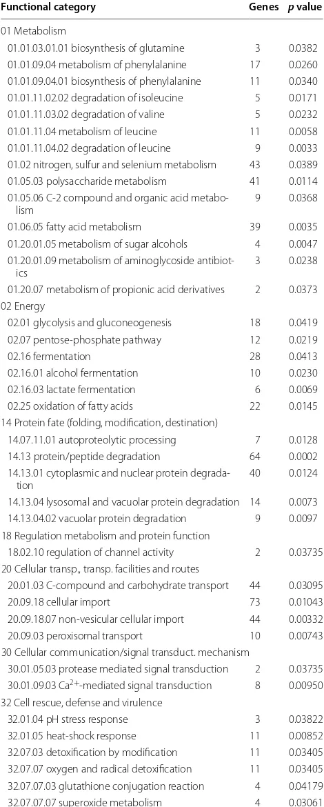
Table 1 MIPS functional catalog category classification of all the genes specifically up-regulated in pkaA deletion strain
Functional category Genes p value
01 Metabolism
01.01.03.01.01 biosynthesis of glutamine 3 0.0382 01.01.09.04 metabolism of phenylalanine 17 0.0260 01.01.09.04.01 biosynthesis of phenylalanine 11 0.0340 01.01.11.02.02 degradation of isoleucine 5 0.0171 01.01.11.03.02 degradation of valine 5 0.0232 01.01.11.04 metabolism of leucine 11 0.0058 01.01.11.04.02 degradation of leucine 9 0.0033 01.02 nitrogen, sulfur and selenium metabolism 43 0.0389 01.05.03 polysaccharide metabolism 41 0.0114 01.05.06 C-2 compound and organic acid
metabo-lism 9 0.0368
01.06.05 fatty acid metabolism 39 0.0035 01.20.01.05 metabolism of sugar alcohols 4 0.0047 01.20.01.09 metabolism of aminoglycoside
antibiot-ics 3 0.0238
01.20.07 metabolism of propionic acid derivatives 2 0.0373 02 Energy
02.01 glycolysis and gluconeogenesis 18 0.0419 02.07 pentose-phosphate pathway 12 0.0219 02.16 fermentation 28 0.0413 02.16.01 alcohol fermentation 10 0.0230 02.16.03 lactate fermentation 6 0.0069 02.25 oxidation of fatty acids 22 0.0145 14 Protein fate (folding, modification, destination)
14.07.11.01 autoproteolytic processing 7 0.0128 14.13 protein/peptide degradation 64 0.0002 14.13.01 cytoplasmic and nuclear protein
degrada-tion 40 0.0124
14.13.04 lysosomal and vacuolar protein degradation 14 0.0073 14.13.04.02 vacuolar protein degradation 9 0.0097 18 Regulation metabolism and protein function
18.02.10 regulation of channel activity 2 0.03735 20 Cellular transp., transp. facilities and routes
20.01.03 C-compound and carbohydrate transport 44 0.03095 20.09.18 cellular import 73 0.01043 20.09.18.07 non-vesicular cellular import 44 0.00332 20.09.03 peroxisomal transport 10 0.00743 30 Cellular communication/signal transduct. mechanism
30.01.05.03 protease mediated signal transduction 2 0.03735 30.01.09.03 Ca2+-mediated signal transduction 8 0.00950
32 Cell rescue, defense and virulence
Table S3). In the wild-type strain, genes encoding proteins of the PDC, kinase activator and anion and ion transport were up-regulated and genes encoding proteins involved in electron transport, transcription and vesicular transport and differentiation were down-regulated after 8 h and 24 h in the presence of avicel (Additional file 2: Table S2; Addi-tional file 3: Table S3).
In summary, deletion of pkaA resulted in the up-regu-lation of degradation pathways and a shift in metabolism to the use of alternative energy sources (e.g., fatty acids), the down-regulation of DNA replication, protein synthe-sis and the PDC. Furthermore, this microarray data show that deletion of pkaA results in increased expression of genes encoding lignocellulosic enzymes, especially after 8 h incubation in cellulose, whereas this response was already reduced after 24 h incubation in cellulose. This indicates a change in the flux of carbon metabolism within the cell.
PkaA is involved in carbon catabolite repression (CCR) To validate the microarray data, the wild-type, ΔpkaA, ΔsnfA and the ΔpkaA ΔsnfA (constructed by genetically crossing ΔpkaA and ΔsnfA) strains were grown in com-plete media for 24 h and then transferred to minimal media supplemented with 1 % avicel or 2 % glucose plus 1 % avicel for 5 days. The growth phenotype of the double mutant was similar to the growth profile of the ΔpkaA strain (Additional file 4: Figure S1). The ΔsnfA strain was included because in S. cerevisiae, the antagonism between PKA and Snf1 regulates carbon utilization [39, 40], while in A. nidulans SnfA has a great influence on cellulase/xylanase induction and CCR [8, 18, 19].
As expected, the wild-type strain showed increased (more than threefold) cellulase (EGL and CBH) produc-tion after 5 days growth on 1 % avicel. Beta-glucosidase (BGL) activity was also measured. Similarly, BGL activ-ity was higher in the ΔpkaA mutant than in the wild-type strain (Fig. 2b). This is in agreement with the microarray data, where during early incubation in avicel (e.g., 8 h), the expression of eglA, eglB and a BGL-encoding gene was ~fourfold higher in the ΔpkaA strain than when
Table 1 continued
Functional category Genes p value
34 Interaction with the environment
34.01.01.01 homeostasis of metal ions (Na, K, Ca, etc.) 29 0.03479 38 Transposable elements, viral, plasmid protein
38.07 proteins necessary for transposon movement 4 0.01412 42 Biogenesis of cellular components
42.19 peroxisome 14 0.00119 42.25 vacuole or lysosome 11 0.02263
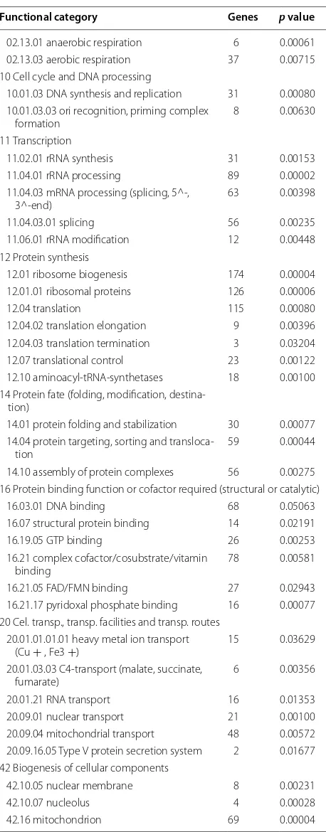
Table 2 MIPS functional catalog category classification of all the genes specifically down-regulated in the pkaA
deletion strain
Functional category Genes p value
01 Metabolism
01.01 amino acid metabolism 107 0.00013 01.01.03 assim. ammonia, metab. glutamate
group 27 0.00201 01.01.03.05 metabolism of arginine 10 0.01657 01.01.03.05.01 biosynthesis of arginine 8 0.00840 01.01.05 metab. urea cycle, creatine and
polyamines 9 0.03639 01.01.06.01 metabolism of aspartate 7 0.00523 01.01.06.01.02 degradation of aspartate 5 0.01900 01.01.06.04 metabolism of threonine 7 0.00359 01.01.06.04.01 biosynthesis of threonine 3 0.03204 01.01.06.05 metabolism of methionine 14 0.00040 01.01.06.05.01 biosynthesis of methionine 7 0.02838 01.01.06.05.01.01 biosynthesis of
homocyst-eine 3 0.05076 01.01.09 metabolism of the cysteine - aromatic
group 43 0.02539 01.01.11.01 metabolism of alanine 3 0.00784 01.01.11.02 metabolism of isoleucine 10 0.00434 01.01.11.02.01 biosynthesis of isoleucine 8 0.00331 01.01.11.02.02 degradation of isoleucine 5 0.02647 01.01.11.03 metabolism of valine 8 0.02220 01.01.11.03.01 biosynthesis of valine 7 0.00523 01.01.11.03.02 degradation of valine 5 0.03558 01.03.01 purin nucleot/nucleoside/nucleobase
metab. 33 0.00029 01.03.07 deoxyribonucleotide metabolism 7 0.01761 01.03.16 polynucleotide degradation 21 0.03254 01.03.16.01 RNA degradation 14 0.02504 01.05.13 transfer of activated C-1 groups 23 0.00011 01.05.13.03 tetrahydrofolate-dependent
C-1-transfer 7 0.00237 01.06.06 isoprenoid metabolism 31 0.02022 01.06.06.11 tetracyclic and pentacyclic
triter-penes (cholesterin, steroids and hopanoids) metabolism
28 0.00044
01.07 metab. vitamins, cofactors, and
pros-thetic groups 53 0.00514 01.07.01 biosyn. vitam, cofactors, prosthetic
groups 33 0.00305 01.20.19 metabolism of secondary products
derived from glycine, l-serine and l-alanine
12 0.00100 01.20.19.01 metabolism of porphyrins 10 0.00134 02 Energy
02.07.01 pentose-phosphate pathway
compared to the wild-type strain. After 24 h though, the expression of these genes was similar between the ΔpkaA and wild-type strains.
To confirm whether carbon catabolite repression was active in the absence of pkaA, the pkaA deletion and wild-type strains were grown in the simultaneous presence of glucose and avicel. Cellulase secretion was repressed in the wild-type strain, whereas the ΔpkaA mutant showed a fourfold increase in cellulase production when com-pared to the wild-type strain in these conditions (Fig. 2a). The ΔsnfA mutant was used as a negative control and showed only a basal level of cellulase production but no clear induction as was observed for the wild-type strain after transfer to avicel for 5 days. BGL activity induction was observed in the ΔsnfA strain; however, the enzyme activity was at similar levels than in the wild-type strain. The double ΔpkaA ΔsnfA deletion mutant behaved like the wild-type strain, secreting a similar amount of cellu-lases in the presence of avicel as the sole carbon source. An increase in the activity of BGL was observed in the double mutant under repressing and de-repressing con-ditions, showing levels of enzyme activity similar to the ΔpkaA mutant under de-repressing conditions. These results suggest that the ΔpkaA mutation can suppress the ΔsnfA mutation and that both genes are somehow geneti-cally interacting.
To check if de-repression also occurs in the presence of xylan, the ΔpkaA, ΔsnfA, ΔpkaA ΔsnfA and wild-type strains were grown in complete media and subsequently transferred to media containing only 1 % xylan or 1 % xylan supplemented with 2 % glucose for 3 days before xylanase and β-xylosidase (BXL) activities were measured in the cul-ture supernatants. Xylanase activity was increased in the presence of xylan and repressed in the simultaneous pres-ence of xylan and glucose in the wild-type strain. In the ΔpkaA mutant, xylanase activity was three times higher than in the wild-type strain in the presence of xylan. This is in agreement with the microarray data where the major xylanase-encoding genes xlnA and xlnC were up-regu-lated ~4.6- and ~4.9-fold, respectively, after 8-h incubation in cellulose in the ΔpkaA strain but not in the wild-type strain (Additional file 2: Table S2). In contrast, after 24-h incubation in cellulose, xlnA and xlnC gene expression was similar between the wild-type and ΔpkaA strains.
Xylanase activity was also increased in the simultane-ous presence of glucose and xylan in the ΔpkaA strain than when compared to the wild-type strain (Fig. 2c). Again, this confirms that deletion of pkaA increases hydrolytic enzyme secretion. BXL activity was measured
Table 2 continued
Functional category Genes p value
02.13.01 anaerobic respiration 6 0.00061 02.13.03 aerobic respiration 37 0.00715 10 Cell cycle and DNA processing
10.01.03 DNA synthesis and replication 31 0.00080 10.01.03.03 ori recognition, priming complex
formation 8 0.00630 11 Transcription
11.02.01 rRNA synthesis 31 0.00153 11.04.01 rRNA processing 89 0.00002 11.04.03 mRNA processing (splicing, 5^-,
3^-end) 63 0.00398 11.04.03.01 splicing 56 0.00235 11.06.01 rRNA modification 12 0.00448 12 Protein synthesis
12.01 ribosome biogenesis 174 0.00004 12.01.01 ribosomal proteins 126 0.00006 12.04 translation 115 0.00080 12.04.02 translation elongation 9 0.00396 12.04.03 translation termination 3 0.03204 12.07 translational control 23 0.00122 12.10 aminoacyl-tRNA-synthetases 18 0.00100 14 Protein fate (folding, modification,
destina-tion)
14.01 protein folding and stabilization 30 0.00077 14.04 protein targeting, sorting and
transloca-tion 59 0.00044 14.10 assembly of protein complexes 56 0.00275 16 Protein binding function or cofactor required (structural or catalytic)
16.03.01 DNA binding 68 0.05063 16.07 structural protein binding 14 0.02191 16.19.05 GTP binding 26 0.00253 16.21 complex cofactor/cosubstrate/vitamin
binding 78 0.00581 16.21.05 FAD/FMN binding 27 0.02943 16.21.17 pyridoxal phosphate binding 16 0.00077 20 Cel. transp., transp. facilities and transp. routes
20.01.01.01.01 heavy metal ion transport
(Cu + , Fe3 +) 15 0.03629 20.01.03.03 C4-transport (malate, succinate,
fumarate) 6 0.00356 20.01.21 RNA transport 16 0.01353 20.09.01 nuclear transport 21 0.00100 20.09.04 mitochondrial transport 48 0.00572 20.09.16.05 Type V protein secretion system 2 0.01677 42 Biogenesis of cellular components
in the same conditions and results were similar to the ones shown for BGL activity. BXL activity was higher in the ΔpkaA strain than when compared to the wild-type strain in the presence of xylan but not in the simultane-ous presence of glucose and xylan (Fig. 2d). There is no difference in BXL activity in the ΔsnfA strain and the double mutant in repressing and de-repressing condi-tions (Fig. 2d).
These enzymatic data validate the microarray and implies that the deletion of pkaA renders the fungus par-tially blind to the presence of glucose, suggesting a role for PkaA in CCR. Additionally, these results also suggest a complex genetic interaction between PkaA and SnfA during cellulase induction and glucose de-repression.
Deletion of pkaA results in reduced CreA nuclear localization upon growth on glucose
The above-mentioned results suggest that PkaA is involved in glucose metabolism and/or CCR. Cellular localization of CreA::GFP in the wild-type and ΔpkaA strains was assessed when the strains were grown in minimal media supplemented with 1 % glucose or 1 % avicel. In the wild-type and ΔpkaA strains, 96 and 25 % of CreA::GFP localized to the nucleus in the presence of glucose whereas 2 and 20 % of CreA::GFP localized to the nucleus in the presence of avicel (Table 3). These results further indicate that PkaA is involved in CCR as CreA is (partially) unable to localize to the nucleus dur-ing repressdur-ing conditions in the absence of pkaA.
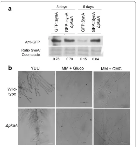
PkaA is involved in protein secretion and hyphal branching As shown by the microarray analysis and enzymatic assays, expression of genes encoding cellulases and xyla-nases was up-regulated and the secretion of cellulases and xylanases was increased in the ΔpkaA strain (Fig. 2). To know whether secretion is specifically responsible for this increase in lignocellulolytic enzyme production or whether it is due to morphological changes, the microarray data were analyzed for the expression of genes involved in pro-tein secretion. Six genes which encode propro-teins involved in the secretion process were up-regulated after 8-h incuba-tion in cellulose in the ΔpkaA strain but not in the wild-type strain. These genes encoded a protein with SNAP receptor activity (AN8488), proteins with transmembrane activity and membrane localization (AN8983, AN5763, AN5559 and AN4019) and a putative transmembrane transporter (AN7295). After 24-h growth in cellulose, the expression of these genes was similar between the ΔpkaA and wild-type strains. To further investigate the influence of pkaA on secretion, a ΔpkaA GFP::SynA strain was generated by crossing the respective parental strains and the amount and fluorescence of GFP::SynA was assessed during growth in cellulose-minimal media. SynA is a V-SNARE synapto-brevin protein involved in the secretion pathway that local-izes to the plasma membrane in actively growing hyphal apex [41, 42]. In agreement with the microarray results, Western blot analysis showed that GFP::SynA levels were about fivefold higher in the ΔpkaA strain than when com-pared to the wild-type strain after 5 days of growth in cel-lulose (Fig. 3a). No differences were observed in GFP::SynA distribution to the hyphal apex of germlings of the parental GFP::SynA and the ΔpkaA GFP::SynA strains after growing for 16 h in cellulose (Additional file 4: Figure S2).
Subsequently, the morphology and number of hyphal tips were assessed and evaluated in the ΔpkaA strain, as
increased protein secretion could correlate with fungal colony morphology. Deletion of pkaA resulted in increased branching (and hence an increased number of tips) when grown in complete media and minimal media supple-mented with glucose or CMC (Fig. 3b). It appears that the deletion of pkaA results in an increased area of secretion which may contribute to the higher amounts of proteins secreted (Additional file 4: Figure S3). At the same time, components of the secretion pathway seem to be up-regu-lated at early and late time points in the ΔpkaA strain, which may also contribute to the observed increase in enzyme secretion. Taken together our results suggest that A. nidu-lans PkaA is important for hyphal branching and secretion.
PkaA translation is triggered under carbon starvation and carbon catabolite de‑repressing conditions
There is very little information about the translation and/ or localization of PkaA in A. nidulans. In S. cerevisiae there are three homologues of pkaA, termed TPK1, TPK2 and TPK3, whose expression after carbon source limita-tion in the stalimita-tionary phase or in the presence of glycerol
Table 3 Percentage of CreA::GFP nuclear localization in different strains under different conditions
Spores were inoculated in 1 % glucose or avicel for 16 h at 22 °C before being transferred to minimal media supplemented with 1 % avicel for 5 h or before 1 % glucose was added to the overnight avicel cultures for 30 min
Strain Grown in Nuclear CreA (%)
creA::GFP 1 % Glucose 96
creA::GFP ΔpkaA 1 % Glucose 25
creA::GFP 1 % Avicel 2
creA::GFP ΔpkaA 1 % Avicel 20
Strain Transfer to Nuclear CreA (%)
creA::GFP 1 % Avicel 17
creA::GFP ΔpkaA 1 % Avicel 24
creA::GFP 1 % Glucose 100
creA::GFP ΔpkaA 1 % Glucose 67
Fig. 3 Deletion of pkaA results in increased hyphal branching. a
was increased [43]. The activity of Tpk1p is controlled by auto-phosphorylation on serine residues. Under starva-tion or glycerol-rich condistarva-tions, Tpk1p is de-phosphoryl-ated [44] and CCR is released. To assess the translation and localization of PkaA in A. nidulans, the correspond-ing PkaA::GFP fusion was constructed. All phenotypes of the PkaA::GFP strain, including growth and conidi-ation, were essentially identical to the wild-type parent. The PkaA::GFP strain was grown in minimal media sup-plemented with glucose and then transferred to minimal media supplemented with avicel or without any carbon source for 0, 15, 30, 60 and 120 min. PkaA translation was assessed by Western blot and fluorescence microscopy. Translation of PkaA increased during the first 30 min after transfer to starvation conditions, as revealed by both microscopy and Western blot, whereas after 60 min translation started to decrease (Fig. 4a, c). In accordance, PkaA activity peaked after 30 min in carbon starvation, before dropping again after 2 h (Fig. 4b). Similar results were observed when cellulose was used as a single car-bon source (Fig. 5a, b). The opposite was observed under the microscope when growing PkaA::GFP in minimal media supplemented with avicel and then transferred to glucose: fluorescence started to decrease after 15 min in glucose (Fig. 6). These results show that the translation of PkaA increases in carbon starvation conditions.
PkaA is involved in glucose uptake and glycolysis in A. nidulans
In mammalian cells, PKA controls the phosphorylation of phosphofructokinase 1 (PFK1), a protein with kinase/ phosphatase bi-functional activity. Under glucose limi-tation, increased levels of cAMP activate PKA which in turn phosphorylates PFK1, hence activating the glycolytic pathway and blocking the gluconeogenesis pathway [45]. This study implicated a role for PkaA in the regulation of CCR and glycolysis in A. nidulans. Subsequently, the wild-type and ΔpkaA strains were grown in complete medium for 24 h before being transferred to minimal medium sup-plemented with glucose for 24 h. The ability to take up glucose was then quantified in both strains. The ΔpkaA strain showed reduced glucose uptake as after 24-h incu-bation in glucose there still remained a small amount of glucose in the minimal medium, whereas the wild-type strain consumed all the glucose after 20 h (Fig. 7a). Fur-thermore, hexokinase/glucokinase activities, the enzymes which convert glucose to glucose-6-phosphate during the first step of glycolysis were reduced in the ΔpkaA strain (32.43 nmol mg−1 min−1) when compared to the wild-type strain (154.07 nmol mg−1 min−1) (Fig. 7b).
To check if other glucose metabolism-related interme-diates were also reduced in the ΔpkaA strain, glycerol and pyruvate levels were quantified in both strains after
growth in the same conditions as described above. Glyc-erol and pyruvate levels were similar in the wild-type and ΔpkaA strains (Fig. 7c, d). These results suggest that dele-tion of pkaA influences glucose uptake and the first step in glycolysis. The reduction in glucose uptake and subse-quent metabolism would lead to the fungus using intra-cellular reserves. To support this hypothesis, the activity of the mitochondrial enzyme alpha-ketoglutarate dehy-drogenase (KGDH) was measured. Indeed, KGDH activ-ity was significantly increased in ΔpkaA strain than when compared to the wild-type strain (Fig. 7e). To verify if the ΔpkaA strain had reduced ability to accumulate osmolytes such as trehalose, an intracellular storage compound required during fungal spore germination [21, 23, 46–48], the two strains were grown in complete media for 24 h and then transferred to minimal media supplemented with glucose plus 1 M sorbitol. After 10 min of incubation in sorbitol-rich conditions, trehalose levels were increased (0.425 and 0.143 mg trehalose.mg protein−1) in both the wild-type and ΔpkaA strains, respectively. After 60 min of incubation, trehalose levels were reduced in both strains with the wild-type having 0.24 mg trehalose.mg pro-tein−1 and ΔpkaA having 0.04 mg trehalose.mg protein−1, indicating almost complete use of trehalose in the latter strain (Fig. 7f). In summary, these results indicate that the ΔpkaA mutant has reduced glucose uptake and metabo-lism due to reduced hexokinase activity and, therefore, increased the utilization of intracellular stores such as trehalose, required for maintaining normal glycerol and pyruvate levels, which are generated by gluconeogenesis.
suggest that PkaA influences glycolysis, subsequently affecting CCR and the energetic status of the cell.
Discussion
The production of bioethanol from plant biomass is eco-nomically dependent on the efficiency of hydrolytic enzyme production. To improve the efficiency of hydrolytic enzyme production in filamentous fungi, it is necessary to under-stand the mechanisms controlling protein synthesis and secretion. This study shows that the deletion of pkaA resulted in increased lignocellulolytic enzyme production. Microarray analysis showed that after 8-h incubation in cel-lulose, cellulase and xylanase gene expression was increased. This increase in gene transcription was not observed in the wild-type strain. Similarly, after 5 days of incubation in cel-lulose, the ΔpkaA strain secreted a higher amount of xyla-nases and cellulases than the wild-type strain. However, microarray data also showed that after 24 h, the expression of some genes (e.g., eglA, eglB, xlnA and xlnC) was similar between both strains. In this study, gene expression was
assessed at early time points (8 and 24 h) whereas enzyme activity assays were carried out at a much later time point (5 days). It is likely that lignocellulolytic enzyme activities and regulation of their responding genes, remained high in the ΔpkaA strain at all the time points tested, whereas in the wild-type strain, there may be fluctuations in enzyme expression/secretion due to CCR. This hypothesis is fur-ther supported by results which showed that lignocellulo-lytic enzyme activities remained high in the simultaneous presence of an inducing (cellulose/xylan) and repressing (glucose) carbon source in the ΔpkaA strain; whereas these enzymes were tightly repressed in the wild-type strain under the same conditions. Furthermore, this work showed that deletion of pkaA resulted in severe defects in glucose metabolism. CreA-mediated CCR tightly controls the
Fig. 5 PkaA is involved in the response to carbon starvation. a West-ern blot pkaA::GFP. The pkaA::GFP strain was grown in minimal media supplemented with 1 % glucose and then transferred to minimal media supplemented with 1 % avicel for the indicated amounts of time. b Fluorescence microscopy of PkaA::GFP grown in the same condition described above
transcription of lignocellulolytic enzymes [8], favoring the usage of preferred carbon sources such as glucose. Deletion of pkaA resulted in the reduced ability of CreA to localize to the nucleus, repressing alternative carbon usage in the presence of glucose. These results indicate that deletion of pkaA renders the fungus partially blind to the presence of glucose. In accordance, the genome-wide microarray analy-ses showed that the deletion of the pkaA caused a quicker response to cellulose in the induction of genes encoding cel-lulases, xylanases, β-glucosidases and β-xylosidases, which are repressed by CreA and are involved in lignocellulose biomass degradation.
Indeed, glucose uptake was reduced in the ΔpkaA strain and more interestingly, the activity of the glucokinase/ hexokinase which catalyzes the first step in glycolysis is also severely reduced in this deletion mutant. This enzyme phosphorylates glucose which serves as a signal for CreA nuclear localization [4]. In addition, CreA may be unable to locate to the nucleus in the presence of glucose. Thus, deletion of pkaA results in a severe defect to the correct uptake and metabolism of glucose, forcing the cell to shift its metabolism to using alternative carbon sources.
Glycolysis results in the production of pyruvate, which can be metabolized by two routes: (1) fermentation, through the pyruvate decarboxylase complex resulting in the production of acetaldehyde which is converted to ethanol [49], or (2) the tricarboxylic acid cycle (TCA). The role of the pyruvate dehydrogenase complex (PDC), which converts pyruvate to acetyl-CoA and directs metabolism towards the TCA cycle is central to determining the fate of pyruvate and reactions triggered by this enzymatic complex are irreversible [50]. Genome-wide microarray analysis showed the down-regulation of genes encoding the PDC, further supporting the hypothesis that the glu-cose pathway is mis-regulated in pkaA deletion mutant. In carbon starvation conditions, the PDC complex is phos-phorylated and inactivated by pyruvate dehydrogenase kinases, thus promoting fatty acid utilization [51]. This is in agreement with the microarray results which showed that genes encoding for proteins involved in fatty acid metabolism are up-regulated in the ΔpkaA mutant. Fatty acid catabolism has been shown to be important for fungal pathogenesis, secondary metabolite production, metabo-lism and development [37]. When faced with a nutrient-poor environment, the fungus switches to using other energy sources, one of which is fatty acids. Fungi are able to solely grow on fatty acids [37]. In fungi, fatty acids are first degraded to C4 compounds via the glyoxylate cycle in peroxisomes before further being converted to acetyl-CoA which enters the TCA cycle in the mitochondria [37]. Fur-thermore, oxidative phosphorylation and ATP production were reduced in the pkaA deletion strain, supporting the hypothesis that the cell is exhibiting a starvation response
in the presence of glucose. This also explains the early and constant up-regulation of genes encoding lignocellulolytic enzymes as these have been proposed to have a “scavenger role” under carbon starvation conditions in A. niger and T. reesei [52, 53].
The SnfA kinase complex, which controls alterna-tive carbon usage, is key for the de-repression of CreA-mediated CCR and hydrolytic enzyme transcription [4]. Extracellular enzymatic levels do not change in the snfA deletion strain under repressing and de-repressing condi-tions (this strain appears to secrete a basal level of ligno-cellulolytic enzymes), whereas in the ΔpkaA ΔsnfA strain cellulase levels were increased. This shows that PkaA and SnfA genetically interact, but the details of this interac-tion remain subject to further investigainterac-tion. Nonetheless, it would appear that PkaA and SnfA have opposing func-tions, i.e., PkaA is important for glucose-mediated-cat-abolite repression while SnfA is important for catglucose-mediated-cat-abolite de-repression. It remains to be investigated the details of the interaction between these protein kinases (for a model, see Fig. 8). In the presence of glucose, PkaA is activated and regulates indirectly the nuclear localization of CreA. This work showed that PkaA controls the activ-ity of hexokinase which catalyzes the first step in glycoly-sis and phosphorylates glucose. Phosphorylated glucose has been shown to be (one of) the signal(s) for CreA localizing to the nucleus in A. nidulans [8]. Furthermore, this work showed that PkaA is involved in the regulation of SnfA by repressing it. SnfA becomes inactive in glu-cose-rich conditions as it is required for the use of alter-native carbon sources such as cellulose [39, 40, 54, 55]. In the presence of non-glucose, complex carbon sources (e.g., cellulose), PkaA is inactive, SnfA becomes activated and subsequently CreA localizes to the cytoplasm.
There is a significant lack of information on the compo-nents of the signaling pathways in which both PkaA and SnfA are involved and Fig. 8 only shows a very prelimi-nary diagram on how both kinases are interacting and their involvement in CreA cellular localization. Further-more, the level of PkaA (and probably SnfA) activity is also likely subject to fluctuations and other regulations as this work showed that it is up-regulated in cellulose the first 30 min after transfer from glucose. The details of the respective pathways remain subject to investigation.
of the stuA transcription factor, which is involved in spa-tial conidial formation, cleistothecia, Hulle cell formation and germination [23]. An increase in the expression of genes encoding transcription factors involved in control-ling development could explain why the pkaA deletion strain exhibited an increase in the number of branches and tip formation when grown in complete media and in minimal media supplemented with glucose or CMC. No difference in the localization of GFP::SynA, a protein which localizes to secretory vesicles, was observed in the wild-type and ΔpkaA strains when examined by fluores-cent microscopy. However, Western blot analysis showed a reduction in GFP::SynA after 5 days of growth in cel-lulose in the wild-type strain but not in the ΔpkaA strain.
Furthermore, microarray analysis identified several genes encoding putative components of secretion to be up-regulated during the first 8 h of incubation in glucose; after 24 h expression levels of these genes were similar to those in the wild-type strain. Again, these results indicate that protein secretion remains high in the ΔpkaA strain throughout all the time points tested here, whereas in the wild-type strain they appear to be subject to fluctua-tions. These results suggest that the increase in enzyme secretion in pkaA deletion strain was due to PkaA play-ing a role in protein secretion and in hyphal morphology. An increase in the number of hyphal tips from which proteins are secreted could contribute to the increased secretion of hydrolytic enzymes by the ΔpkaA strain.
Conclusion
In summary, PkaA is involved in controlling the cel-lular metabolic and energetic status. PkaA is directly involved in glycolysis and deletion of this protein kinase resulted in CCR mis-function, forcing the cell into a state of starvation, where hydrolytic enzyme secretion is increased, expression of genes encoding mitochondrial components as well as genes involved in fatty acid utilization were up-regulated. This works implies that PkaA regulates the secretion of enzymes required for plant biomass degradation. The absence of PkaA leads to the cell secreting higher amounts of hydrolytic enzymes.
This work further contributes to unraveling the meta-bolic pathways governing different states of the cell. In addition, this study highlights how fungal morphology can impact on enzyme secretion. Furthermore, deletion of pkaA resulted in reduced glucose uptake and a defect in glycolysis, which as a consequence, reduces CCR, result-ing in hydrolytic enzyme induction in the presence of inducer molecules. Deletion of pkaA also results in the inability to perform glycolysis which puts the cell in a state of carbon starvation. This work shows that PkaA is specif-ically induced in carbon starvation conditions, as has also been reported in other organisms. Together, this study identified various roles in carbon metabolism for PkaA within the cell and shows how a tightly interconnected metabolic network governs lignocellulolytic enzyme pro-duction and secretion. This work provides a basis for fur-ther research which aims at elucidating cellular regulatory pathways which can be useful for the further engineer-ing of fungal strains, which are highly efficient in protein secretion with the aim to use these enzymes in various industrial processes. Up to this point, the direct and indi-rect targets of PkaA are unknown and uncovering these proteins could provide a platform for the engineering of fungal strains with improved plant biomass degradation capabilities.
Methods
Strains and culture conditions
The two A. nidulans strains TN02A3 (pyrG89; pyroA4; nkuA::argB) and A4 were used as reference strains (wild type). The ΔpkaA and ΔsnfA null mutants used in this work were obtained from the protein kinase deletion col-lection [59] and are publicly available at the Fungal Genet-ics Stock Center (http://www.fgsc.net). A list of all strains used in this study is found in Table 4. All strains were grown at 37 °C in either liquid (without agar) or solid (with 20 g/l agar) minimal medium [MM: 1 % (w/v) carbon source, 50 mL of a 20× salt solution (120 g/l NaNO3, 10.4 g/l KCl, 30 g/l KH2PO4, 10.4 g/l MgSO4), 1 mL of 5× trace elements [22.0 g/l ZnSO4, 11 g/l boric acid, 5 g/l MnCl2, 5 g/l FeSO4, 1.6 g/l CoCl2, 1.6 g/l CuSO4, 1.1 g/l (NH4)2MoO4, 50 g/l ethylenediaminetetraacetic acid (EDTA)], pH 6.5 or in liq-uid complete medium complete [2 % w/v glucose, 0.5 % w/v yeast extract, trace elements (same as described above)]. Depending on the auxotrophy of the strain, uridine (1.2 g/l), uracil (1.2 g/l) or pyridoxine (0.005 mg/μL) were added.
Strain construction
The construction of the Pka::GFP strain was performed according to Colot et al. (2006) [60]. Standard molecular techniques were performed according to Sambrook and Russel [61]. The pkaA 5′ and 3′ untranscribed regions (UTR), ORF (open reading frame) plus the pkaA gene (minus the stop codon), gfp gene and spacer region and the pyrG gene were co-transformed into S. cerevi-siae SC9721 strain (MATα his3-Δ200 URA3-52 leu2Δ1 lys2Δ202 trp1Δ63) obtained from the Fungal Genetic Stock Center (FGSC). Primer sequences are described in Table 5. The pkaA 5′UTR and ORF were amplified
using the primers “pRS426-5′ PKA UTR F’” and “PKA
Spacer GFP R”; the gfp gene was amplified using primers “Spacer GFP Fw” and “GFP Ve 3 Afu Rv”; the pyrG frag-ment was generated using primers “GFP-PyrG Fw” and “Afu PyrG Rv FGSC” and the pkaA 3′ UTR region was
Table 4 A. nidulans strains used in this study
The genotypes of each strain are also shown
Strain Genotype References
TN02A3 pyroA4 pyrG89; chaA1; ΔnKuA::argB [73]
R21 pabaA1; yA2 FGSC
ΔpkaA pyrG89; wA3; argB2; ΔnkuAku70::argB pyroA4; sE15nirA14 chaA1 fwA1; ΔpkaA::pyrG+ [59, 74]
ΔsnfA pyrG89; wA3; argB2; ΔnkuAku70::argB pyroA4; sE15 nirA14 chaA1 fwA1; ΔsnfA::pyrG+ [73, 74]
ΔsnfA PABA− pyrG89; wA3; argB2; ΔnkuAku70::argB pyroA4; sE15 nirA14 chaA1 fwA1; pabaA1; ΔsnfA::pyrG+ [8]
ΔpkA ΔsnfA pyrG89; wA3; argB2; ΔnkuAku70::argB pyroA4; sE15 nirA14 chaA1 fwA1; pabaA1; ΔpkaA::pyrG+; ΔsnfA::pyrG+ This study
GFP::SynA PIRO− pyrG89, GFP::synA::pyrGAf, nkuA::bar, pyroA4 [41]
GFP::SynA PABA− pyrG89, gfp::synA::pyrGAf, nkuA::bar, pyroA4 pabaA1 This study
amplified using primers “PyrG 3 UTR PKA F” and “PKA 3 UTR-pRS426″. Homologous recombination within S. cerevisiae created the construct, which was subsequently amplified from pooled S. cerevisiae DNA, and 20 μg was subsequently transformed into TN02a3 according to Osmani et al. (1987) [62]. Transformants were selected via their ability to grow on solid MM supplemented with pyridoxine in the absence of uridine and uracil.
Homologous integration was confirmed by PCR using a forward primer that anneals out of the recombina-tion locus (F_pka_checkinsert_A) and a reverse primer (GFP Ve 3 Afu R) that anneals at the end of the GFP gene (Table 5). The ∆pkaA ∆snfA and GFP::synA ΔpkaA strains were generated by sexually crossing the parental strains and the genotype of the double mutant was con-firmed by PCR (Table 5) and in case of the GFP construc-tion, by microscopy. The deletion of pkaA and snfA were confirmed by PCR using primers pRS426-5′ PKA UTR F and pRS426-5′ snfA UTR F, respectively, with Afu PyrG RV FGSC as the reverse primer for both constructions.
Microarray analysis
Initially 1 × 10⁷ conidia A. nidulans were inoculated in complete media at 37 °C in a rotatory shaker (180 rpm) for 24 h. Subsequently, mycelia were washed with sterile water and then incubated in minimal media supplemented with 1 % cellulose at 37 °C for an additional 8 and 24 h. At each step the mycelia from three biological replicates were collected by vacuum filtration and immediately frozen in liquid nitrogen. Agilent custom-designed oligonucleotides arrays [63] were used to identify the transcriptional differ-ences during growth on complete media (Cy3 reference)
and cellulose (Cy5) for the wild-type and ΔpkaA strains. Total RNA was extracted and RNA integrity confirmed as described in the section “RNA extraction”. Array hybridi-zation and data analysis were performed according to De Assis et al. (2015) [50]. The dataset was deposited in the
Gene Expression Omnibus (http://www.ncbi.nlm.nih.
gov/geo/query/acc.cgi?acc=GSE70917) under the num-ber GSE70917. Genes were determined as differentially expressed between carbon sources by applying a t test (p < 0.01) performed within the Mev software [64]. The functional profile and identification of overrepresented GO terms within the differentially expressed gene sets from each strain under the two nutritional conditions were identified using the GO Slim mapper ( http://www.asper-gillusgenome.org/cgi-bin/GO/goTermMapper) and Fun-Cat (http://mips.helmholtz-muenchen.de/funcatDB/).
RNA extraction
Mycelia were harvested by vacuum filtration and imme-diately frozen in liquid nitrogen. Mycelia were ground to a fine powder under liquid nitrogen and total RNA was extracted using TRIZOL, according to manufactur-er’s instructions (Invitrogen), before being treated with RNAse-free DNAse (Promega) and purified with the RNeasy® Mini Kit, according to manufacturer’s instruc-tions (Qiagen). RNA integrity was confirmed using the Bioanalyser Nano Kit (Agilent Technologies) and the Agilent Bioanalyser 2100, considering RIM value 8.0 as the RNA quality threshold. The SuperScript III First Strand Synthesis system (Invitrogen) and oligo(dT) primers were used for cDNA synthesis according to manufacturer’s instructions. All the primer sequences used in this work are described in the Additional file 5: Table S4.
Xylanase and cellulase assays
Xylanase xylanase) and cellulase (endo-1,4-β-glucanase) assays were performed using Azo-Xylan (Birch-wood) and Azo-CM-Cellulose (both from Megazyme International, Bray, Ireland) as substrates, according to man-ufacturer’s instructions. All enzyme assays were carried out on the supernatants of biological triplicates. Technical tripli-cates were carried out on each biological replicate.
β‑Glucosidase and β‑xylosidase assays
β-Glucosidase (BGL) and β-xylosidase (BXL) activities were measured in 20 μL culture supernatants. ρ-Nitro phe-nyl glucopyranoside (ρ-PNG) and 4-β-D-xylopyranoside (ρ-PNG) were used as substrates for BGL and BXL activi-ties in 50 mM buffer citrate pH 6.0 as previously described [65]. Enzyme activities were calculated using the slope of the linear curve generated during 30 min of reac-tion at 37 °C. All enzyme assays were carried out on the
Table 5 List of the primer pair used in this work
Primer Sequence
pRS426-5′ PKA UTR F TAACGCCAGGGTTTTCCCAGTCACGACGTTCT-GAAGCCCGATACAACC
PKA Spacer GFP R AAAGTTCTTCTCCTTTACTCATTCCCCGTGTTC-CGAAATCGGGGAACAGGTGACCG
PyrG 3 UTR PKA F AAGAGCATTGTTTGAGGCGAATTCACCCTCTAAC-GAGTGATG
PKA 3 UTR-pRS426 R GCGGTTAACAATTTCTCTCTGGAAACAGCTCTAA-GGCAGGCAGTTCTCG
GFP PyrG FW GCATGCAAGCTTGGCGTATTCTGTCTGAGAG-GAGGC
Afu PyrG RV FGSC GAGCAGCGTAGATGCCTCGACC
Spacer GFP FW GGAACACGGGGAATGAGTAAAGGAGAAGAACT GFP Ve 3 Afu RV CTCAGACAGAATACGCCAAGCTTG
pRS426-5′ snfA UTR F GTAACGCCAGGGTTTTCCCAGTCACGACGTGGA-GATGGAAGTCGAAAGG
supernatants of biological triplicates. Technical triplicates were then carried out on each biological replicate.
Fluorescence microscopy
Strains were inoculated in 3 mL MM supplemented with 1 % glucose, cellulose or CMC in a small Petri dish with a cover slip and incubated for 16 h at 22 °C. For the trans-fer experiments, coverslips were washed three times with water and then transferred to minimal media supple-mented with a different carbon source for 5 h.
Secretome and cellulase assay on plate
Mycelia were grown from 107 spores in 50 mL media in the specified conditions. Culture supernatants were sepa-rated from the mycelia and centrifuged at 1500 g at 4 °C for 5 min. Supernatants (40 mL) were transferred to new clean tubes and freeze-dried before being re-suspended in 3 mL buffer containing 50 mM Tris–HCl pH 7.0, 1 mM DTT and protease inhibitors (EDTA-free Complete mini, Roche). About 20 μL of the re-suspended supernatants were run on a 10 % SDS-PAGE gel. The gel was then silver stained as described previously [66]. The cellulase assay on plate was carried out as previously described [67, 68].
Western blots
Mycelia were grown from spores in the specified condi-tions before being harvested and ground to a fine powder under liquid nitrogen. Mycelial powder were re-suspended in extraction buffer [50 mM Tris–HCl pH 7.0, 50 mM NaF, 1 mM Na3VO4, 1 mM DTT, phosphatase inhibitor cocktail P0044 (Sigma) and the complete mini EDTA-free protease inhibitor cocktail (Roche)], prior to centrifuga-tion for 5 min at 14000×g. The concentration of the pro-tein extracts was measured using the Bio-Rad Bradford protein assay, according to manufacturer’s instructions. Proteins were precipitated with 0.25 M NaOH and 1 % β-mercaptoethanol and incubated on ice for 10 min before trichloroacetic acid was added to a final concentration of 6 % for 10 min on ice. Samples were pelleted by centrifu-gation for 5 min at 4°, maximum speed. Pellets were re-suspended in electrophoresis Bolt (Life Technologies) LDS sample buffer and reducing agent (according to manufac-turer’s instructions). Samples were run on Bolt® 4–12 % Bis–Tris Gels (Life Technologies) before proteins were transferred to membranes with the iBlot® 2 Gel transfer device, according to manufacturer’s instructions. Mem-branes were incubated with a 1:10,000 dilution of anti-GFP antibody (AbCam ab290) overnight at 4 °C. The next day membranes were washed and incubated with a 1:5000 dilu-tion of secondary antibody Rabbit (Cell Signaling). Proteins were detected using the Super Signal West Pico (Thermo Scientific) chemiluminescent substrate according to manu-facturer’s instructions.
Glucose uptake
Mycelia were grown from 3 × 10⁶ spores in 30 mL
com-plete media for 24 h at 37 °C in a rotary shaker (180 rpm). Mycelia were then transferred to minimal medium sup-plemented with 1 % glucose as a carbon source for an additional 24 h. The amount of free glucose remaining in the culture supernatants was measured using the Glucose GOD-PAP Liquid Stable Mono-reagent kit (LaborLab Laboratories Ltd. Guarulhos, São Paulo, Brazil), accord-ing to the manufacturer’s instructions. Glucose uptake was calculated via determining the difference in glucose present in the initial media and at different time points during the 24-h incubation in MM.
Hexokinase/glucokinase activities
Proteins were extracted from mycelia grown in the specified conditions and protein content was measured as described in the section “Western blots”. The hexokinase/glucokinase activity was measured in protein extracts according to [69] with some modifications. 3 μg of total protein extract was mixed with reaction buffer (50 mM Hepes pH 7.5, 50 mM KCl, 5 mM MgCl2, 2 mM ATP, 1 mM phosphoenolpyruvate, 0.4 mM NADH, 5 U pyruvate kinase, 15 U lactate dehydro-genase) to a final volume of 200 μL. Samples were first incu-bated for 30 min at 37 °C to stabilize the reactions and get an even reaction curve. Enzymatic reactions were triggered with the addition of 100 mM glucose and the decline in absorbance at 340 nm was measured during 15 min at 37 °C in the SpectraMax I3 spectrometer (molecular devices).
Pyruvate and glycerol measurement
Proteins were extracted from mycelia grown in the specified conditions and protein content was measured as described in the section “Western blots”. Glycerol and pyruvate con-centrations were measured in 5 and 10 μg, respectively, of total extracted protein as described previously [50].
Alpha‑ketoglutarate assay
Proteins were extracted from mycelia grown in the speci-fied conditions and protein content was measured as described in the section “Western blots”. Alpha-ketoglu-tarate activity was measured in 30 μg of total extracted protein as described in [70–72] with modifications. The reaction buffer consisted of 100 mM Tris–HCl pH 7.0,
5 mM 2-mercaptoethanol, 1 mM MgCl2, 2 mM thiamine
pyrophosphate (TPP), 5 mM alpha-ketoglutarate acid
disodium salt, 1 mM NAD+ and 0.2 mM Coenzyme A.
The reduced form of NADH was measured at 340 nm in the SpectraMax I3 spectrometer (molecular devices).
PKA Activity
as described in the section “Western blots”. PKA activ-ity was measured using PepTag Assay Non-radioactive Detection of cAMP-dependent Protein Kinase assay Ref. V5340 (Promega) according to manufacturer’s instructions.
Trehalose assay
Proteins were extracted from mycelia grown in the speci-fied conditions and protein content was measured as described in the section “Western blots”. The trehalose content was measured in 10–20 μg protein using the Tre-halose Assay kit K-TREH 11/12 (Megazyme) according to manufacturer’s instructions. A standard curve of 0–4 μg trehalose dehydrate was also prepared.
ADP/ATP ratio
Proteins were extracted from mycelia grown in the specified conditions and protein content was measured as described in the section “Western blots”. The ADP/ ATP ratio was measured in 10 μg of total protein extract using the ADP/ATP ratio assay kit MAK135 (Sigma) fol-lowing the manufacturer’s instructions. Luminescence was read in the SpectraMax I3 spectrometer (Molecular Devices).
Statistical analysis
Statistical analyses were performed for all reactions of three biological replicates using a one-tailed t test (Prism, GraphPad) with *p < 0 0.05, **p < 0.01 and ***p < 0.001.
Additional files
Additional file 1: Table S1. List of CAZymes. Additional file 2: Table S2. MIPS FunCat 8 h. Additional file 3: Table S3. MIPS FunCat 24 h.
Additional file 4: Figure S1. Deletion of pkaA results in a severe growth defect. Strains ΔpkaA, ΔacyA, R21, ΔsnfA and ΔpkaA snfA were grown in a concentration from 105 to 102 (left to right) from spores on different car-bon sources [YUU (Complete media), Glu (glucose), Casa (casaminoacids), Gly (glycerol), Xyl (Xylose), Fru (fructose) and Trybut (trybutirin)]. Figure S2. Expression of GFP::SynA. A. Microscopy picture (GFP) of GFP::SynA grown for 16 h at 22 °C in 1 % cellulose. B. Fluorescence in mycelia which were grown from spores in minimal media supplemented with 1 % cellulose at 22 °C for 16 h. Fluorescence was then assessed using ImageJ freeware. An average of 50 pictures were taken and evaluated for each strain. Figure S3. The ΔpkaA strain secretes more proteins. A) Cellulase secretion in dif-ferent strains. Strains were grown (upper row) for 48 h on minimal media supplemented with 1 % CMC (carboxymethylcellulose) as sole carbon source. Plates were then strained with congo red (lower row) and the halo/mycelia was measured. B) Secretion of proteins by the wild-type and ΔpkaA strains. Strains were grown in triplicates from spores in complete media for 16 h and then transferred to minimal media supplemented with 1 % cellulose for 5 days. Culture supernatants were harvested, dried and re-suspended before proteins were run on a SDS-PAGE gel and silver stained. Arrows indicate proteins secreted by the pkaA mutant but not by the wild-type strain.
Additional file 5: Table S4. List of the primer pair used in this work.
Abbreviations
AA: auxiliary activities; ADP: adenosine diphosphate; ATP: adenosine triphosphate; BGL: β-glucosidase; BXL: β-xylosidase; cAMP: cyclic adenosine monophosphate; CAZy: carbohydrate-active enzymes; CBH: cellobiohydro-lases; CCR: carbon catabolite repression; CE: carbohydrate esterases; CMC: carboxymethyl cellulose; DTT: dithiothreitol; EDTA: ethylenediaminetetraacetic acid; FAD: flavin adenine dinucleotide; FMN: flavin mononucleotide; GFP: green fluorescent protein; GH: glycoside hydrolases; GO: gene ontology; GPCR: G protein-coupled receptor; GT: glycosyltransferases; KCl: potassium chloride; KGDH: alpha-ketoglutarate dehydrogenase; MgCl2: magnesium chloride; MM:
minimal media; Na3VO4: sodium orthovanadate; NADH: nicotinamide adenine
dinucleotide reduced form; NaF: sodium fluoride; ORF: open read frame; PDC: pyruvate dehydrogenase complex; PKA: cAMP-dependent protein kinase catalytic subunit A; PL: polysaccharide lyases; TCA: tricarboxylic acid cycle; TPP: thiamine pyrophosphate; V-SNARE: synaptobrevin protein involved in the secretion pathway; YG: complete media (yeast extract and glucose); ρ-PNG: ρ-nitro phenyl glucopyranoside; ρ-PNX: 4-β-d-xylopyranoside.
Authors’ contributions
LJA performed enzymatic assays, construction of the strains, and contrib-uted to design, acquisition and analysis of data. MS performed the PCR and Southern analysis of the mutants, genetical crossing analysis and experiments of molecular genetics. TFR performed the statistical analysis, experiments with GFP::SynA and GFP microscopy analysis. LNAR performed microar-ray hybridizations and analysis, and contributed to design, acquisition and analysis of data. NAB contributed to the interpretation of gene ontology, design, acquisition and analysis of data. GHG contributed to the concept and design of the investigation in addition to the preparation of the manuscript. All authors were involved in the drafting of the manuscript, contributed to the discussion and approved the final manuscript. All authors read and approved the final manuscript.
Author details
1 Departamento de Ciências Farmacêuticas, Faculdade de Ciências
Farmacêu-ticas de Ribeirão Preto, Universidade de São Paulo, Av. do Café S/N, CEP 14040-903, Ribeirão Preto, São Paulo, Brazil. 2 Plant Biology and Crop Science,
Rothamsted Research, Harpenden, Herts AL5 2JQ, UK.
Acknowledgements
We would like to thank the Fundação de Amparo a Pesquisa do Estado de São Paulo (FAPESP) and Conselho Nacional de Desenvolvimento Científico e Tecnológico (CNPq), Brazil, for providing financial support. We also thank the two reviewers and the editor for their suggestions and comments.
Competing interests
The authors declare that they have no competing interests.
Received: 6 August 2015 Accepted: 30 November 2015
References
1. Ochiai A, Itoh T, Kawamata A, Hashimoto W, Murata K. Plant Cell Wall Degradation by Saprophytic Bacillus subtilis strains: gene clusters respon-sible for Rhamnogalacturonan depolymerization. Appl Environ Microbiol. 2007;73:3803–13.
2. DeBoy RT, Mongodin EF, Fouts DE, Tailford LE, Khouri H, Emerson JB, Mohamoud Y, Watkins K, Henrissat B, Gilbert HJ, Nelson KE. Insights into plant cell wall degradation from the genome sequence of the soil bacte-rium Cellvibrio japonicus. J Bacteriol. 2008;190:5455–63.
3. Boutard M, Cerisy T, Nogue P-Y, Alberti A, Weissenbach J, Salanou-bat M, Tolonen AC. Functional diversity of carbohydrate-active enzymes enabling a bacterium to ferment plant biomass. PLoS Genet. 2014;10:e1004773.
4. Rytioja J, Hilden K, Yuzon J, Hatakka A, de Vries RP, Makela MR. Plant-polysaccharide-degrading enzymes from basidiomycetes. Microb Mol Biol Rev. 2014;78:614–49.