Gynecol Surg (2006) 3: 18–22 DOI 10.1007/s10397-005-0153-1
O R I G I N A L A RT I C L E
C. Altgassen . S. Kuss . U. Berger . W. Michels . K. Diedrich . A. Schultze-Mosgau
Learning laparoscopic myomectomy
Published online: 26 November 2005 #Springer-Verlag Berlin/Heidelberg 2005
Abstract To describe and to analyse the learning effect in laparoscopic myomectomy. Hospital chart records of 351 patients were retrospectively evaluated. Myomectomy was performed in a standardised fashion. To assess a potential learning effect, only cases presenting a singular intramural fibroid were analysed if a surgeon had done more than ten laparoscopic myomectomies of this type. Cases were analysed according to the order of surgery for the individual surgeon: group A comprised the first ten cases of each surgeon compared with group B, which comprised the subsequent cases. Eighty cases were performed by five surgeons. Group A comprised 50 cases and group B comprised 30 cases. No difference was found with regard to patients' age, BMI, and size of the fibroid. Rate of intra-and postoperative complications according definition were lower in group B but failed to be of statistical significance. Duration of surgery improved after ten cases and no severe complications were noted beyond the tenth case. A learning experience of at least ten laparoscopic myomec-tomies was necessary in our institution to improve duration of surgery and to reach a low level of severe complications.
Duration of the surgical procedure and rate of severe complication were adequate study end points to assess a learning effect.
Keywords Laparoscopic . Myomectomy . Learning curve
Introduction
Pedunculated fibroids were the first to be removed lap-aroscopically [1]. While the technique developed women’s self perception changed and the procedure was not only regarded as a means to improve fertility but also to relieve symptoms. Today, laparoscopic myomectomy has become an accepted technique [2,3] and nearly all fibroids can be removed endoscopically independent of their size or their position within the uterus [4–6]. In a randomised study intensitivity of postoperative pain was found to be sig-nificantly lower, and full recuperation was reached earlier, after laparoscopic myomectomy than after abdominal myomectomy [7]. Other well designed studies showed that compared to abdominal myomectomy laparoscopic myo-mectomy is more cost effective, associated with shorter hospital stays, less blood loss, less postoperative complica-tions, and lower frequency of adhesions, though rate of recurrence is higher and operative time is longer [7–12]. Laparoscopic myomectomy is regarded as a procedure of medium severity [13–15], because it demands complete laparoscopic haemostasis and closure of the operative site to avoid uterine rupture in a potential future pregnancy. Little is known about the learning aspect of this surgical method. In this study, our objective was to evaluate the factors con-sidered for proficiency and to estimate the number of procedures necessary in a teaching setting to achieve competence.
Materials and methods
Surgical data of all women who underwent laparoscopic myomectomy between August 1994 and November 2002
The study complies with the current laws of the Federal Republic of Germany.
S. Kuss . W. Michels
Department of Gynaecology, Friedrich-Schiller-University, Bachstrasse 18,
07740 Jena, Germany
U. Berger
Institute for Medical Psychology, Friedrich-Schiller-University, Bachstrasse 18,
07740 Jena, Germany
C. Altgassen (*) .K. Diedrich . A. Schultze-Mosgau Department of Obstetrics and Gynaecology,
University of Schleswig-Holstein, Campus Luebeck, Ratzeburger Allee 160, 23538 Luebeck, Germany
e-mail: [email protected] Tel.: +49-451-5002134
at the Department of Gynaecology at the Friedrich-Schiller-University, Jena, Germany, were analysed. Data regarding patients’ history, characteristics, and outcome were available from chart reviews of the hospital, out-patients records, and questionnaires sent out, and returned in May 2003, by the patients. The internal review board approved the study.
All women routinely underwent preoperative pelvic ul-trasound. Patients received perioperative antibiotics and kept an indwelling catheter until the first postoperative morning. Low molecular heparin was administered daily for prophylaxis of thromboembolism after surgery in all patients. All procedures were video documented. Myo-mectomy was performed in a standardised fashion by all surgeons under general endotracheal anaesthesia. No uter-ine manipulator was used and no dye was injected to facilitate myomectomy. For myomectomy, three 5-mm trocars were placed in the lower abdomen. The uterine serosa and myometrium were coagulated with a bipolar forceps and incised to expose the surface of the fibroid. The lower middle 5-mm trocar was replaced by a 10-mm trocar and the fibroid was grasped with a 10-mm tenaculum grasping forceps and extracted from of the surrounding myometrium by blunt dissection with the activated bipolar forceps step-by-step. Connective tissue bridges were co-agulated additionally and cut with scissors to reduce blood loss. Fibroids were stored in the pouch of Douglas. The myometrium was bipolar coagulated to achieve near-com-plete haemostasis. The uterus was closed with interrupted intracorporeal sutures (0-Vicryl, sharp curved needle) in one or two layers depending on the depth of the defect to prevent adhesion formation and to achieve complete hae-mostasis. Fibroids were removed after morcellation with an electric morcellator (Morcellator 26200M; Karl Storz, Tuttlingen, Germany) and extracted through the midline 10-mm trocar. Clots and debris were removed through repeated irrigation and suctioning. An intraabdominal Robinson drain was placed on day 1 after surgery.
Size, position of the fibroid, time for the whole surgical procedure, number and type of intra- and postoperative complications, haemoglobin difference, consecutive order of the procedure for each individual surgeon, and pre- and postoperative endovaginal sonographic assessment of the uterus and the pouch of Douglas were registered. Duration of surgery was calculated from insertion of the Verress needle until skin closure of the trocar sites. The hae-moglobin level was checked preoperatively and on day 2 after surgery. All patients were evaluated clinically and by ultrasound on the day of discharge. Complications were defined as follows:
Intraoperative complications: bleeding requiring trans-fusion, bladder, bowel, and ureteral injury, subcutaneous emphysema, and unintended conversion from laparoscopic myomectomy to abdominal myomectomy or hysterectomy. Postoperative complications: (a) major complications— haemorrhage requiring treatment, fistula, thrombosis, em-bolism or re-operation within 8 weeks and (b) minor— fever including unexplained, related to operative site, or not related to operative site (e.g. urinary tract infection),
wound complications, and others. In case of more than one postoperative complication, the causative problem was taken into account, e.g. urinary tract infection and as-sociated fever was considered as urinary tract infection.
All laparoscopic myomectomies in our teaching institu-tion were analysed. To assess a potential learning curve, we only considered procedures by surgeons experienced in general and laparoscopic surgery. They should have under-taken more than 20 laparoscopic myomectomies (multiple fibroids and/or different presentations) within the study period. Out of these 20 laparoscopic myomectomies, more than ten women must have had a singular fibroid lying intramurally or a fibroid that did not bulge subserously more than 50%. Cases were analysed according to the order of surgery for the individual surgeon. During the study period, all surgeons undertook laparoscopic myomec-tomies in women presenting with multiple fibroids or even more complex laparoscopic procedures.
For comparison, patients were grouped. Group A com-prised the first ten cases of each individual surgeon, whereas group B comprised the subsequent cases.
Data were collected in an ACCESS data base (Microsoft, Oregon) and statistics were analysed with SPSS (SPSS, Illinois). Fisher’s exact test was used for comparison of complications. Duration of surgery and haemoglobin dif-ference was evaluated with Student’st-test. Mann-Whitney
U-test was used to compare age, and BMI. AP-value <0.05 was considered to be of statistical significance.
Results
Three-hundred and fifty-one women underwent laparo-scopic myomectomy at the Department of Gynaecology at the Friedrich-Schiller-University, Jena, Germany, during the study period.
A total of 654 fibroids were removed by 24 surgeons. One-hundred and twenty-eight patients presented a singu-lar fibroid that was located intramurally or to less than 50% subserously. Of these 128 patients, 80 were operated on by
Table 1 Characteristics and results in 80 laparoscopic myomectomies
Group A (n=50)
Group B (n=30)
Age (years) 35 34
Median−IQ 11 7.8
BMI, kg/m2 21.9 21.9
Median−IQ 4.6 4.3
Size in cm (mean±SD) 5.7±1.9 5.6±2.2 Time in min (mean±SD) 117.9±48.8 90.8±35.4 Haemoglobin difference
g/dl (mean±SD)
−0.5±1.3 −0.2±1.5
Intraoperative complications (%) 8.0 3.3 Postoperative complications (%) 12.0 6.7
five surgeons qualifying for this analysis. Group A com-prised 50 cases (first ten cases of each qualifying surgeon) and group B comprised 30 cases (the subsequent cases of these five surgeons).
In 21 cases, additional surgery was undertaken: tubal coagulation (8.75%), chromopelvicopy (8.75%), adnexal surgery (7.50%), or appendectomy (1.25%). Apart from additional surgery in 11.3% of the women, additional ad-hesiolysis became necessary to succeed in myomectomy.
Median age was 34 years (range 19–60) and median BMI was 23.1 kg/m2(range 18.0–37.3). Mean fibroid size was 5.6±2.0 cm. Mean operating time was 107.6±44.9 min, and mean drop in haemoglobin was 0.4±1.4 g/dl. Five intraop-erative complications (according to definition) were re-ported (6.25%): in a 39-year-old patient presenting a 53 mm fibroid, emphysema occurred after an operating time of 150 min (surgeon’s first case); in a 40-year-old woman, injury of the broad ligament was caused by the electric morcellator and provoked bleeding that was stopped by bipolar cautery when an 80 mm fibroid was removed (surgeon’s third case); in a 26-year-old woman presenting a 24 mm fibroid, a serosal lesion of the sigmoid occurred (surgeon’s third case); and in a 37-year-old nulliparous woman 2 units of blood had to be given 1 day after surgery because haemoglobin was 4.8 g/dl after enucleating a 112 mm fibroid (surgeon’s fourth case). One conversion occurred (surgeon’s 12th case). The 40-year-old patient presented a 80 mm diameter fibroid. During the operation, no cleavage plane was found and no fibroid could be located during intraoperative ultrasound scan. Surgery was aban-doned and 2 days later the patient underwent a vaginal hysterectomy. The histopathologic evaluation revealed a fibroid associated with severe adenomyosis.
Seven postoperative complications occurred (8.8%): infection of the trocar sites (n=3), lower abdominal pain (n=1), cystitis (n=1), pain within the shoulder girdle (n=1), and laparoscopic re-evaluation in a 30-year-old woman (n=1) who had undergone surgery 3 months before because of a fibroid 70 mm in diameter (surgeon’s fourth case). Sonographic follow-up revealed a persisting suspicious area of 27 mm and re-laparoscopy showed a liquid hae-matoma at the former site of the fibroid. It was drained and the patient recovered uneventfully. All complications were managed endoscopically.
Comparing group A and group B, the only significant difference that could be found was operating time (see Table 1). Operating time dropped from 117.9 min to 90.8 min after ten cases, although no difference could be seen for each individual surgeon. Complications are listed in Table2.
In total, three complications confirmed as related to myomectomy occurred (3.8%): lesion of intestinal serosa; lesion of the broad ligament by the electric morcelator; and blood transfusion on the day after surgery after enucleation of a fibroid of 112 mm in diameter. All complications happened within the operating surgeons’ first ten cases. The subsequent cases for each surgeon were without severe complications, apart from one, where a fibroid was not found during surgery despite ultrasonographic reassess-ment in the theatre.
Discussion
At our teaching institution, laparoscopic myomectomy has reached a low level of surgery-associated morbidity.
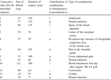
Table 2 Type of complications
in 80 laparoscopic myomectomies Consecutive order (for the individual surgeon)
Size of fibroid (cm)
Duration of surgery (min)
Classification of complication (i=intraoperative p=postoperative)
Type of complication
1 53 150 i Emphysem
2 77 152 p Wound infection
3 80 135 i Injury of the broad
ligament
3 58 74 i Lesion of the intestinal
serosa
4 57 70 p Re-aparoscopy because of sonographic
suspicious area of the uterine scar
4 60 245 p Pain in the shoulder
girdle
5 33 100 p Lower abdominal pain
9 67 90 p Wound infection
10 112 280 i Blood transfusion first day
after surgery, Hb 4.8 g/dl
12 80 n/a i Conversion
13 24 55 p Cystitis
Though large series on laparoscopic myomectomy exist [15–18], little attention has been given to the learning aspects of laparoscopic myomectomy so far [17]. Mean fibroid size was 5.6 cm, comparable with other studies where mean fibroid size was within the range of 4.6–5.2 cm [15,17]. Mean operating time in our series was 107.9 min and is within the range of 100–130 min quoted in the literature, where time depended on the number and size of fibroids [5, 15,16]. Mean decrease in haemoglobin was 0.4±1.4 g/dl and is comparable with 1.4 g/dl observed in larger trials [5, 15]. Intraoperative complication rate was 6.3% compared to 3.3% [15] and rate of postoperative complication was 10.0% comparable with larger trials where intraoperative complication rate was 5.8–11.7% [15,
16]. Differences are due to the specific definition of com-plications. In our series all fibroids were intramurally located and no pedunculated fibroid eased the procedure. Therefore, we believe that our study population may serve as a reference for discussion.
Any discussion of a learning curve should first identify factors that constitute measurable endpoints that signify some form of improvement [19]. Reduction of operating time and complications are the factors most quoted in the literature [20,21].
In most studies cited, conversion was not considered to be a complication. Rate of conversion can be as high as 41% [5,11,15,17,18,22]. The fact that conversion rate could be reduced by more than half after gaining experi-ence [22], and that the rate of conversion increases with growing population size and increasing number of sur-geons performing the procedure, indicates the existence of a learning effect [5, 17]. One larger series investigated surgeon’s experience and its relation to conversion [17]. Seventeen surgeons performed laparoscopic myomectomy in 426 women. Four surgeons performed 11–50 proce-dures, 12 surgeons performed less than 11 procedures whereas the author performed more than 50 procedures within a period of 10 years. In this study, risk of conversion to an open procedure was related neither to the study period nor to surgeon’s experience [17]. More than half of the conversions were due to difficulties in achieving satisfac-tory uterine suture and in an additional one third conver-sion was indicated because of intraoperative haemorrhage [5]. In laparoscopic myomectomy rate of conversion could serve as a measurable endpoint to assess a learning effect, but in our study only one conversion occurred, for a common reason: lack of a cleavage plane. In this particular case, ultrasound was not of help in localising the fibroid.
We decided to investigate duration of surgery, haemo-globin difference, and rate and type of complications. Initially, we thought to verify significant changes between cases 1–5 and cases 6–10, but we failed to show any significant differences. When we compared each surgeon’s first ten cases with their subsequent cases duration of surgery differed significantly. Therefore we chose a cut-off of ten cases, where in consequence only five surgeons qualified. Aside from duration of surgery, complications strictly related to laparoscopic myomectomy occurred within the first ten cases only.
Surgeons participating in our study were involved in the daily surgical routine of our teaching hospital. Lessons learned in one case were naturally adopted in subsequent cases and might have accelerated the learning process. This is a realistic setting. The major limitation of our study is that it is retrospective in nature. However, given this, we were able to evaluate factors important to assess and identify a level for competence in laparoscopic myomec-tomy where the cardinal points are: removing the fibroid, suturing of the defect and extracting the fibroid with the help of an electric morcellator.
Conclusion
A learning experience of at least ten laparoscopic myomec-tomies was necessary in our institution to improve duration of surgery and to reach a low level of severe complications. Duration of the surgical procedure and rate of severe complication were adequate study end points to assess a learning effect.
References
1. Semm K (1977) Pelviskopische Chirurgie in der Gynäkologie. Geburts u Frauenheilk 37:909–920
2. Dubuisson J-B, Fauconnier A, Babaki-Fard K, Chapron C (2000) Laparoscopic myomectomy: a current view. Hum Reprod Update 6(6):588–594
3. Nezhat F, Seidman D, Nezhat C, Nezhat C (1996) Laparoscopic myomectomy today. Why, when and for whom? Hum Reprod 11(5):933–937
4. Varasteh N, Neuwirth R, Levin B, Keltz M (1999) Pregnancy rates after hysteroscopic polypectomy and myomectomy in infertile women. Obstet Gynecol 94:168–171
5. Dubuisson J-B, Chapron C, Levy L (1996) Difficulties and complications of laparoscopic myomectomy. J Gynecol Surg 12:159–165
6. Tulandi T, Al-Took S (1999) Endoscopic myomectomy. Gynaecol Oper Endosc 26:135–148
7. Mais V, Ajossa S, Guerriero S, Mascia M, Solla E, Melis G (1996) Laparoscopic versus abdominal myomectomy: a pro-spective, randomized trial to evaluate benefits in early outcome. Am J Obstet Gynecol 174:654–658
8. Stringer N, Walker J, Meyer P (1997) Comparison of 49 lapa-roscopic myomectomies with 49 open myomectomies. J Am Assoc Gynecol Laparosc 4(4):457–464
9. Silva B, Falcone T, Bradley L et al (2000) Case-control study of laparoscopic versus abdominal myomectomy. J Laparoendosc Adv Surg Tech 10(4):191–197
10. Subramanian S, Clark M, Isaacson K (2001) Outcome and resource use associated with myomectomy. Obstet Gynecol 98 (4):583–587
11. Kolmorgen K (1995) Zur laparoskopischen Myomektomie. Zent bl Gynaekol 117:659–662
12. Fauconnier A, Capron C, Babaki-Fard K, Dubuisson J-B (2000) Recurrence of leiomyomata after myomectomy. Hum Reprod Update 6(6):595–602
13. Darai E, Dechaud H, Benifla J-L, Renolleau C, Panel P, Madelenat P (1997) Fertility after laparoscopic myomectomy: preliminary results. Hum Reprod 12(9):1931–1934
15. Landi S, Zaccoletti R, Ferrari L, Minelli L (2001) Laparoscopic myomectomy: technique, complications, and ultrasound scan evaluation. J Am Assoc Gynecol Laparosc 8(2):231–240 16. Nezhat C, Nezhat F, Silfen S, Schaffer N, Debra E (1991)
Laparoscopic myomectomy. Int J Fertil 36(5):275–280 17. Dubuisson J-B, Fauconnier A, Fourchotte V, Babaki-Fard K,
Coste J, Chapron C (2001) Laparoscopic myomectomy: predicting the risk of conversion to an open procedure. Hum Reprod 16(8):1726–1731
18. Salfelder A, Lueken R-P, Gallinat A, Möller C, Busche D, Nugent W (1999) Pelviskopische Myomoperation und Schwangerschaft—Ergebnisse. Geburts u Frauenheilk 59:57–61
19. Rosen DMB, Cario GM, Carlton MA, Lam AM, Chapman M (1998) An assessment of the learning curve for laparoscopic and total laparoscopic hysterectomy. Gynaecol Endosc 7(6):289–293 20. Aubard Y, Piver P, Grandjean MH, Baudet J (1996) Lapa-roscopically assisted vaginal hysterectomy for non- malignant disease of the uterus. Report on a personal series of 126 cases. Eur J Obstet Gynecol Reprod Biol 68(1–2):147–154
21. Ikhena SE, Oni M, Naftalin NJ, Konje JC (1999) The effect of the learning curve ion the duration and peri-operative complications of laparoscopically assisted vaginal hysterecto-my. Acta Obstet Gynecol Scand 78(7):632–635