Iranian Biomedical Journal 13 (3): 179-183 (July 2009)
Short Report
Human Coagulated Plasma as a Natural and Low Cost Matrix for
in vitro Angiogenesis
Kamran Mansouri
*1, Ali Mostafaei
1, Manochehr Mirshahi
2, Hamidreza Mohammadi
Motlagh
1, Ali Maleki
1and Maryam Keshavarz
11
Medical Biology Research Center, Kermanshah University of Medical Sciences, Kermanshah, Iran; 2Faculty of Basic
Sciences, Tarbiat Modarres University, Iran
Received 8 October 2008; revised 1 June 2009; accepted 8 June 2009
ABSTRACT
Background: Angiogenesis, the development of new blood vessels, is an important process in tissue development and wound healing, but becomes pathologic when associated with solid tumor growth, proliferative retinopathies, and rheumatoid arthritis. Accurate and reliable qualification of neovascular (angiogenic) response, both in vitro and in vivo, is an essential requirement for the study of new blood vessel growth. The complexity of currently used three-dimensional in vitro angiogenesis systems makes it difficult to approve material in its models. Capillary-like structure occurs on basement membrane components such as collagen and/or laminin, while in other models, CLS formation occurs on transitional matrices such as fibrin. To solve this problem, we were interested in developing an angiogenesis system which allows rapid and
reliable quantification of three-dimensional neovessel formation in vitro. Methods: Human bone marrow
endothelial cells were seeded on gelatin-coated microcarriers and suspended in a solution of platelet-poor plasma which was induced to polymerize by addition of calcium chloride. In this way, microcarriers were
entrapped in three-dimensional coagulated plasma. Results: Within a few hours, endothelial cells begin to
leave these supporting microcarries and migrate into the coagulated-plasma matrix and formed CLS within
48-72 hours. Conclusion: We developed a convenient angiogenesis in vitro system which allows reliable
quantification of capillary formation in a three-dimensional environment. Iran. Biomed. J. 13 (3): 179-183, 2009
Keywords: Angiogenesis, Endothelial cells (EC), Human coagulated-plasma, Microcarriers (MC)
INTRODUCTION
ngiogenesis is characterized by the formation of new capillary from pre-existing vessels. The process consists of a number of steps, beginning with activation of endothelial cells (EC) by growth factors, followed by enzymatic degradation of basement membrane, detachment of EC from adhesion proteins, EC migration into the perivascular spaces and proliferation, and final new vessel formation. The event is highly regulated by various growth factors and cytokines [1].
Angiogenesis, the formation of new vessels from existing microvasculature, is a tremendously complex and intricate process, essential for
embryogenesis and development of multi-cellular organism, but it rarely occurs only in, adult tissues in a tightly controlled manner during normal wound healing and the female reproductive cycle (corpus luteum, placenta and uterus). When tight controls are breached, it results in unchecked angiogenesis, which implicated in the development and progression of a variety of diseases including rheumatoid arthritis, psoriasis, tumor growth and metastasis, diabetic retinopathy, obesity and age-related macular degeneration. The prevalence of pathologic angiogenesis in human disease, and the significant mortality associated with these disorders,
underscores the importance and emergence of anti-angiogenesis therapy as a major clinical tool [2-4].
A
180 Mansouri et al. Iran. Biomed. J., July 2009
http://IBJ.pasteur.ac.ir
The classical assays for studying angiogenesis in
vivo include the hamster cheek pouch, the rabbit ear chamber, dorsal skin and air sac, the chick embryo chorioallantoic membrane and the iris and avascular cornea of the rodent eye. In each system, an angiogenic substance must be implanted. These assays suffer from requiring artificially induced angiogenesis, the requirement for a sustained release polymeric vehicle for the angiogenic substance and inhibitor, and the technical complexities associated with setting up the assay and measuring the outcome. Because of these disadvantages, there is a
great need for physiologically relevant in vitro
assays for angiogenesis, particularly human
angiogenesis [5, 6]. Studies indicate that the
biological substrates can drastically modify the behavior of EC in culture. These findings demonstrate that the macromolecules of the extracellular matrix play a major role in microvascular morphogenesis and suggest that extracellular substrates and diffusible factors may be cooperating in the angiogenic process.
Subsequent models of in vitro angiogenesis
showed EC form capillary-like structure (CLS) in two dimensions on matrix of collagen and basement membrane constituents, or in three dimensions when the EC are sandwiched in collagen or fibrin gel. In addition to collagen, other basement membrane components such as laminin have been shown to play a crucial role in the differentiation of
EC into CLS in vitro. Kubota and colleagues
[7] demonstrated the importance of laminin in CLS formation on matrigel, a basement membrane-like extract composed of lamin, collagen, heparin sulphate proteoglycan and nidogen/entactin [8-10]. In addition to basement membrane components, matrices are derived from the coagulation system. Nicosia et al. [11] made use of a matrix formed by clotted chick plasma in rat aortic model and showed the importance of fibrin
in a three-dimensional model of in vitro
angiogenesis shown to provide scaffolding for
CLS formation [12]. The above mentioned in vitro
assays have usually entailed establishing long-term cultures of EC or inducing formation of microvessels by placing the cells on extracellular matrices, or exposing the cells to various angiogenic stimuli. Preparation of artificial
matrices is time-consuming, and some of them are
expensive.
In the present study, we report CLS formation by human bone marrow EC on a matrix-derived from
human plasma to mimic in vivo situation. Moreover,
we morphologically characterized this novel three-dimensional in vitro angiogenesis model.
MATERIALS AND METHODS
Materials. Cytodex3 microcarriers (MC, Pharmacia Sweden), DMEM, FCS (Gibco Germany), calcium chloride (Merck), human bone marrow endothelial cell line (provided by Dr. Manochehr Mirshahi, Tarbiat Modarres University, Tehran, Iran).
Platelet-poor plasma (PPP). Freshly obtained blood from 10-20 healthy donors, who had not taken any drugs for 14 days prior to the study, was pooled
and stored at 4°C. The whole blood unit was first
centrifuged using light spin (2000 ×g) for 3 minutes, yielding platelet-rich plasma (PRP) in the upper portion and red blood cells (RBC) in the lower portion. The PRP was expressed into an attached bag, leaving RBC in the primary bag. The two attached bags were re-centrifuged using a heavy spin (3210 ×g) for 10 minutes to produce an aggregated platelet button from the PRP. Approximately 200 ml of citrated PPP was removed [13]. For sterilization, the PPP was filtered (0.2 µm pore size) and then stored at -20ºC for subsequent uses.
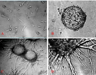
Microcarrier cell culture. Gelatin-coated cytodex-3 MC were prepared according to the recommendations of the supplier. Freshly, autoclaved MC were suspended in DMEM + 10% FCS, 100 IU/ml penicillin, 100 µg/ml streptomycin, and EC were added. The cells were allowed to attach to the MC during 10 hours of incubation at 37ºC. Then, the MC were suspended in a larger volume of medium and cultivated in a 5% CO2 atmosphere at 37ºC for 1 to 2 days [14] and MC were embedded in coagulated plasma when the whole surface of MC had been covered with EC (Fig. 1B).
Capillary tube formation in three-dimensional
human coagulated plasma. In vitro angiogenesis assays were done in which HBMEC (Fig.1A) were coated on MC and then, added to PPP (24-well plates) with different volumes and clotting was induced by addition of different volumes (40, 60, 80,
100, 110, 120, 130 and 150 µl) by different
concentrations (10, 20, 30, 40 and 50 mM) for any volume per well CaCl2. After clot formation, 1,000 µl of complete culture medium containing 10% FCS was added. Two or three days after polymerization
Iran. Biomed. J., July 2009 Natural Matrix for in vitro Angiogenesis 181
Fig. 1. Human coagulated plasma matrix for in vitro angiogenesis. (A) Human bone marrow endothelial cells (magnification 40×); (B) micro carriers seeded with human bone marrow endothelial cells were embedded in coagulated platelet-poor plasma matrix (magnification 100×); (C) total explants area (magnification 40×) and (D) more details of outgrowth area, in which endothelial cells are seen to form tube-like structures (magnification 400×).
of plasma, the formation of capillary tubes which arise from the periphery of microcarrier beads was observed. These capillaries were photographed with a camera connected to an inverted microscope. Also, two or three days after polymerization of the plasma, the number of micro carriers with cellular processes or with whole cells invaded the surrounding coagulated-plasma matrix (Fig. 1C and D).
RESULTS
The results of our assay (Fig. 1) showed that the PPP can be used as a suitable matrix for designing an angiogenesis model. Regarding this finding, we understood that the ratio of 2:3 plasma to 1:3 calcium chloride (270 µl plasma to 130 µl CaCl2) was a good ratio for providing suitable matrix as cells simply could proliferate and migrate and CLS occurred. Optimum concentration of calcium chloride for activation of coagulation cascade with no cytotoxic effect could activate gel formation determined to be 30 mM.
Thickness of matrix by which EC could degrade matrix and serve as a three-dimensional malleable scaffold such as that supporting EC migration, proliferation and survival was well established by
addition of 400 µl mix plasma and calcium chloride per well in a 24-well plate. In providing durable and suitable thickness, we could see angiogenesis phenomena by an inverted microscope and we were able to take a photograph from it (Fig. 1C and D).
DISCUSSION
Angiogenesis was first observed in vitro by
Folkman and Haudenschild [15] 28 years ago. During vascular morphogenesis, the extracellular matrix (ECM) serves as a three-dimensional malleable scaffold in which individual EC and clusters of EC can transduce mechanical forces to other EC at a considerable distance. Thus, by generation of mechanical, contractile forces within ECM, EC are able to establish tension-based guidance pathways that allow them to form interconnected cords after long-term culture of capillary EC. These authors observed the spontaneous organization of these cells into CLS. The presence of a lumen within these CLS was confirmed by phase contrast microscopy and transmission electron micrography. This report of angiogenesis in culture dish provided a basis for definition of in vitro endothelial angiogenesis. All
182 Mansouri et al. Iran. Biomed. J., July 2009
http://IBJ.pasteur.ac.ir
the subsequently published assays referred to the presence of a lumen in the CLS as a criterion for the
validation of in vitro model. From a physiological
point of view, an ideal in vitro model would take
into account all the representative steps of in vivo
angiogenesis, from detachment of EC from vascular wall to final tubular morphogenesis, maturation, and connection to a functional vascular network. During angiogenesis, proliferating and migrating EC organize to form new three-dimensional capillary network. Furthermore, it should be rapid and easy to use reproducibly and be easily quantifiable (e.g. CLS length, area covered by capillary-like network, number of tubes, and complexity of the network). Depending on the ways the cells reorganize, the assays are described as data classified in two categories: two-dimensional (when the cells develop tubular structures on the surface of the substrate) and three-dimensional (when the cells invade the surrounding matrix consisting of a biogel) assays. Since the first reports of CLS formation by EC in culture, a number in vitro angiogenesis models have been described [16-20].
In some models, CLS formation occurs on basement membrane components such as collagen
and/or laminin, while in other models, capillary
formation occurs on transitional matrices such as fibrin. These models often require a high thrombin or reptilase concentration to convert fibrinogen to fibrin and do not yield a fibrin matrix. Since biogels are polymers, the concentration and the biochemical conditions of the matrix polymerization must be carefully defined because they may affect the density and the mechanical properties of the substrate, leading to proliferative, migratory or tubular EC phenotypes [21-23]. Another studies use plasma as matrix, but in these models, plasma clot matrices were prepared by addition of thrombin to citrated PPP and seeding EC onto thick coagulated-plasma matrix using stimulator such as phorbol
myristate acetate or growth factors that cause
two-dimensional conditions in in vitro angiogenesis
model [24, 25].
However, in our model, CaCl2 as coagulant factor 4 was used because the coagulation system has a crucial role in this phenomenon. Since the above mentioned models have used artificial matrices and these matrices have not all of the extracellular matrix components and coagulation factors, the angiogenesis is not similar to angiogenesis in body. Nevertheless, in our model, plasma is used that includes all of the coagulant factors and other proteins such as fibrinogen: therefore, it is very
similar to extracellular matrix and to the
angiogenesis in the body. The study of the
angiogeneic process and the search for a novel therapeutic agent to inhibit or stimulate angiogenesis have employed a wide range of in vitro angiogenesis assay. The difference between them is greatly in their difficulties, quantitative nature, rapidity and the cost.
In this study, we described a novel in vitro
angiogenesis system which allows quantification of angiogenic responses of EC in a three-dimensional matrix. We designed this model with PPP by mixing accurate ratio of CaCl2 and PPP and provided three-dimensional malleable scaffold supporting EC migration, proliferation and survival. The advantages of our angiogenesis assay lies in its technical simplicity, making use of small amounts of human plasma and closely fulfilling the optimal condition for an in vivo model because it allows the preservation of the vessel architecture during in vitro
assay. This model allows us the investigation of putative role of the hemostatic system in angiogenesis because of using human coagulated plasma. Therefore, our model is an inexpensive and rapid tool for screening angiogenic and angiostatic molecules.
ACKNOWLEDGMENTS
We extend special thanks to Dr. Yadollah Shakiba, Shahram Parvaneh and Hadi Mozafari for their comments and technical assistances.
REFERENCES
1. Folkman, J. (2006) Angiogenesis. Annu. Rev. Med. 57 (1): 1-18.
2. Carmeliet, P. and Jain, R.K. (2000) Angiogenesis in cancer and other disease. Nature 407 (6801): 249-257.
3. Lijnen, H.R. (2007) Angiogenesis and obesity.
Cardiovas. Res. J. 78 (2): 286-293.
4. Board, R.E., Thistlethwaite, F.C. and Hawkins, R.E. (2007) Anti-angiogenic therapy in the treatment of advanced renal cell cancer. Cancer Treat. Rev. 33 (1): 1-8.
5. Staton, C.A. and Lewis, C.E. (2005) Angiogenesis Assays: A Critical Appraisal of Current Techniques. John Wiley & Sons Ltd., PO19 8SQ, England. 6. Ribatti, D., Vacca, A., Roncali, L. and Dammacco, F.
(1996) The chick embryo chorioallantoic membrane as a model for in vivo research on angiogenesis. Int. J. Dev. Biol. 40 (6): 1189-1197.
Iran. Biomed. J., July 2009 Natural Matrix for in vitro Angiogenesis 183
7. Kubota, Y., Kleinman Martin, G.R. and Lawley, T.J. (1988) Role of laminin and basement membrane in the morphological differentiation of human endothelial cells into capillary-like structures. J. Cell Biol. 107 (4): 1589-1598.
8. Nicosia, RF. and Villaschi, S. (1999) Autoregulation of angiogenesis by cells of the vessel wall. Int. Rev. Cytol. 185: 1-43.
9. Nicosia, R.F. and Ottinetti, A. (1990) Modulation of microvascular growth and morphogenesis by reconstituted basement membrane gel in three-dimensional cultures of rat aorta: a comparative study of angiogenesis in matrigel, collagen, fibrin and plasma clot. In vitroCell Dev. Biol. 26 (2): 119-128. 10. Hanahan, D. and Folkman, J. (1996) Patterns and
emerging mechanisms of the angiogenic switch during tumorigenesis. Cell 86 (3): 353-364.
11. Berbers, G. and Benjamin, L.E. (2003) Tumorgenesis and angiogenic switch. Nat. Rev. Cancer 3 (6): 401-410.
12. Vailhe, B., Vittet, D. and Feige, J.J. (2001) In vitro
models of vasculogenesis and angiogenesis. Lab. Invest. 81(4): 439-452.
13. Victor Hoffbrand, A., Daniel, C. and Edward, G.D. (2005) Post graduated hematology book, Fifth edition, Wiley-Blackwel Ltd., Massachusetts, USA. pp. 249-277.
14. Nehls, V. and Drenckhahn D. (1995) A novel, microcarrier-based in vitro assay for rapid and reliable quantification of three-dimensional cell migration and angiogenesis. Microvasc. Res. 50 (3): 311-322.
15. Folkman, J. and Haudenschild, C. (1980)
Angiogenesis in vitro. Nature 288: 551-556.
16. Auerbach, R., Lewis, R., Shinners, B., Kubia, L. and Akhtar, N. (2003) Angiogenesis assays: a critical overview. Clin. Chem. 49 (1): 32-40.
17. Jain, R.K., Schlenger, K., Hockel, M. and Yuan, F. (1997) Quantitative angiogenesis assays: progress and problems. Nat. Med. 3 (11): 1203-1208.
18. Friedl, P. and Brocker, E.B. (2000) The biology of cell locomotion within three-dimensional extracellular matrix. Cell. Mol. Life Sci. 57 (1): 41-64.
19. Plunkett, M.L. and Hailey, J.A. (1990) An in vivo
quantitative angiogenesis model using tumor cell entrapped in alginate. Lab. Invest. 62 (4): 510-517. 20. Hlatky, L., Hahnfeldt, P. and Folkman, J. (2002)
Clinical application of antiangiogenesis therapy: microvessel density, what it does and does not tell us.
J. Natl. Cancer Inst. 94 (12): 883-893.
21. McDonald, D.M. and Choyke, P.L. (2003) Imaging of angiogenesis; from microscope to clinic. Nat. Med. 9 (6): 713-725.
22. Davis G.E. and Senger, D.R. (2005) endothelial extracellular matrix: Biosynthesis, Remodeling and functions during vascular morphogenesis and neovessel stabilization. Circ. Res. 97: 1093-1107. 23. Taraboletti, G. and Giavazzi, R. (2004) Modeling
approaches for angiogenesis. Eur. J. Cancer 40 (6): 881-889.
24. Jurasz, P., Santos-Martinez, M.J., Radomska, A. and Radomski, M.W. (2007) Development and pharmacological characterization of a novel plasma-based in vitro angiogenesis assay. Kardiochirurgia i Torakochirurgia Polska 4 (2): 126-130.
25. Guimarães, A.H.C., Laurens, N., Weijers, E.M., Koolwijk, P., Hinsbergh, V.W.M. and Rijken, D.C. (2007) TAFI and pancreatic carboxypeptidase B (CPB) modulate in vitro capillary tube formation by human microvascular endothelial cells. Arterioscler. Thromb. Vasc. Biol. 27: 2157-2162.