Introduction
Invasive cancer of the cervix is the major cause of death from gynecologic cancer world-wide [1]. It remains not only the most common gynecologic malignancy, but also the third most frequently diagnosed cancer in women (after breast and colorectal cancer). With the
in-troduction of the Papanicolaou (Pap) smear in the 1940s, early detection and treatment of preinvasive disease became possible. Conse-quently, both the incidence and mortality rates owing to invasive cervical cancer have de-clined. In developing countries, cervical cancer accounts for 15% of female malignancies.
In contrast, in developed countries, cervical cancer accounts for only 4.4% of female malig-Medical Journal of the Islamic Republic of Iran.Vol. 21, No.1, May 2007. pp. 1-10
Screening for cervical cancer and precancerous lesions in Tabriz
M. Jafari Shobeiri, MD.1, M. Halimi, MD.2, A. Dastranj MD.3, and, J. Shahamphar, MD.4
Departments of Oncology, Gynecology, Pathology and Social Medicine of Tabriz University of Medical Sciences, Tabriz, Iran
Abstract
Background:Cervical cancer is the most common female genital tract malignancy and is the major cause of death from gynecologic cancer worldwide. The majority of cervical cancers develop through a series of gradual, precancerous lesions. Screening asymptomatic women with regular Pap smears allows diagnosis of the readily treatable preinvasive phase. We performed this study to determine the prevalence of precancerous lesions and assess the effect of demographic, pathologic and family economic factors on developing lesions.
Methods:This cross-sectional, descriptive, analytic study was carried out on 6024 women under coverage of Tabriz health care centers. Pap smear was done in all subjects and questionnaires were filled by health providers. The data were analyzed with SPSS statistical software (version 12) and statistical methods such as chi-square and t-test.
Results:Of 6024 Pap smears, 62 (1.02%) demonstrated precancerous lesions of which 41(0.68%) were atypical squamous cell of undetermined significance (ASC-US), 11 (0.18%) were low grade squamous intraepithelial lesion (LSIL) and 10 (0.16%) were high grade squamous intraepithelial lesion (HSIL). No invasive cancer case was observed in this study. According to our observation, the following factors increased the risk of precancer-ous lesions: multiparity >_ 3, abortion >_ 1, gravida >_3, husband marriage >1. The protective effect of condom as a barrier contraceptive was observed.
Conclusion:According to this study it is imperative to make readily available facilities for screening asymptomatic women all over Iran.
Keywords: screening, precancerous cervical lesions, cervical cancer, risk factors, Pap smear
1.Corresponding author, Department of Oncology- Gynecology, Alzahra Hospital, Tabriz University of Medical Sciences, South Artesh Ave, Tabriz, Iran. Fax: +98 411 5566449, email:jafarimsh@yahoo.com
nancies [2]. Invasive cancer of the cervix has been considered a preventable cancer because it has a long preinvasive state, because cervical cytology screening programs are available, and because the treatment of preinvasive lesions is effective [3]. Screening asymptomatic women with regular Pap smears allows diagnosis of the readily treatable preinvasive phase. Today, Pap smear is a cost-effective and fundamental test for cervical cancer and precancer screening [1,2,3,4,5,6,7].
There are numerous risk factors for cervical precancer and cancer: young age at first inter-course (<16 years), multiple sexual partners, cigarette smoking, race, high parity, low so-cioeconomic status, sexually transmitted agents, and immunosuppression from human immun-odeficiency virus (HIV) [2,3].
Unfortunately there are few investigations in the field of cervical cancer screening in Iran. In regard of screening programs, the importance in reduction of both incidence and mortality of cervical cancer and as there are no health care centers for formal cervical cancer screening in Iran, this study was carried out on women to de-termine the prevalence of cervical cancer and precancerous lesions and assess the effect of risk factors on developing lesions.
Methods
This cross-sectional, descriptive, analytic study was carried out on 6024 women under coverage of 86 health care centers in Tabriz city (one of the major cities located in northwest of Iran) during 2003 to 2005 to determine cervical cancer and precancerous lesion prevalence and assess the correlation of demographic, patho-logic and family economic factors with develop-ing lesions. The study population with any range of age had at least one marriage history, and was selected by cluster sampling. Ten midwives were selected after pretest exam and trained for vaginal examination, cervical anatomy, normal cervical distinction from abnormal and provid-ing correct Pap smear tests durprovid-ing a 2 month
pe-riod. After passing a posttest exam and reassur-ance of their ability in research, they were re-sponsible for selecting subjects and inviting them to health care centers, doing Pap tests and filling out questionnaires on demographic, pathologic and family economic factors. To ob-tain an optimal specimen, subjects were re-quired to avoid having intercourse and use of any type of intravaginal products for 48 hours before the test. The ectocervical and endocervi-cal samples were obtained with plastic spatula and cervical brush respectively. Each sample was rapidly applied on a glass slide. The slides were fixed immediately in 95% ethyl alcohol for 20 minutes. Slides were allowed to air dry before sending to the laboratory where they were stained by Papanicolaou staining. The slides were studied by two pathologists with power field of 40 and 100.
According to the Bethesta III system (2001), precancerous lesions were classified into three groups: Atypical cells were regarded as ASC-US and ASC-H (atypical squamous cells in which high-grade lesions must be excluded), mild dysplasia as LSIL and moderate, severe and in situ carcinoma as HSIL. After determin-ing the lesions prevalence, the population study were classified into control and patient groups according to having or not having lesions and assessed the relation between demographic, pathologic and family economic factors be-tween the two groups.
Factors such as age of women, number of de-livery above 5 months, abortion and gravida, marriage age of women, number of women and husbands’ marriage, women’s age at first deliv-ery, number of husband’s wives, use of any con-traceptive methods and age of husbands were classified into groups and considered as demo-graphic factor. Smoking by woman, husband and relatives who live together were considered as a pathologic factor. To assess family economic factor, women and husbands’ literacy, occupa-tion and income were considered.
All statistical calculations were performed
by using the SPSS software package version 12. Chi-square and T-Test statistics were used to compare of the study variables. Significant dif-ferences were accorded at P <0.05.
Results
6024 women under coverage of 86 Tabriz health care centers were screened for precan-cerous lesions and cancer of the cervix by Pap smear test.
Demographic characteristics of the study population are shown in Table 1.
The pathologic factor results of the study population were as follows:
Of a total 6024 screened women, 36 subjects (0.6%) were smoking and the remaining 5988 subjects (99.4%) were not. The frequency of smoking in the husbands was 32.4%. 686 sub-jects (11.4%) were at the exposure of smoking by relatives living with them.
The family economic characteristics of the population were as follows: 26.2% of women and 14.4% of husbands were illiterate (have not attended school), 34.1% of women and 35.3% of husbands had primary school literacy, 17.8% of women and 23.7% of husbands had guidance school literacy, 20.1% of women and 20.1% of husbands had high school literacy, 0.6% of women and 2.4% of husbands had a two year college certification, 1.1% of women and 3.5% of husbands were bachelors and none of the women and 0.6% of husbands had master de-gree or higher.
96% of the population studies were house-wives and only 4% were employed. 98% of the husbands were employed. The most common occupation of husbands were self-employed (45.3%), labor worker (22.6%) and driver (8.3%).
The monthly mean income of women and husbands was 78.38± 57.71 and 116.30±67.06 thousand Tomans respectively.
The frequency of cervical precancerous le-sions [cervical intraepithelial neoplasia (CIN)] was 1.02% (62) and there was no invasive
can-cer of the can-cervix. The frequency of the different types of precancerous lesions was as follows: 41 ASC-US (0.68%), 11 LSIL (0.18%) and 10 HSIL (0.16%). Of ten HSIL, the frequency of moderate dysplasia, severe dysplasia and in situ carcinoma were 7 (0.11%), 2 (0.02%) and 1 (0.01%) respectively.
5962 subjects with no precancerous lesions (control) and 62 subjects with precancerous le-sions (case) were compared by different risk factors and assessed for their effect on develop-ing precancerous lesions. The case and control groups correlation with different age groups of women and husbands are shown in Table 2.
The mean age of women in case and control groups was 36.01±9.73 (range 18-63) and 33.17±9.08 (range 16-76) respectively, t= -2.409, df=6004 and P=0.016. The most common age groups of husbands frequency which belonged to age group 36-50 years was 71.2% and 53.3% in case and control groups respectively. The age group differences between the two groups were significant, χ2=7.50, df=2, P=0.023. In this
re-gard, the precancerous lesions frequency was increased by rising of the husbands’ age.
Obstetrical status of case and control groups is given in Table 3. The mean gravida numbers in case and control groups was 4.14±2.89 (range 0-13) and 3.05±2.25 (range 0-16) re-spectively, t= -3.796, df=6022, P<0.0005.
According to different gravida groups, the most common gravida group in case and con-trol groups was 3-4(45.9%) and 1-2 (51.1%) re-spectively, χ2=16.721, df=3, P=0.001.
The precancerous lesions frequency in-creased by increasing of the gravida to 3 and more.
The mean deliveries above five months in case and control groups was 3.50±2.36 (range 0-10) and 2.61±1.92 (range 0-13) respectively, t= -3.571, df=6022, P<0.0005. The most com-mon groups in case and control groups were 3-4 (39%) and 1-2 (58.4%) respectively, P=0.008. The higher the number of deliveries above 3 to 5 and more, the higher the frequency of
Table 1. Demographic characteristics of the study population (n=6024).
(yrs)
Women’s age
Yes No
cerous lesions.
The mean abortion numbers in case and con-trol groups was 0.72±1.02 (range 0-3) and 0.44±0.86 (range 0-10) respectively, t= -2.482, df=6022, P=0.013. The abortion groups
differ-ences between case and control groups were statistically significant, χ2=13.658, df=2, P=0.001.
In this regard the precancerous lesions frequency rises with increasing abortion numbers. The minimum, maximum and mean±Std of age at
Table 2. Case and control groups correlation by different age groups of women and husbands.
Table 3. Obstetrical status in case and control groups.
first delivery in case and control groups were 14, 26, 19.09±3.05; and 11, 40 and 19.96±3.20 years respectively. The most common age groups frequency which belonged to group 19-24 years was 47.5% and 57.6% in case and con-trol groups respectively. The differences were not significant.
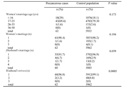
Case and control differences according to women’s age at marriage, marriage number of women and husbands and husbands’ wives numbers are shown in Table 4. The minimum, maximum and mean±Std of marriage age of women in case and control groups were 9, 27, 17.67±3.31; and 9, 48, 18.21±3.13 respectively. The results of t-test analysis indicate that the differences were not significant. The most com-mon age group frequency at marriage which be-longed to 17-25 years, was 69.4% and 79.3% in case and control groups respectively.
The marriage age group differences between the two groups were not significant. The mean marriage number of women in case and control
groups were 0.98±0.22 (range 1-2) and 1.01± 0.16 (range1-3) respectively; the differences were not significant. The marriage numbers dif-ferences between the two groups were not sig-nificant,χ2= 4.717, df=3, P=0.194.
The minimum, maximum and mean±Std of husbands’ marriage numbers in case and con-trol groups were 1, 3, 1.10±0.35; and 1, 4, 1.03±0.19 respectively.
The husbands’ marriage number differences between the two groups were significant,
χ2=8.389, df=3, P=0.039.
By increasing husbands’ marriage number, the precancerous lesions prevalence was in-creased. The minimum, maximum and mean ±Std. of husbands’ wives numbers in case and control groups were 1, 2, 0.96± 0.31; and 1, 3, 0.99± 0.14 respectively.
The husbands’ wives numbers differences between case and control groups were signifi-cant,χ2 =21.136 df=3, P<0.0005. In this
man-ner, the precancerous lesions prevalence was
Table 4. Marriage characteristics in case and control groups.
Women’s marriage (n) Women’s marriage age (yrs)
Husband’s marriage (n)
Husband’s wives (n)
higher in women whose husbands have had two or more wives at the same time.
Concerning the pathologic factor effect in-cluding (smoking) on lesions, the results were as follows:
The frequency of smoking in the case group was 1.6% while the corresponding figure in the control group was 0.6%. There was no signifi-cant difference in the smoking frequency of two groups.
The frequency of husbands’ smoking in the case and control groups was 37.1% and 32.4% respectively. The results of chi-square analysis, indicate that the differences were not signifi-cant,χ2=0.630, df=1, p=0.427. The frequencies
of smoking by relatives who live together in case and control groups were 12.5% and 11.3% respectively. The differences were not signifi-cant.
Concerning the effect of family economic factors on lesions, the results were as follow: the most common level of women’s literacy in case and control groups was illiteracy (39.3%) and primary school level (34.1%) respectively. The difference was not significant (Table 5). The most common husbands’ literacy status frequency which belonged to primary school level was 36.1% and 35.3% in case and control groups respectively.
The husbands’ literacy status differences be-tween the two groups was not significant. Num-ber and percent of the most common husbands’ occupational status were as follows:
govern-ment employee, worker, self employee and driver in case and control groups were 16 (26.7%) and 1035 (17.5%), 11 (18.3%) and 1340 (22.7%), 22 (36.7%) and 2726 (46.1%), and 5 (8.3%) and 492 (8.3%) respectively.
The occupational type differences between the two groups were not significant. Mean in-come of husbands in case and control group was 122.03 ± 77.53 and 116.25 ± 66.95 thou-sand Tomans respectively.
There was no significant difference in the mean husbands’ income of the two groups, P=0.528.
Mean income of women in case and control groups was 125.00±106.06 and 77.85± 57.26 thousand Tomans, respectively. Mean income differences between the two groups were not significant.
As results show, the literacy, income and oc-cupational status differences of women and husbands in case and control groups were not significant, and we concluded that family eco-nomic factors did not have any effect on devel-oping of cervical precancerous lesions.
The association between oral contraceptive, intrauterine device, preejaculatory withdrawal, injectable progestin, Norplant and cervical pre-cancerous lesions did not turn out to be statisti-cally significant. But the relation between oral progestin, condom use and precancerous lesions with 0.047 and 0.046 P-values, was significant.
Table 5. Literacy status of women in case and control groups.
Conclusion
Concerning the epidemiologic information among the study population, most women (43.9%) were in the age group of 26-35 yr. (mean of 33.20 yr± 9.09) and 50.8% of them had one to two pregnancies. The results are sim-ilar to those of Thistle et al [8] who found that 62.6% of women had zero to three pregnancies. N’Golet et al [9] in their cervical precancerous screening research found that the majority of the women were 21 to 30 years old, These re-sults are similar to ours.
Mehdizadeh et al [10] described their study on 3000 Pap smear tests. They did not find any invasive cervical cancer case. Our results are similar to them. Krivak et al [3] in their study found an incidence of intraepithelial lesions of 5 to 6 %. The incidence of ASC, LSIL and HSIL was 50-66 to , 1-2% and 0.5-1% respectively. These results are dissimilar to our results which show the intraepithelial lesions incidence of 1.02% and ASCUS, LSIL, HSIL incidence of 0.68%, 0.18% and 0.16% respectively.
Our results are similar to those of Mehdizadeh et al [10] and Allameh [11] who found 0.5% and 0.7% incidences respectively. The most accept-able explanation for these differences is different sexual behaviors and socio cultural status. This-tle et al [8] reported an intraepithelial incidence of 15.5% in the rural population of Zimbabwe. It seems that the higher incidence of lesions might be due to their study which was limited to women of reproductive age.
Harahap[12] in his study did not find signifi-cant influence of the patients’ age on the devel-opment of CIN. Reis et al[13] did not find any association between age and severity of the dis-ease either. These results are similar to ours which show that age groups differences in case and control groups were not significant. In a study by N’Golet et al [9], in contrast to our da-ta, the most common CIN frequency belonged to age group 20-29 yr. However, we in concor-dance with N’Golet et al [9] found the most common frequency in the 26-35 age group.
Women marriage age differences of case and control groups were not statistically significant. The results are similar to Harahap [12] and Wright et al [14] who concluded no significant influence of the age at first sexual contact on the development of precancerous lesions and dis-similar to those of Kohler et al [15] who found early sexual contact <14-17 years increased the risk of CIN or invasive cervical carcinoma. Ac-cording to our observation, age at first child-birth was not a risk factor for lesions. Cuzick et al [16] in their case-control study to examine known risk factors for CIN, showed cases of CIN III had a risk profile similar to that seen for invasive disease whereas CIN1 cases were sim-ilar to the controls in all risk factors examined. As 84% of lesions in our study were mild, the results of our two studies are similar.
Multiparity increased the risk of precancer-ous lesions.The results are similar to those of Kohler et al [15] who showed an association be-tween multiparity and risk of the lesions. Hinkula et al [17] in their study concluded that the increased incidence of cervical cancer and CIN among grand multiparous women (at least five children) suggests a causal association to human papillomavirus (HPV) 16 and Chlamydia trachomatis infections. As there is a potent rela-tion between HPV and precancerous lesions and as our population study were not examined for HPV, It seems that the increase in the inci-dence of precancerous lesions in multiparous women might be related to HPV infection. Studies in this field are necessary for revealing this relation.
Parazzini et al [18,19] did not observe a con-sistent association between the risk of CIN and spontaneous or induced abortions. In our study, a consistent association emerged between the risk of precancerous lesions and increased number of abortions. As induced abortion is il-legal in Iran, a lot of unwanted pregnancies are terminated by intrauterine instrumentations. It might have an effect on the development of le-sions. Studies to evaluate the relation between
illegal abortions (intrauterine instrumenta-tions) and development of precancerous and cancer lesions are necessary.
The husband marriage and wife number dif-ferences between the two groups were signifi-cant. In this way the incidence of precancerous lesions is increased by elevation of the number of husbands’ marriages and wives. In several studies, multiple sexual partners was the only factor incriminated to significantly increase the risk of CIN occurrence [15,20]. The similarity of the results shows that increased number of husbands’ wives and marriages in our study acts as multiple partners on the occurrence of CIN.
We did not find any association between the risk of precancerous lesions and cigarette smoking (direct and indirect). The results are dissimilar to Wu et al [21] who concluded that life time indirect tobacco exposure is a major determinant for contracting cervical neo-plasm’s among nonsmoking women in Taiwan. No consistent association emerged between the risk of intraepithelial cervical neoplasm and oral contraceptive use.
Kjellberg et al [22] concluded that oral con-traceptive use was associated with HSIL, but these associations lost significance after taking HPV into account. The result differences need further studies in the field of HPV. Parazzini et al [23] found that use of barrier methods low-ered the risk of intraepithelial neoplasia. The re-sults are similar to those of our study which of-fered the significant relation between condom use and the risk of precancerous lesions.
By this study we found 62 cases of intraep-ithelial lesions. So for decreasing the incidence rate of invasive cervical cancer, the establish-ment of strict cervical cancer screening centers including a central registry, central cytology laboratory and well-trained clinicians to obtain the smears, and a network of colposcopy follow up clinics where women with abnormal results are evaluated and treated is necessary. As awareness and education are important
prereq-uisites to efforts aimed at screening programs, it is recommended to assess knowledge, attitudes about Pap smear and cervical cancer among women.
Acknowledgement
This research was supported by a grant from Tabriz University of Medical Sciences. The authors would like to thank Dr. Nikniaz and Dorostkar for accessing of health centers, Dr. Sadagat for statistical advice and Mr. Oshaghi for providing type and print of this article and all reviewers for useful suggestions.
References
1. Hacker N. Cervical cancer. In: Berek J, Hacker N, edi-tors. Practical Gynecologic Oncology. 4th ed., Philadelphia: Lippincott, Williams & Wilkins; 2005.pp. 337-386.
2. Chi D, Abu-Rustum N, Hoskins W. Cancer of cervix. In: Rock J, Jones H, editor. Telinde’s Operative Gynecology. 9th ed., Philadelphia: Lippincott, Williams & Wilkins; 2003. pp.1373-1444.
3. Krivak TH, Mc Broom J, Elkas J. Cervical and vagi-nal cancer. In: Berek J, editor. Novak’s Gynecology. 13th ed., Philadelphia: Lippincott, Williams & Wilkins; 2002. pp. 1199-1244.
4. Krebs H. Premalignant lesions of the cervix. In: Copeland L, editor. Textbook of Gynecology. 2nd ed., Philadelphia: W. B. Saunders Company; 2000. pp.1225-1259.
5. Braly P, Sedlacek T, Kinney W, Sheets E, Walton L, Farber F, et al. Reporting the potential benefits of new technologies for cervical cancer screening. Journal of Lower Genital Tract Disease 2001; 5(2): 73.
6. Liu S, Semenciw R, Probert A, Mao Y. Cervical can-cer in Canada: Changing patterns in incidence and mor-tality. International Journal of Gynecology Cancer 2001; 11(1): 24.
7. MacGregor JE, Campbell MK, Mann EM, Swanson KY. Screening for cervical intraepithelial neoplasia in north east Scotland shows fall in incidence and mortality from invasive cancer with concomitant rise in preinva-sive disease. BMJ 1994; 308(6941): 1407-11.
8. Thistle PJ, Chirenje ZM. Cervical cancer screening in a rural population of Zimbabwe. Cent Afr J Med 1997; 43(9): 246-51.
9. N’Golet A, Koutoupot BR, Lubuele L, Moukassa D, Etoka SE. Cervical intraepithelial neoplasia (CIN) in Brazzaville, Congo. A situation analysis. Ann Pathol 2004; 24(4): 324-8.
10. Mehdizadeh A, Akbarian A, Magazeei T. Cervical cancer screening: Pap smear of 3000 women in south of Tehran. Ministry of Health, Treatment and Education 2002. pp. 23-25.
11. Allameh T. Cervical cancer screening: Pap smear of women in Isfahan. Ministry of Health, Treatment and Education 2002. pp. 23-25.
12. Harahap RE. Influence of sexual activity on devel-opment of cervical intraepithelial neoplasia (CIN). Can-cer Detect Prev 1986; 9(3-4): 237-41.
13. Reis FM, Oliveira LL, Brito MF. The association of age and sexual history with the severity of the histolog-ical findings in women with cervhistolog-ical intraepithelial neo-plasia. Gynecol Obstet Invest 1996; 42(4): 258-60.
14. Wright VC, Riopelle MA. Age at beginning of coitus versus chronologic age as a basis for Papanicolaou smear screening: an analysis of 747 cases of preinvasive disease. Am J Obstet Gynecol 1984; 149(8): 824-30.
15. Kohler U, Wuttke P. Results of a case-control study of the current effect of various factors on risk of cervix cancer. Factors in reproduction; sex behavior and infectious genital disease. Zentralbl Gynecol 1994; 116(6): 318-24.
16. Cuzick J. Singer A, De stavola BL, Chomet J. Case-control study of risk factors for cervical intraep-ithelial neoplasia in young women. Eur J Cancer 1990; 26(6): 684-90.
17. Hinkula M, Pukkala E, Kyyronen P, Laukkanen P, Koskela P, Paavonen J, et al. A population-based study on the risk of cervical cancer and cervical intraepithelial neoplasia among multiparous women in Finland. Br J Cancer 2004; 90(5): 1025-9.
18. Parazzini F, La Vecchia C, Negri E, Cecchetti G, Fedele L. Reproductive factors and the risk of invasive and intraepithelial cervical neoplasia. Br J Cancer 1989; 59(5): 805-9.
19. Parazzini F, La Vecchia C, Negri E, Fedele L, Fran-ceschi S, Gallotta L.Risk factors for cervical intraepithe-lial neoplasia. Cancer 1992; 69(9): 2276-82.
20. Audu BM, EL Nafaty AU, Khalil M, Otubu JA.Se-xual attitudes and their relation to cervical intraepithelial neoplasia in Maiduguri, Nigeria. J Obstet Gynecol 1999;19(4): 412-6.
21. Wu MT, Lee LH, Ho CK, Liu Cl, Wu TN, Wu SC, et al. Lifetime exposure to environmental tobacco smoke and cervical intraepithelial neoplasms among nonsmok-ing Taiwanese women. Arch Environ Health 2003; 58(6): 353-9.
22. Kjellberg L, Hollmans G, Ahren AM, Johansson R, Bergman F, Wadell G, et al. Smoking, diet, pregnancy and oral contraceptive use as risk factors for cervical intraep-ithelial neoplasia in relation to human papillomavirus in-fection. Br J Cancer 2000; 82(7): 1332-8.
23. Parazzini F, Negri E, la Vecchia C, Fedele L.
Barri-er methods of contraception and the risk of cBarri-ervical neo-plasia. Contraception 1989;40(5): 519-30.