The Role of Ultrasonography and Fine Needle Aspiration
Cytology in the Diagnosis of Neck Swellings - Our
Experience
Afshan Fathima, Shubhi Tyagi, Borlingegowda Viswanatha*
Department of ENT, Bangalore Medical College and Research Institute, Bangalore, India
Abstract
Introduction: Palpable masses in the head and neck region can arise from various structures such as thyroid gland, lymph nodes, salivary glands, soft tissues, blood vessels and neural structures. Evaluation of these swellings clinically, sonologically and cytologically increases the probability of making the correct diagnosis and planning the appropriate management. Materials & Methods: The present study was carried out between February 2018 and January 2019. 60 consecutive patients with neck swellings attending the outpatient department of ENT, Bangalore Medical College & Research Institute, Bangalore were included in the present study. The study population was clinically evaluated in the outpatient department followed by subjecting them to ultrasonography (USG) and fine needle aspiration cytology (FNAC) of the neck swelling. The results were tabulated and correlated in terms of age, gender, frequency of occurrence, and the anatomical location. The sensitivity, specificity and accuracy of USG and FNAC was evaluated. Results: The most common neck swelling in the present studywasthyroid swellings (51.67%).Thiswasfollowedbycervicallymphadenopathy (33.33%). The final diagnosis was made by histopathology. The swellings were evaluated clinically, sonologically & cytologically. In our study, USG was 87.20% sensitive, 78.15% specific, positive predictive value (PPV) of 85.42%, negative predictive value of 80.50% and accuracy of 78.33%. FNAC showed a sensitivity of 93.67%, specificity of 88.80%, positive predictive value (PPV) of 91.62%, negative predictive value (NPV) of 89.23% and an accuracy of 88.23%. Conclusion: Neck swellings are encountered more frequently by surgeons in their day to day practise. These swellings can arise from various structures in the neck. A thorough evaluation of these neck swellings is mandated. Though histopathology remains the gold standard in arriving at a diagnosis, evaluation of these swellings on a clinical, USG and FNAC basis gives a comprehensive understanding of the nature of these swellings and helps the surgeons in their better management. Using all three i.e. clinically, USG and FNAC in the evaluation of neck swellings will help in better diagnosis and decision making for need of surgery.Keywords
Neck swellings, Ultrasonography, Fine needle aspiration cytology1. Introduction
Neck swellings are frequently encountered by surgeons in the day to day practice. The swellings can arise from various structures in the neck such as the thyroid gland, major and minor salivary glands, lymph nodes, soft tissues, blood vessels and neural structures adding to the ambiguity of the diagnosis. These swellings can present as inflammatory or non-inflammatory lesions. The age and gender of the patient, location, size, onset, duration and progression of the swelling gives important clues in making the differential diagnosis from the clinical point of view.
* Corresponding author:
drbviswanatha@yahoo.co.in (Borlingegowda Viswanatha) Published online at http://journal.sapub.org/otolaryn
Copyright © 2019 The Author(s). Published by Scientific & Academic Publishing This work is licensed under the Creative Commons Attribution International License (CC BY). http://creativecommons.org/licenses/by/4.0/
Neck sonography was first introduced in 1966-1967 [1]. It has been widely practiced since the 1970 and is now one of the most popular radiological methods of diagnosing neck disease [2]. Sonography is commonly the first imaging modality after clinical examination. It is an easily accessible, non-invasive way to image the neck mass and its pathology. It helps to pin point a possible abnormality at an early stage and includes the elements of differential diagnosis that result in subsequent thorough examination and timely treatment in appropriate cases. On the basis of the sonographic findings selection of additional imaging modalities including CT and MRI of neck can be applied more judiciously.
the thyroid [4-6]. However, the success of FNAC is contingent upon several important contributing factors including aspirator experience [7-9], skilful cytological interpretation and a rational analysis based upon a synthesis of cytological and clinical information in the context of an individual patient [8, 9].
Evaluation of the neck mass must be approached in a thorough and disciplined manner [10]. Especially in the adult population, these masses can present as the only manifestation of a serious and potentially malignant pathology. The common pathologies are usually from enlargement of lymph nodes (lymphoproliferative disorders, inflammatory process or infiltration by metastatic malignant cells), thyroid gland (goitre, thyroiditis, benign and malignant tumours), salivary glands (sialadinitis, cysts, benign and malignant tumours) and various other lesions like thyroglossal cyst, epidermoid cyst, dermoid cyst, lipoma etc. Neoplasia makes a significant differential diagnostic consideration because neck mass is often the first and sole presentation of the metastatic process. It also helps in detecting recurrences or emergence of new tumours after treatment, obviating the need for surgical intervention in most cases.
Hence there is a need to evaluate the neck masses thoroughly in order to make an accurate diagnosis which helps in planning the further line of treatment.
Aims & Objectives:
1) To classify the neck swellings based on anatomical sites, age group, gender and nature of lesion.
2) To evaluate the neck swellings based on clinical, sonological and cytological findings.
2. Materials & Methods
The present study was carried out in the department of ENT, Bangalore Medical College & Research Institute, Bangalore between February 2018 and January 2019.
Sample size: 60 patients
Mode of selection: 60 consecutive patients presenting with clinically palpable neck swellings, to our outpatient department of ENT, were selected for the present study.
Inclusion criteria: Patients consenting for the study, clinically palpable neck swellings, patients willing to undergo ultrasonography (USG) and fine needle aspiration cytology (FNAC) of the neck swellings.
Exclusion criteria: Patients with clinically undetectable neck swellings, previous history of neck trauma or neck surgeries and patients with neck abscesses.
Study design: Hospital based case series.
Statistical Analysis: Data was collected and tabulated in an excel sheet. Results presented as percentages and proportions. Diagnostic tests such as sensitivity, specificity, positive predictive value, negative predictive value and accuracy were calculated for USG and FNAC.
Method of data collection: After taking an informed written consent, a total of 60 consecutive patients presenting with neck swellings were included in the present study. Each patient was thoroughly examined in the department of ENT and details regarding history, clinical examination and probable diagnosis were noted. Data regarding age, gender, anatomical location and presenting complaints were documented. The patients were then subjected to undergo an ultrasonography of the neck to study the swellings from sonological point of view in terms of the size, shape, site, margins, appearance, echo texture and vascularity and relation with the adjacent structures. This helped in making a diagnosis from the USG point of view. This was followed by subjecting the patients to undergo fine needle aspiration cytology of the neck swelling which provided us diagnosis in terms of cytology. The diagnosis was ultimately confirmed by histopathological examination of the neck swellings.
The neck swellings were evaluated by clinical methods, USG and FNAC. These findings were documented accordingly. A battery of diagnostic tests of sensitivity, specificity, positive predictive value (PPV), negative predictive value (NPV) and accuracy were calculated for ultrasonography and fine needle aspiration cytology accordingly.
3. Results
The present study titled “The role of ultrasonography and fine needle aspiration cytology in the diagnosis of neck swellings - our experience” was undertaken at the department of ENT, Bangalore Medical College & Research Institute, Bangalore from February 2018 to January 2019.
Of the 60 patients in our present study, 22(36.67%) were males and 38(63.33%) were females. The female patients outnumbered the males. (Table 1). The patients were assessed according to age distribution. The most common age group presenting in our study was 21-30 years which included 16 patients (26.67%). This was followed by 31-40 years (18.33%). This showed that the younger age group of 21-40 years was affected in the majority (45%). The mean age in our study was 28.35 years. (Table 2).
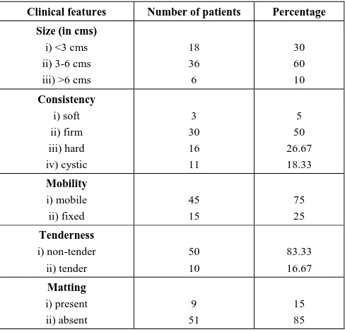
Mean age noted in our study population was 28.35 years. The neck swellings were assessed clinically in the outpatient department in terms of size, consistency, mobility, association with pain and presence/ absence of matting. Most of the swellings were between 3-6cms (60%) in size. Clinically most of the swellings were firm in consistency which was 30(50%) in number. (Table 3).
Table 1. Showing age distribution
Gender Number of Patients Percentage
Male 22 36.67
Female 38 63.33
Table 2. Showing Age Group Distribution
Age (in years) Number of Patients Percentage
1-10 3 5
11-20 9 15
21-30 16 26.67
31-40 11 18.33
41-50 5 8.33
51-60 6 10
>60 10 16.67
Total 60 100
Table 3. Showing clinical features of neck swellings
Clinical features Number of patients Percentage
Size (in cms) i) <3 cms ii) 3-6 cms iii) >6 cms
18 36 6
30 60 10
Consistency i) soft ii) firm iii) hard iv) cystic
3 30 16 11
5 50 26.67 18.33
Mobility i) mobile ii) fixed
45 15
75 25
Tenderness i) non-tender ii) tender
50 10
83.33 16.67
Matting i) present ii) absent
9 51
15 85
Clinically the most common neck swelling in our study were thyroid swellings which were 28(46.67%) in number. This was followed by cervical lymph node swellings which were 18(30%). Based on the anatomical location and other physical characteristics such as size, consistency, mobility, pain etc it was only possible to make a broad diagnosis of the neck swelling clinically as shown in Table 4. The most common neck swelling noted in our study was thyroid swellings. This was followed by cervical lymphadenopathy and major and minor salivary gland swellings respectively.
Table 4. Showing clinical diagnosis of neck swellings
Site of lesion Number of patients Percentage
Thyroid Swellings 28 46.67
Cervical Lymphadenopathy 18 30
Major and Minor Salivary
Gland Swellings 10 16.66
Miscellaneous 4 6.67
Inconclusive 10 16.66
On performing a USG of neck, the most common swellings were thyroid swellings which were 28(46.67%) in number. Among these thyroid swellings, the most common were solitary thyroid nodules which were 15(25%) in number. This was followed by multinodular goitres in 6(10%) patients. Thyroglossal cysts and malignancy were 4(6.67%) and 3(5%) in number respectively. Among the cervical lymphadenopathy, tubercular lymphadenopathy was found to be most common in 9(15%) patients. This was followed by metastatic lymph nodes in 7(11.67%) patients. Among the 60 patients, on ultrasonography, 6(10%) were inconclusive. (Table 5).
The swellings on evaluation with the FNAC yielded the following results. Thyroid swellings were most dominant swellings in 30(50%) of patients. Among these solitary thyroid nodules were seen in 15(25%), multinodular goitres in 6(10%), thyroglossal cysts in 5(8.33) and malignant thyroid swellings in 4(6.67%) were noted. Among the cervical lymphadenopathy tubercular lymph nodes were noted in 10(16.67%), metastatic lymph nodes in 7(11.67%) and non specific nodes were noted in 2(3.33%) patients. The details of other neck swellings are given in Table 5. The numbers of inconclusive swellings noted on FNAC were in 2(3.33%) patients. The FNAC diagnosis was correlated with histopathological evaluation.
On histopathology, the number of thyroid swellings noted was 31(51.66%). Of these one swelling which was considered a thyroglossal cyst and another neck swelling which was inconclusive on FNAC turned out to be papillary and follicular thyroid carcinomas respectively. Similarly in cervical lymphadenopathy, one inconclusive neck swelling was diagnosed to be a squamous cell carcinoma of neck swelling with an unknown primary. Overall the total number of neck swellings as cervical lymphadenopathy were 20(33.33%) in number. Swellings involving major and minor salivary glands were 10(16.67%) in number. One parotid swelling diagnosed as pleomorphic adenoma on FNAC was rediagnosed as mucoepidermoid carcinoma of parotid on histopathology. (Table 5).
Table 5. Showing ultrasonography, fine needle aspiration cytology and histopathology diagnosis of neck swellings
ULTRASONOGRAPHY FINE NEEDLE
ASPIRATION CYTOLOGY HISTOPATHOLOGY
NECK SWELLINGS NUMBER OF
PATIENTS %
NUMBER OF
PATIENTS %
NUMBER OF
PATIENTS %
THYROID SWELLINGS
15 6 4 3
25 10 6.67
5
15 6 5 4
25 10 8.33 6.67
15 6 4 6
25 10 6.67
10 i) solitary thyroid nodule
ii) multinodular goitre iii) thyroglossal cyst iv) malignancy
CERVICAL
LYMPHADENOPATHY
9 7 2
15 11.67
3.33
10 7 2
16.67 11.67 3.33
10 8 2
16.67 13.33 3.33 i) tubercular lymphadenopathy
ii) metastatic lymphadenopathy iii) non-specific
MAJOR AND MINOR SALIVARY GLAND SWELLINGS
2 6 2
3.33 10 3.33
2 6 2
3.33 10 3.33
2 5 3
3.33 8.33 5 i) inflammatory
ii) benign swellings iii) malignant swellings
MISCELLANEOUS (dermoid/
paraganglioma/lipoma etc) 4 6.67 5 8.33 5 8.33
INCONCLUSIVE 6 10 2 3.33 0 0
Table 6. Showing sensitivity, specificity, positive predictive value (PPV), negative predictive value (NPV) and accuracy of USG and FNAC
SENSITIVITY SPECIFICITY POSITIVE
PREDICTIVE VALUE
NEGATIVE
PREDICTIVE VALUE ACCURACY
ULTRASONOGRAPHY 87.20% 78.15% 85.42% 80.50% 78.33%
FINE NEEDLE
ASPIRATION CYTOLOGY 93.67% 88.80% 91.62% 88.23% 88.23%
4. Discussion
A thorough knowledge of the neck and its structures such as fascia, muscles, glands, lymphatics, vessels etc. is vital as it helps in understanding the nature and origin of the neck swellings. Among the several methods available today for evaluating the neck swellings clinical examination, ultrasonography and fine needle aspiration cytology forms the preliminary array of modalities. These are easily available, simple to perform and minimally invasive in nature. It forms an important triad in evaluating the neck swellings.
Clinical evaluation of neck swellings is the first line of screening available to the clinician. It is a subjective method which is solely operator dependant and follows a learning curve with experience. It requires a keen sense of observation and assessment on the part of the examiner.
Ultrasonography has become the first line of imaging modality for evaluation of the neck swellings due to excellent visualization of the internal parenchyma. It is highly sensitive in detecting small swellings, calcification, septations and cysts. The use of real time ultrasonography with high frequency transducers can significantly improve the evaluation of patients with various types of head and
neck swellings. In our present study, USG showed an overall sensitivity of 87.20%, specificity of 78.15%, PPV of 85.42%, NPV of 80.50% and accuracy of 78.33%. In a study done by Chandak et.al. [11] in which USG showed a sensitivity and accuracy of 98.5%, which were higher in comparison to our findings. In another study done by Venkatachalapathy et.al. [12] the sensitivity and specificity of USG was 73% and 85.3% respectively which was comparable to our findings.
thyroid swellings were more predominant. FNAC provides a more direct and clear diagnosis of neck swellings. With respect to thyroid swellings, the use of FNAC helps in reducing the number of thyroidectomies by approximately 50% [16, 17], roughly doubles the surgical yield of carcinoma and reduces the overall cost of medical care in these patients by 25%. [17]
5. Conclusions
The head and neck region encompasses a wide range of lesions especially of thyroid, lymph nodes, major and minor salivary glands with a variety of differential diagnosis ranging from inflammatory to neoplastic. Depending on the nature of neck swellings, most of these may require surgical intervention. In order to make an accurate and effective surgical management, it is essential to make a clear preoperative assessment of the nature of these lesions [18]. Though histopathology remains the gold standard for the final diagnosis of neck masses, a combined approach of ultrasonography and FNAC of neck swellings gives a sensitive, specific and accurate diagnosis of these lesions, thereby aiding the surgeon in planning the treatment protocol.
REFERENCES
[1] Fujimoto F, Oka A, Omoto R, Hirsoe M. Ultrasound scanning of the thyroid gland as a new diagnostic approach. Ultrasonics, 1967; 5: 177-80.
[2] Bruno A., Policeni Wendy, R.K Smoker, Deborah L. Reede. Anatomy and Embryology of the thyroid and parathyroid glands. Semin ultrasound CT MRI, 2012; 33: 104-14.
[3] Martin HE, Ellis EB (1930). Biopsy by needle puncture and aspiration. Ann Surg 92: 169-81.
[4] Asp AA, Georgitis W, Waldron EJ, Sims JE, Kidd GS 2nd (1987). Fine needle aspiration of the thyroid--`d. Use in an average health care facility. Am J Med 83: 489-93.
[5] Bottles K, Miller TR, Cohen MB, Ljung BM (1986). Fine needle aspiration biopsy. Has its time come? Am J Med 81: 525-31.
[6] Burch HB (1995). Evaluation and management of the solid thyroid nodule. Endocrinol Metab Clin North Am 24: 663-710.
[7] Guiffrida D, Gharib H (1995). Controversies in the management of cold, hot and occult thyroid nodules. Am J Med 99: 642-50.
[8] de Vos tot Nederveen Cappel RJ, Bouvy ND, Bonjer HJ, van Muiswinkel JM, Chadha S (2001). Fine needle aspiration cytology of thyroid nodules: how accurate is it and what are the causes of discrepant cases? Cytopathology 12: 399-405.
[9] La Rosa GL, Belfore A, Guiffria D, Sicurella C, Ippolito O et al. (1991). Evaluation in the fine needle aspiration biopsy in the preoperative selection of cold thyroid nodules. Cancer 67: 2137-41.
[10] Chitumalla PK. Study of cervical lymphadenitis, correlation between clinical features, FNAC and histopathology of cervical lymphadenitis. Int J Contemporary Med Res. 2016; 3(8): 2231-4.
[11] Chandak R, Degwekar S, Bhowte RR, Motwani M, Banode P, Chandak M, et al. An evaluation of efficacy of ultrasonography in the diagnosis of head and neck swellings. Dentomaxillofac Radiol 2011; 40: 213-21.
[12] Venkatachalapathy TS, Sreeramulu PN, Ramesh Krishna M (2012). A Prospective Study of Clinical, Sonological and Pathological Evaluation of Thyroid Nodule. Thyroid Disorders Ther 1:109.
[13] Lokhande, R., Gedam, B., Shah, Y., Kale, V., Tandon, M., & Ansari, I. (2015). The accuracy of ultrasonography and fine needle aspiration cytology in the diagnosis of nodular goitre: A prospective analysis of fourty two cases. International Journal of Biomedical and Advance Research, 6(1), 43-6.
[14] Basista H, Modwal A, Prasad B. Clinicopathological evaluation of neck masses. Sch J App Med Sci 2015; 3(9B): 3235-41.
[15] Soni S, Pippal SK, Yashweer B, Srivastava P; Efficacy of fine needle aspiration cytology in diagnosis of neck masses. World article in ear, nose and throat, 2010; 3(2).
[16] Korun N, Ascii C, Yilmazlar T, Duman H, Zorluoglu A, Tuncel E et al. Total thyroidectomy or lobectomy in benign nodular disease of thethyroid: changing trends in surgery. International Surgery. 1997; 82:417-19.
[17] Mazzaferri EL. Management of a solitary thyroid nodule. The New England Journal of Medicine. 1993; 328: 5539.