Paweł Tabakow
1, Marcin Czyż
1, Włodzimierz Jarmundowicz
1,
Ewa Lechowicz-Głogowska
2Surgical Treatment of Pituitary Adenomas Using
Low-Field Intraoperative Magnetic Resonance Imaging
Operacyjne leczenie gruczolaków przysadki z użyciem niskopolowego
śródoperacyjnego rezonansu magnetycznego
1 Department and Clinic of Neurosurgery, Wroclaw Medical University, Wrocław, Poland
2 Department and Clinic of Anesthesiology and Intensive Therapy, Wroclaw Medical University, Wrocław,
Poland
Abstract
Background. Intraoperative magnetic resonance imaging (iMRI) is a new technique for imaging of the brain and is used with increasing frequency during neurosurgical operations, enabling the surgeon to make decisions based on real-time images.
Objectives. This paper presents the technique for the surgical treatment of pituitary adenomas using low-field iMRI, evaluates the safety of iMRI usage in pituitary surgery and examines the influence of iMRI on the extent of tumor removal.
Material and Methods. From October 2008 to December 2010, 18 patients were treated for pituitary adenomas using the low-field iMRI system Polestar N20. The procedures were conducted via the transsphenoidal approach, using the microscopic technique in 15 cases and endoscopically in three cases. The patients’ mean age was 56 ± 15 years; their mean American Society of Anesthesiologists (ASA) score was 2; 67% of them were male. Most of the patients were operated on for macroadenomas, 83% of which were hormonally inactive. The analysis concerned the technical aspects of iMRI usage, such as preparation and surgery time and the quality of the iMRI-scans performed. The safety of iMRI and its influence on decisions regarding further tumor resection.
Results. The operations on pituitary adenomas using iMRI were safe. Only two hemorrhagic complications were noted, and they were not related to iMRI usage. The mean preparation and surgery times were 109 ± 37 minutes and 238 ± 188 minutes, respectively. The iMRI images of sella turcica were of satisfactory quality in 16 patients. In 50% of the cases, iMRI conducted when the surgeon believed that the desired extent of tumor resection had been attained showed that there were still tumor remnants to be resected. In 67% of these cases, continued tumor removal lead to achievement of the desired degree of resection.
Conclusions. Low-field iMRI-guided operations on pituitary tumors are safe and feasible, and they ensure an increased radicality of tumor resection (Adv Clin Exp Med 2012, 21, 4, 495–503).
Key words: intraoperative magnetic resonance, low-field, pituitary adenomas.
Streszczenie
Wprowadzenie. Śródoperacyjny rezonans magnetyczny jest nową techniką obrazowania mózgu i jest coraz częś-ciej stosowany podczas operacji neurochirurgicznych, pozwalając chirurgowi na podejmowanie decyzji na podsta-wie obrazów uzyskanych w czasie rzeczywistym.
Cel pracy. Przedstawienie techniki operacji gruczolaków przysadki z wykorzystaniem niskopolowego śródopera-cyjnego rezonansu magnetycznego (iMRI), ocena bezpieczeństwa zabiegów oraz wpływu zastosowania iMRI na stopień uzyskanej doszczętności resekcji guza.
Materiał i metody. Analizie poddano 18 pacjentów operowanych z powodu gruczolaków przysadki z użyciem niskopolowego rezonansu śródoperacyjnego Polestar N20 od października 2008 do grudnia 2010 r. Operacje wyko-nywano z dostępu przezklinowego, w 15 przypadkach metodą mikrochirurgiczną, a w 3 techniką endoskopową. Średni wiek pacjentów wynosił 56 ± 15 lat, mediana ASA-2, mężczyźni stanowili 67% operowanych. W większości przypadków operowano pacjentów z makrogruczolakami przysadki, w tym w 83% przypadkach z powodu hormo-nalnie nieczynnych gruczolaków. Poddano analizie aspekty techniczne związane z zastosowaniem iMRI, takie jak:
Adv Clin Exp Med 2012, 21, 4, 495–503 ISSN 1899–5276
ORIGINAL PAPERS
The transsphenoidal approach is currently the preferred method of surgical treatment for more than 90% of pituitary tumors [1]. For several decades these tumors have been removed using the microscopic technique. The main obstacle to transsphenoidal microsurgery was incomplete vis-ualization of sellar and suprasellar spaces, which often led to incomplete tumor resection. The in-troduction of endoscopy provided the neurosur-geon a better view into the sellar, parasellar and suprasellar regions and thus improved the radical-ity of the operations performed [2, 3]. In addition to neuroendoscopy, another recent development enabling better visualization of pituitary tumors is the use of intraoperative magnetic resonance imag-ing (iMRI). Several iMRI systems are used in brain surgery. They mainly differ in the strength of the magnetic field produced and can be divided into high-field and low-field iMRI systems [4, 5]. The use of iMRI has been shown to be extremely useful in transsphenoidal microsurgery, enabling reliable monitoring of tumor resection and thus decreas-ing the likelihood of repeat surgeries [3, 6–8].
This paper presents the authors’ experience us-ing the low-field iMRI Polestar N20 system durus-ing transsphenoidal surgery for pituitary tumors. The article examines the safety, the technical aspects and the impact of this modality on the degree of tumor resection.
Material and Methods
Patient Population
Between October 2008 and December 2010, 18 patients diagnosed with pituitary tumors were treated surgically at the Wroclaw Medical Univer-sity Department of Neurosurgery with the use of low-field intraoperative magnetic resonance imag-ing. The patients’ mean age was 56 ± 15 years. Their median American Society of Anesthesiologists (ASA) score was 2. The group included 12 men and 6 women. The majority of the patients had been diagnosed with a macroadenoma; in 83% of
the cases it was hormonally inactive. The modified Hardy’s grading system classification was used to assess the extent of suprasellar invasion of the tu-mor, as follows: grade I (n = 3): intrasellar tumor; grade II (n = 7): tumor with suprasellar extent within 10 mm of the planum sphenoidale; grade III (n = 4): tumor with suprasellar extent up to 30 mm elevating and filling the anterior third ven-tricle; and grade IV (n = 4): tumor extending far beyond the sellar space [9].
Invasion of the cavernous sinus was also as-sessed, using the Knosp Steiner classification; it was observed in 6 patients [10]. Each patient com-pleted a standardized questionnaire concerning metallic implants used before the planned diag-nostic examinations using high-field MR and gave written consent for an operative procedure with iMRI.
Intraoperative Imaging
and Surgical Procedure
The patients were operated on using a Pole-Star N20 low-field iMRI imager (Medtronic Navi-gation, Louisville, CO, USA). This mobile iMRI apparatus generated a constant magnetic field of an intensity of 0.15 T, allowing examinations in T1, T2 and FLAIR sequences with a 16 × 9 cm field of view (FOV) to be conducted. All the procedures took place in a shielded operating room adapted for iMRI application. The general principles of pa-tient preparation in the operating theater, anesthe-sia management and the performance of the iMRI studies have been described in a previous publi-cation [11]. Briefly, after patient positioning and the preparation of the operating field, the patient's head was registered in the optical system for cranial navigation (StealthStation, Medtronic Navigation, Louisville, CO, USA) based on the preoperative high-field MRI study. In the next step, a position-ing coronal iMRI scan was performed to assess the optimal position of the magnet during the op-eration (e-steady time-reversed fast imaging with steady-state precession 24 s, 8 mm). This study
czas przygotowań, czas operacji, jakość wykonanych badań iMRI oraz bezpieczeństwo procedury i wpływ wyniku badania iMRI na decyzję operatora o dalszej resekcji operowanego guza.
Wyniki. Operacje gruczolaków przysadki z użyciem iMRI były bezpieczne. Stwierdzono jedynie 2 przypadki powik- łań krwotocznych, które nie były związane z użyciem iMRI. Średni czas przygotowań pacjenta do operacji wynosił 109 ± 37 min, średni czas operacji 238 ± 188 min. U 16 pacjentów uzyskano satysfakcjonujący obraz diagnostycz-ny siodła tureckiego w badaniu iMRI. U 50% pacjentów badanie iMRI, wykonane, gdy operator uznał, że uzyskał zakładaną doszczętność resekcji guza przysadki, wykazywało resztki guza. W 67% przypadkach udało się dzięki kontynuacji operacji uzyskać planowany stopień doszczętności zabiegu.
Wnioski. Śródoperacyjny niskopolowy rezonans magnetyczny jest narzędziem przydatnym i bezpiecznym w przez-klinowych operacjach gruczolaków przysadki. Pozwala na zwiększenie radykalności resekcji gruczolaków przysadki (Adv Clin Exp Med 2012, 21, 4, 495–503).
was followed by a diagnostic iMRI scan after in-travenous administration of gadolinium contrast at a dose of 0.4 mL/kg (T1 3-dimensional gradient echo sequence, slice thickness 4 mm, acquisition time 7 min). The T1-weighted iMRI scans were compared with the preoperative high-field MRI scan to assess whether the quality of the intraop-erative diagnostic study was satisfactory. Before starting the operation, the scanner was draped and lowered to the home position under the operat-ing table. The pituitary tumor resection operations were conducted in the typical manner, using the endonasal transsphenoidal technique and classi-cal ferromagnetic microsurgiclassi-cal instruments. In 15 cases the tumor was resected using an operat-ing microscope (OPMI NC4/Pentero, Carl Zeiss Meditec AG, Jena, Germany) and in three cases using a neuroendoscope (Aesculap, Tuttlingen, Germany). The iMRI study of the sella region was performed when the neurosurgeon considered the planned tumor resection was complete, based on a microscopic view of the surgical field (e.g. a col-lapse of the diaphragma sellae). A hemostasis of the sella turcica was performed using gadolinium-soaked cotton pledgets according to the method described by Ahn et al. [12]. The intraoperative images obtained were transferred automatically to the optical neuronavigation system.
Statistical Analysis
Statistical analysis was conducted using Statis-tica 9.0 software. The χ2 test with Yates’s
correc-tion was used to assess the differences between the tested groups. The accepted significance level was p < 0.05.
Results
Technical Considerations
The patient preparation time (mean time: 109 min) and the duration of the operation (mean: 238 min) were significantly longer than the authors’ standard approach, e.g. when only optical track-ing navigation was used for image guidance [11]. Very clear visualization of the pituitary adenoma was obtained in 16 of the operations (89%). The best quality iMRI images was obtained in cases when hemostasis in the sella and sphenoid sinus was good and the image was taken 5 to 15 minutes after administering the intravenous contrast. Un-satisfactory tumor visualization occurred in cases when the MR scanner was run by doctors who had not had professional iMRI training. The average number of iMRI scans was five, including three e-steady scans and three diagnostic scans. The
optimal scenario of the operative procedure was one e-steady scan followed by two diagnostic T1-weighted scans – one performed at the beginning and one at the end of the operation.
The Safety of iMRI-guided
Transsphenoidal Surgery
Using the low-field iMRI scanner was safe both for the patient and the operating team. There were two cases of intraoperative hemorrhage from the sellar region that required interruption of the procedure, but they were not directly related to the use of iMRI. The magnetic field generated by the iMRI did not affect the microsurgical instru-ments, the neuronavigation system or the surgical microscope when they were kept at an appropriate distance from the magnets when the iMRI appara-tus had been positioned under the operating table. In some cases transient magnetization of the fer-romagnetic instruments and difficulties with ad-justing the focus of the microscope occurred when these devices were too close to the magnets. Dur-ing the performance of the iMRI study all electri-cally driven devices in the operating theater were switched off and all ferromagnetic instruments were taken away from the operating table. It took only several minutes to prepare the operating field for the iMRI study and to position the magnets of the scanner in the appropriate position.
Intraoperative Resection
Assessment
Radical removal of the pituitary tumor was possible in 12 cases (66.7%). These cases included three microadenomas (Grade I according to Har-dy’s classification) and nine macroadenomas clas-sified as follows: Grade II: n = 7, Grade III: n = 4 and Grade IV: n = 4.
The quality of the 0.15-T iMRI studies per-formed was in most cases satisfactory enough to permit the achievement of reliable control of the tumor resection. There were generally two surgical scenarios. In the first one, which involved nine pa-tients (50%), the iMRI study performed when the neurosurgeon thought the surgical goals had been achieved confirmed those expectations (Fig. 1). In the remaining nine cases (50%) the iMRI re-vealed unexpected tumor remnants, to the sur-geon’s surprise, and continued tumor removal was necessary. In 67% of these cases (33% of the total number of cases in the study) the desired radicality of the operation was then achieved (Fig. 2).
Fig. 1. A case of gross total resection of a Grade III pituitary macroadenoma. A) Preoperative high-field T1-weighted enhanced sagittal (left) and coronal (right) 1.15 T MRI scans showing a tumor compressing the optical chiasm and elevating the third ventricle. B) Low-field 0.15 T T1-weighted enhanced coronal iMRI scans. The image on the left shows the first diagnostic scan, performed at the beginning of the operation. The image on the right was performed at the end of surgery, showing complete removal of the tumor with effective optical chiasm decompression. C) Post-operative high-field MR scans performed 3 months post surgery confirming the effect of gross total resection of the macroadenoma shown by the iMRI study
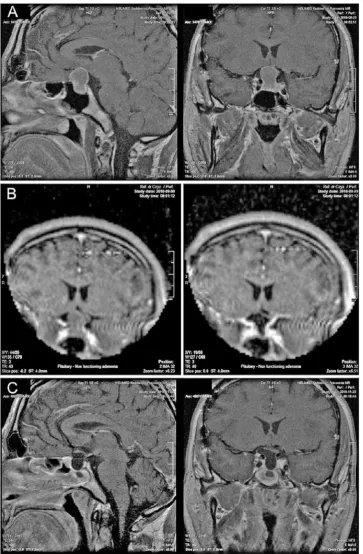
Fig. 2. A case of successful debulking of an invasive hormonally inactive Grade IV macroadenoma. A) Preoperative high-field T1-weighted enhanced sagittal (left) and coronal (right) MRI scans showing a giant tumor compressing and elevating the optical chiasm, filling the suprasellar cisterns and invading the right cavernous sinus and the clivus. B) Low- field T1-weighted enhanced coronal iMRI scans. The first image (on the left) shows the diagnostic scan per-formed at the beginning of the operation. The image in the middle was perper-formed when the surgeon believed the sur-gical goal had been achieved. To his surprise the iMRI showed several tumor remnants that required further removal. The image on the right shows the final effect of tumor debulking resulting in chiasmal decompression. The arrow shows the optical chiasm. C) Postoperative high-field MR scans performed 3 months post surgery showing effective decompression of the optic chiasm. The tumor was removed from the sellar and suprasellar space. Residual tumor remnants invading the cavernous sinus and the clivus were left for observation
low-field iMRI system seemed to be especially use-ful in cases of operations on macroadenomas of Grades III and IV (n = 8). The use of iMRI contrib-uted to increasing the number of cases of achieved surgical goals from 1 to 5; however, this result was not statistically significant (p = 0.12). In cases of Grade I and II adenomas, the influence of iMRI on the decision of the surgeon was not statistically significant (p = 1.0). More detailed information concerning the results of surgical treatment for pi-tuitary tumors is summarized in Table 1.
Discussion
Technical Considerations
of Usage of iMRI System
Theapplication of iMRI in brain surgery has been shown very helpful as a tool for near real-time navigation and for resection control. The achieve-ment of these main advantages of using both high-field and low-high-field iMRI is connected with a sub-stantial increase in the patient preparation time and the duration of the surgery. This prolongation of time did not increase the complication rate, but required a greater effort from the surgical and anesthesiological team [3, 6, 7, 11]. The question which of the two modalities – high- or low-field
iMRI – is more useful in brain surgery has not been answered yet and is beyond the scope of this article. The authors’ opinion about the use of the Polestar N20 ultra-low-field iMRI system is that it can be implemented relatively easily in operations for brain pathologies, including transsphenoidal pituitary surgery. This low-field magnetic imager is small and mobile, so it can be placed under the operating table during surgery. In cases when it is needed it can be positioned within several min-utes at the level of the patient’s head to perform diagnostic scans, without the need for any special preparation of the operating field and without the support of any technical staff [3, 11]. The patient does not need to be transferred to another operat-ing room to conduct MR-imagoperat-ing, as in the case of high-field iMRI. The MR-imager is recognized as an active tool by the system for cranial navigation. This allows the correct positioning of the magnet in relation to the reference frame for navigation, and also the use of images for real time naviga-tion.
The quality of the performed low-field MR-images, although not as good as in the case of high-field iMRI, has been found to be satisfactory in many studies [3, 6–8, 11, 12]. In the presented series of patients operated on for pituitary ad-enomas, very clear visualization of the tumor was obtained in 16 operations (89%). The suprasellar
Ryc. 2. (cd.) B) Śródoperacyjne niskopolowe badania MRI w sekwencji T1-zależnej w projekcji wieńcowej. Pierwsze zdjęcie (po stronie lewej) pokazuje diagnostyczne badanie iMRI wykonane na początku operacji. Zdjęcie środkowe zostało wykonane, gdy chirurg uznał, że osiągnął zaplanowany stopień resekcji guza. Ku jego zaskoczeniu, badanie iMRI pokazało kilka fragmentów guza wymagających dalszej resekcji. Zdjęcie po stronie prawej wykonane pod koniec operacji pokazuje końcowy rezultat pomniejszenia guza ze skuteczną dekompresją skrzyżowania wzrokowego. Strzałka wskazuje na skrzyżowanie wzrokowe. C) Pooperacyjne wysokopolowe badanie MRI wykonane 3 miesiące po zabiegu pokazujące skuteczną dekompresję skrzyżowania wzrokowego. Guz został usunięty z części siodłowo-nad-siodłowej. Pozostałe części guza znajdujące się w zatoce jamistej i stoku zostały pozostawione do dalszej obserwacji
Table 1. Tumor grade of and extent of tumor resection. GTR: gross total resection; TB: tumor debulking with successful optic chiasm decompression; PR: partial resection without appropriate chiasm decompression; a: grading according to the modified Hardy’s classification; b: resection conducted before conducting the iMRI; c: resection conducted after the iMRI study
Tabela 1. Stopień inwazyjności guza oraz doszczętność jego resekcji. GTR - doszczętne usunięcie guza; TB – subtotalne usunięcie guza z właściwą dekompresją skrzyżowania wzrokowego; PR – częściowa resekcja bez dekompresji skrzyżowania wzrokowego; a – zmodyfikowana skala Hardiego inwazyjności gruczolaków przysadki; b – zakres resekcji guza przed wyko-naniem badania iMRI; c – zakres resekcji guza po wykonaniu iMRI
Grade of tumora
(Stopień inwazyjności guza) Extent of first resection
b
(Doszczętność pierwszej resekcji) Extent of second resection
c
(Doszczętność drugiej resekcji)
GTR TB PR GTR TB PR
Grade I (n = 3) 2 (66.6%) 1 (33.4%) 0 3 (100%) 0 0 Grade II (n = 7) 5 (71.4%) 0 2 (28.6%) 6 (85.7%) 0 1 (14.3%) Grade III (n = 4) 1 (25%) 0 3 (75%) 3 (75%) 0 1 (25%)
Grade IV (n = 4) 0 0 4 (100%) 0 2 (50%) 2 (50%)
region had the best visibility on the iMRI scans, followed by the sella turcica and the cavernous si-nus. The sphenoid sinus was almost always filled with blood and was not suitable for analysis.
There are several tips that the authors find im-portant for achieving better quality low-field iMRI images. First, the central parts of the magnets as well as the receiving coil need to be positioned at the level of the brain pathology. Second, there should be minimal electromagnetic noise in the operating theater during the process of scanning, which al-lows the achievement of a high signal-to-noise ra-tio. In the cases presented here this was achieved by adapting the operating room for iMRI use. The room was equipped with filters and screens that reduced the influence of external interference with the magnetic field. Another less expensive option that can be applied in a standard (not adapted) op-erating room is the use of a portable cage to block radio-frequency interference from affecting the in-traoperative MRI apparatus [7].
Another crucial step enabling good visualiza-tion of the sellar region during iMR imaging is the performance of proper hemostasis in the surgical field. This can be achieved by several methods, such as gadolinium-soaked cotton pledgets [12], bone wax balls covered with a piece of a rubber glove [8], introduction of a suction catheter into the resection cavity [6 ] or leaving a titanium nasal speculum with an indicator introduced in the the resection bed during the MR scanning [7]. After testing most of these methods the authors decided to use contrast-filled cotton pledgets, as described by Ahn et al. [12]. The area where the pledgets were placed appeared dark on contrast-enhanced T1-weighted sequences because highly concentrat-ed contrast agents show dark signal intensity, as opposed to contrast agents that are administered intravenously.
The quality of T1-weighted 0.15 T MR-images of the pituitary region can be also improved when a double dose of contrast agent is administered (0.4 ml Gadolinium/kg body weight) [6, 7, 12].
Intraoperative Resection
Assessment
The surgical goal for the transsphenoidal ap-proach was gross total resection (GTR) for non-in-vasive pituitary tumors (Grades I and II and some Grade III tumors according to Hardy’s classifica-tion) and debulking with optic chiasm decompres-sion for invasive tumors (some Grade III tumors and all Grade IV tumors). For safety reasons the authors did not plan to use the transsphenoidal approach to remove parts of the tumor that had
invaded the cavernous sinus or were localized far away from the sellar region, e.g. in the clivus, the interpeduncular cistern, the planum sphenoi-dale or the Sylvian fissure. The plan of the opera-tions was to remove the pituitary tumor using the micro-or endoscopic trassphenoidal technique as radically as possible and to perform iMRI scans at the point when the surgeon was convinced that the surgical goal established preoperatively had been achieved. The criterion for failure of the surgical plan was finding a residual tumor along a non-in-vasive margin that is accessible via the transsphe-noidal approach.
The results of the current authors’ operations on pituitary adenomas using iMRI imaging are similar to those achieved by other authors who used this modality, and they show an evident im-provement in the effectiveness of treatment of these tumors.
The current authors’ initial rate of GTR of pi-tuitary adenomas was 44.4%, which was compara-ble the GTR rate of Wu et al. (58.2%) [7] and Nim-sky et al. (58%) [13]. Similarly to other studies, the current study had a surprisingly high rate of cases (50%) of iMRI scans identifying accessible tumor remnants that required further removal. Apart from the study by Gerlach et al. [6], in which the rate of identification of unexpected tumor rem-nants was 17.5%, other groups have reported a rate from 40% up to as high as 66% [3, 7, 13]. In the current study, after diagnosing an accessible tu-mor remnant, continued tutu-mor removal led to the achievement of the desired radicality of the opera-tion in 67% of the cases. As a result, the use of the low-field iMRI system led to an increase in the rate of GTR from 44.4% to 66.7%, and – more impor-tantly – increased the rate of achievement of surgi-cal goal (including cases of GTR and effective optic chiasm decompression) from 50% to 83.3%. The 22% increase in the rate of GTR obtained by the current authors is similar to the results of Wu et al. and Nimsky et al., who reported an increase of the rate of GTR from 58.2% to 83.6%. and from 58% to 82% respectively [7, 13]. In the present study the rate of achievement of surgical goals after the application of low-field iMRI was relatively high (83.3 %), but in other studies it was even higher and reached values of 96% for 0.15 T iMRI [7] or even 100% for 0.3 T iMRI [3]. Interestingly, these results are comparable to the results of using high-field iMRI [13].
adenoma (n = 10), iMRI studies led to further re-section only in 3 cases (Table 1). Thus the authors believe that while the usefulness of low-field iMRI in the surgical treatment of Grade III and Grade IV macroadenomas is evident, its influence on the result of surgical treatment of microadenomas or Grade II macroadenomas is debatable.
In the present study, in three casesof patients with Grade IV macroadenomas the endoscopic-surgical technique was combined with iMRI. De-spite the superior visualization ensured by the en-doscope compared to that offered by a microscope, in all three cases performing iMRI changed the surgeon’s decision by revealing unexpected tumor remnants. The surgical goals could be achieved only in one of these patients. As these were the authors’ first attempts to combine endoscopy with iMRI and the cases were more difficult than in the microscopic group, no definite conclusions can be drawn about the reliability of the combination of these two techniques. Nevertheless, other authors have shown that iMRI offers additional benefits in
resection control when compared to pure endo-scopic technique [14].
The authors concluded that the use of low-field iMRI system in transsphenoidal pituitary surgery was safe and feasible. Although it substantially pro-longed the duration of the procedure and required the operative team to master some new skills to ensure the acquisition of images of good quality, several important advantages have been shown to support the validity of its use. These include a 22% increase in the rate of GTR and an increase in the percentage of achieved surgical goals from 50% to 83.3%. The low-field iMRI system seemed to be especially useful in cases of operation on Grade III and Grade IV macroadenomas. In these cases the surgeon’s adjustment to the results of the iMRI was the main factor determining the success of the operation. A prospective controlled randomized study is needed to assess the influence of low-field iMRI on the longterm outcome of patients oper-ated on for pituitary adenomas.
References
[1] Tindall GT, Barrow DL: Tumours of the sellar and parasellar area in adults. In: Neurological Surgery: A Comprehensive Reference Guide to the Diagnosis and Management of Neurosurgical Problems. Ed.: Youmans JR, W.B. Saunders Co., Philadelphia 1996, 4th ed., 2935–2969.
[2] Jho HD, Carrau RL: Endoscopic endonasal transsphenoidal surgery: Experience with 50 patients. J Neurosurg 1997,87, 44–51.
[3] Bohinski RJ, Warnick RE, Gaskill-Shipley MF, Zuccarello M, van Loveren HR, Kormos DW, Tew JM Jr:
Intraoperative magnetic resonance imaging to determine the extent of resection of pituitary macroadenomas dur-ing trassphenoidal microsurgery. Neurosurgery 2001, 49, 1133–1144.
[4] Black PM, Moriarty T, Alexander E 3rd, Stieg P, Woodard EJ, Gleason PL, Martin CH, Kikinis R, Schwartz RB,
Jolesz FA: Development and implementation of intraoperative magnetic resonance imaging and its neurosurgical applications. Neurosurgery 1997, 41, 831–845.
[5] Sutherland GR, Kaibara T, Louw D, Hoult DI, Tomanek B, Saunders J: A mobile high-field magnetic resonance system for neurosurgery. J Neurosurg 1999,91, 804–813.
[6] Gerlach R, de Rochemont RM, Gasser T, Marquardt G, Reusch J, Imoehl L, Seifert V: Feasibility of PoleStar N20, an ultra-low-field intraoperative magnetic resonance imaging system in resection control of pituitary mac-roadenomas: lessons learned from the first 40 cases. Neurosurgery 2008, 63, 272–285.
[7] Wu JS, Shou XF, Jao CJ, Wang YF, Zhuang DX, Mao Y, Li SQ, Zhou LF: Transsphenoidal pituitary macroad-enomas resection guided by Polestar N20 low-field intraoperative magnetic resonance imaging: comparison with early postoperative high-field magnetic resonance imaging. Neurosurgery 2009, 65, 63–71.
[8] Baumann F, Schmid C, Bernays RL: Intraoperative magnetic resonance imaging-guided transsphenoidal surgery for giant pituitary adenomas. Neurosurg Rev 2010, 33, 83-90.
[9] Knosp E, Steiner E, Kitz K, Matula C: Pituitary adenomas with invasion of the cavernous sinus space: A magnetic resonance imaging classification compared with surgical findings. Neurosurgery1993, 33, 610–618.
[10] Hardy J: Transphenoidal microsurgical treatment of pituitary tumours, In: Recent Advances in the Diagnosis and Treatment of Pituitary Tumours. Linfoot J (ed.). New York, Raven Press, 1979, 375–388.
[11] Czyż M, Tabakow P, Jarmundowicz W: Prospective study on efficacy of the low field intraoperative magnetic resonance imaging (iMRI) application in neurosurgical operations. Neurol Neurochir Pol 2011, 45(3), 226–34.
[12] Ahn JY, Jung JY, Kim J, Lee KS, Kim SH: How to overcome the limitations to determine the resection margin of pituitary tumours with low-field intra-operative MRI during trans-sphenoidal surgery: usefulness of Gadolinium-soaked cotton pledgets. Acta Neurochir 2008, 150, 763–771.
[13] Nimsky C, von Keller B, Ganslandt O, Fahlbusch R: Intraoperative high-field magnetic resonance imaging in transsphenoidal surgery of hormonally inactive pituitary macroadenomas. Neurosurgery 2006, 59, 105–114.
Address for correspondence:
Paweł Tabakow
Department and Clinic of Neurosurgery Wroclaw Medical University
Borowska 213 50-556 Wrocław Poland
E-mail: [email protected]
Conflict of interest: None declared Received: 21.02.2012