Seyithan Taysi
1, Seydi Okumus
2, Sinan Ezirmik
3, Naim Uzun
4, Adnan Yilmaz
5,
Mehmet Akyuz
6, Umit Tekelioglu
7, Ahmet Dirier
8,
Behcet Al
9The Protective Effects of L-Carnitine and Vitamin E
in Rat Lenses in Irradiation-Induced Oxidative Injury
Ochronne działanie L-karnityny i witaminy E na soczewki szczura
w uszkodzeniu oksydacyjnym wywołanym przez promieniowanie
1 Department of Biochemistry and Clinical Biochemistry, Gaziantep University,
Medical School, Gaziantep, Turkey
2 Department of Ophthalmology, Gaziantep University, Medical School, Gaziantep, Turkey 3 Department of Radiation Oncology, Ataturk University, Erzurum, Turkey
4 Biochemistry, Medical School, Ataturk University, Erzurum, Turkey 5 Department of Biochemistry, Rize University, Rize, Turkey
6 General Directorate of Pharmaceuticals and Pharmacy, Quality Control Department,
Turkish Ministry of Health, Dıskapı-Ankara, Turkey
7 Department of Anesthesiology, Birecik State Hospital, Sanliurfa, Turkey
8 Department of Radiation Oncology, Gaziantep University, Medical School, Gaziantep, Turkey 9 Emergency Department, Gaziantep University, Medical School, Gaziantep, Turkey
Abstract
Objectives. The aim of this study was to evaluate the antioxidant role of L-carnitine (LC) and vitamin E against radiation-induced cataracts in rat lenses after total cranial irradiation with a single 5 Gray (Gy) dose of gamma irradiation.
Material and Methods. Thirty two Sprague-Dawley rats were used for the experiment. The control group did not receive LC and vitamin E or irradiation but received both 0.1 ml physiological saline intraperitoneally and sham irradiation. The irradiation (IR) group received 5 Gy gamma irradiation to the total cranium as a single dose plus 0.1 ml physiological saline intraperitoneally. The IR plus vitamin E group received irradiation to total cranium plus 10 mg/kg/day vitamin E intraperitoneally. The IR plus LC group received irradiation to total cranium plus 100 mg/kg/day LC intraperitoneally. Biochemical parameters measured in murine lenses were carried out using spectrophotometric techniques.
Results. Total superoxide scavenger activity (TSSA), non-enzymatic superoxide scavenger activity (NSSA), gluta-thione-S-transferase (GST) and glutathione reductase (GRD) activities, significantly increased in the control, IR plus vitamin E and LC plus IR groups when compared with the IR only group. Lens TSSA and NSSA activities in the control group were significantly increased compared to that of the IR only group, but decreased compared to those of the IR plus vitamin E and IR plus LC groups. Lens xanthine oxidase (XO) activity in the IR group signifi-cantly increased compared to those of other groups.
Conclusions. Results show that LC and vitamin E prevented oxidative stress by scavenging free radicals generated by ionizing radiation in rat lenses (Adv Clin Exp Med 2011, 20, 1, 15–21).
Key words: carnitine, vitamin e, antioxidant enzymes, irradiation, oxidative stress, lens.
Streszczenie
Cel pracy. Ocena antyoksydacyjnego działania L-karnityny (LC – L-carnitine) i witaminy E przeciw zaćmie wywo-łanej promieniowaniem u szczurów po całkowitym napromieniowaniu czaszki pojedynczą dawką 5 Gy promie-niowania gamma.
Materiał i metody. Badaniem objęto 32 szczury Sprague-Dawley. Grupa kontrolna nie otrzymała LC i witaminy E ani napromieniowania, otrzymała natomiast 0,1 ml soli fizjologicznej dootrzewnowo oraz symulowane napro-mieniowanie. Grupa napromieniana (IR – irradiation) otrzymała pojedynczą dawkę 5 Gy na całą czaszkę oraz 0,1 ml soli fizjologicznej dootrzewnowo. Grupa IR plus witamina E otrzymała poza napromieniowaniem 10 mg/
Adv Clin Exp Med 2011, 20, 1, 15–21 ISSN 1230-025X
ORIGINAL PAPERS
Radiation therapy is a common and important tool for cancer treatment [1]. Eighty percent of cancer patients need radiotherapy at some time or other, either for curative or palliative purposes. The radiosensitivity of normal tissue adjacent to the tu-mor limits therapeutic gain. The responses of nor-mal tissues to therapeutic radiation exposure range from those that cause mild discomfort to others that are life threatening. The speed at which a response develops varies widely from one tissue to another and often depends on the dose of radiation that the tissue receives [2–4]. Ionizing radiation is known to generate reactive oxygen species (ROS) in irradi-ated tissue. Because human tissues contain 80% wa-ter, the major radiation damage is due to aqueous free radicals, generated by the action of radiation on water. Advanced glycation end products (AGEs) are known to be generated in foods and biological systems. Recently, these AGEs were reported to be harmful to biological systems. They were also con-sidered to generate reactive oxygen species (ROS) under the presence of transition metals in vitro and
in vivo [5]. These free radicals react with cellular macromolecules, such as deoxyribonucleic acid (DNA), ribonucleic acid (RNA), proteins, mem-branes, etc., and cause cell dysfunction and mortal-ity. These reactions take place in tumors as well as normal cells when exposed to radiation [6].
A cataract is opacity of the eye lens that inter-feres with vision. Cataracts are formed in response to a variety of different agents and environmen-tal stresses, and this damage seems in almost all cases to have an oxidative damage component. Although cataract of the eye lens is a known late effect of ionizing radiation exposure, most of the experimental work to date has concentrated on single, acute, high doses or multiple, fractionated, and chronic exposures [7, 8].Radiation cataracts are expressed after latency. The duration of the latency depends inversely on dose: the higher the dose, the more rapidly the cataract develops. For a single treatment, the lowest cataractogenic dose was reported to be 2 Gy [9, 10].
Vitamin E not only acts as an effective lipo-philic antioxidant and radical scavenger but also stabilizes cellular membranes [11]. The protective role of vitamin E against radiation-induced oxida-tive damage was demonstrated in vitro [12]. Using an injectable form of vitamin E (α-tocopherol), there was a clear improvement in post-irradiation survival compared with the results for dietary ad-ministration of vitamin E [7, 12].
L-carnitine (LC) is a natural substance that acts as a carrier for fatty acids across the inner mi-tochondrial membrane for subsequent beta-oxida-tion, and for the removal of potentially toxic me-tabolites from the inner aspect of mitochondrion as acyl-CoA and acylcarnitines. LC and its short chain esters, propionyl-LC and acetyl-L-carnitine, are endogenously synthesized in man and also found in diet. Carnitines are essential factors of several enzymes necessary for the transformation of long-chain fatty acids, and act also as scavengers of oxygen free radicals in mammalian tissues [6]. LC prevents oxidative stress and regulates nitric oxide, cellular respiration and the activity of en-zymes involved in defense against oxidative dam-age [13]. It has been shown that acetyl-L-carnitine has an antioxidant activity towards oxidative stress and that the improvement in cognitive ability seen with acetyl-L-carnitine may occur through an amelioration of cellular dysfunction via an inhibi-tion of the increase in lipid hydroperoxidainhibi-tion ob-served in the brain tissue of untreated senescence- acceleration-prone mice [14].
Cells have developed different antioxidant sys-tems and various antioxidant enzymes to defend themselves against free radical attacks. Superoxide dismutase (SOD) catalyses the dismutation of the O2•– into hydrogen peroxide (H2O2]. The gluta-thione-dependent antioxidant system consisting of reduced glutathione and an array of function-ally related enzymes plays a fundamental role in cellular defense against reactive free radicals and other oxidant species.Of these enzymes, glutathi-one peroxidase (GSH-Px) is a selenoprotein that kg/dzień witaminy E dootrzewnowo. Grupa IR plus LC otrzymała poza napromieniowaniem 100 mg/kg/dzień LC dootrzewnowo. Pomiary wskaźników biochemicznych w soczewkach gryzoni były przeprowadzone techniką spektrofotometryczną.
Wyniki. Całkowita aktywność wymiataczy nadtlenków (TSSA – total superoxide scavenger activity), nieenzyma-tyczna aktywność wymiataczy nadtlenków (NSSA – non-enzymatic superoxide scavenger activity), aktywności trans-ferazy glutationowej (GST – glutathione-S-transferase) i reduktazy glutationu (GRD – glutathione reductase) zwięk-szyły się istotnie w grupie kontrolnej, grupie IR plus witamina E i plus LC w porównaniu z grupą IR. Aktywności TSSA i NSSA w soczewkach w grupie kontrolnej były istotnie większe w porównaniu z grupą IR, ale mniejsze w porównaniu z grupami IR plus witamina E i IR plus LC. Aktywność oksydazy ksantynowej (XO – xanthine oxi-dase) w soczewkach w grupie IR była istotnie większa w porównaniu z innymi grupami.
Wnioski. Uzyskane wyniki przemawiają za tym, że LC i witamina E zapobiegają stresowi oksydacyjnemu przez wymiatanie wolnych rodników powstających w soczewkach szczurów na skutek promieniowania jonizującego (Adv Clin Exp Med 2011, 20, 1, 15–21).
reduces hydroperoxides as well as H2O2 while oxi-dizing glutathione. A number of potentially toxic electrophilic xenobiotics are conjugated to nucleo-philic glutathione by glutathione-S-transferases (GSTs) present in high amounts in cell cytosol. GST can also catalyze reactions reducing perox-ides like GSH-Px. Reduction of oxidized glutathi-one (GSSG) to GSH is mediated by the widely dis-tributed enzyme glutathione reductase (GRD) that uses NADPH as the reductant [15, 16]. Xanthine oxidase (XO) functions in purine and free radi-cal metabolism. It also catalyses the conversion of xanthine and hypoxanthine to uric acid and the production of superoxide radicals (O2•–), which are potentially toxic to cellular structures [17].
To the best of our knowledge, there are no studies on the simultaneous effects of both LC and vitamin E on total (enzymatic plus non-enzymatic) superoxide scavenger activity (TSSA), non-enzy-matic superoxide scavenger activity (NSSA, GRD, GST, XO) activities in the lenses of rats with ion-izing-induced cataracts. Therefore, in the present study, we aimed to investigate the effects of these antioxidant substances on antioxidant (TSSA, NS-SA, GRD, GST) and oxidant parameters (XO) in the lenses of rats with or without exposure to total cranium irradiation with a single dose of 5 Gy of gamma rays.
Material and Methods
Rats and Experiments
Thirty-two Sprague-Dawley rats, 10–14 weeks old, weighing 195 ± 18 g at the time of radiation, were used for the experiment. All procedures in-volving the Sprague-Dawley rats adhered to the ARVO Resolution on the Use of Animals in Re-search. The rats were quarantined for at least 3 days before gamma irradiation and fed standard labora-tory chow and water ad libitum. The laboratory was windowless with automatic temperature [22 ± 1°C) and lighting controls [14 h light/10 h dark). We divided the rats into four equal groups of eight animals each and housed them in different cages. The control group did not receive LC, vitamin E or irradiation but received both 0.1 ml physiological saline intraperitoneally and sham irradiation. The irradiation (IR) group received 5 Gy gamma irra-diation to the total cranium as a single dose plus 0.1 ml physiological saline intraperitoneally. The IR plus vitamin E received total cranium irradia-tion plus 10 mg/kg/day vitamin E intraperitone-ally. The IR plus LC group received irradiation to total cranium plus 100 mg/kg/day (0.1 ml for a day) LC (Carnitine, ampule, Sigma-tau, Rome,
Italy) intraperitoneally every day starting 1 day before irradiation and ending 10 days after irra-diation (total 11 days). The rats in the the IR plus vitamin E group received 10 mg/kg/day (0.1 ml for a day) vitamin E (containing 300 mg di-alpha-to-copherol acetate, Evigen ampule, Erasilac, Istanbul, Turkey) daily by intramuscular injection starting from 3 days before irradiation and ending 7 days after irradiation (total 10 days). Both the control group and the IR group were administered 0.1 ml physiological saline intraperitoneally daily starting 1 day before irradiation and ending 10 days after irradiation.
Prior to total cranium irradiation, the rats were anesthetized with 80 mg/kg ketamin HCl (Pfizer Ilac, Istanbul, Turkey) and placed on a plexiglas tray in the prone position. While the rats in the control group received sham irradiation, the rats in the IR, the IR plus LC and the IR plus vitamin E groups were irradiated using a cobalt-60 telether-apy unit (Picker, C 9, Maryland, NY, USA) from a source-to-surface distance of 80 cm by 5 × 5 cm anterior fields with 5 Gy to the total cranium as a single fraction. The dose rate was 0.49 Gy/min. To insure the lens received a maximal dose, a wax bolus material 0.5 cm thick, was placed over the rat eyes. The central axis dose was calculated at a depth of 0.5 cm. The maximum dose is normal-ized to 95% on the lens.
Biochemical Analysis
Ten days after irradiation, all animals were killed by decapitation, their eyes were enucleated, and the lenses were dissected immediately. Lenses were homogenized in physiological saline solution (Omni Accessory Pack International Homogeniz-er, Warrenton, VA, USA). The homogenate was centrifuged at 10,000 g for 1 hr to remove debris. The clear upper supernatant was collected and all assays were carried out on this fraction. All the procedures were performed at +4°C.
the sample with 20% (w/v) TCA solution (to re-moved all enzymes and proteins), and centrifuging at 5000 × g for 30 min. After the elimination of proteins by this procedure, NSSA activity assay is performed in the supernatant fraction.
GRD activity was determined by coupled spec-trophotometric registration at 340 nm, using GSSG as substrate and NADPH at 37 oC [19]. Glutathione S-transferase (GST) activity of the cell supernatant was measured by using 1-chloro-2,4-dinitroben-zene (CDNB) and GSH as described [20]. XO ac-tivity was measured spectrophotometrically by the formation of uric acid from xanthine [21] . The protein content was determined by using the Brad-ford method [22]. Results were expressed in U/mg protein for TSSA, NSSA; mU/mg protein for GRD, GST and XO activities. One unit of TSSA, NSSA was defined as the amount of enzyme protein caus-ing 50% inhibition in nitrobluetetrazolium reduc-tion rate. Biochemical measurements were carried out using a spectrophotometer (CECIL CE 3041, Cambridge, UK).
Statistical Analyses
Statistical and correlation analyses were un-dertaken using a one-way variance analysis and Spearman’s rank correlation test, respectively. Least significant difference (LSD) multiple range tests were used to compare the mean values. Ac-ceptable significance was recorded when P values were < 0.05. Statistical analysis was performed with Statistical Package for the Social Sciences for Win-dows (SPSS, version 10.0, Chicago, IL, USA).
Results
Antioxidant Parameters
All parameters are shown in Table 1. Lens TSSA, NSSA, GST and GRD activities significantly increased in the control, IR plus vitamin E and LC plus IR groups when compared with the IR group. Lens TSSA and NSSA activities in the IR plus vi-tamin E and IR plus LC groups were significantly increased compared to those of the control group.
Oxidative Stress Parameters
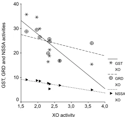
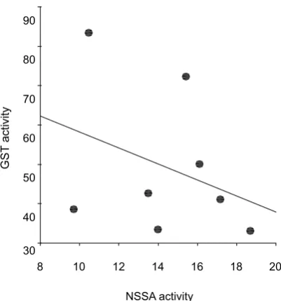
Lens XO activity in the IR group significantly increased compared to those of all other groups.Correlation analyses were shown in Table 2. As seen in Table 2, correlation analysis revealed a sig-nificant negative correlation between XO and NSSA (r = –0.893, p < 0.001), XOand GST (r = –0.83, p < 0.01), XO and GRD (r = –0.79, p < 0.05) in the IR group (Fig. 1). There were significant posi-tive correlations between GSTand NSSA (r = 0.78, p < 0.05), and GST and GRD (r = 0.71, p < 0.05] in the IR plus vitamin E group (Fig. 2) and IR plus L-carnitine group (Fig. 3), respectively. However, no correlation could be found among the param-eters in other groups.
Discussion
As the world’s population ages, cataract-in-duced visual dysfunction and blindness are on the
Table 1. Antioxidant (TSSA, NSSA, GRD, GST) and oxidative stress (XO) parameters in rat lenses
Tabela 1. Wskaźniki antyoksydantów (TSSA, NSSA, GRD, GST) i stresu oksydacyjnego (XO) w soczewkach szczurów
Control group
(Grupa kontrolna) IR group(Grupa IR) IR + Vitamin Egroup (Grupa IR plus witamina E)
IR + L-carnitine group
(Grupa IR plus LC)
TSSA – U/mg protein
(U/mg białka) 50.6 ± 11.6
a 35.2 ± 7.8d 64.3 ± 16.6c,d 63.5 ± 11.7c,d
NSSA – U/mg protein
(U/mg białka) 9.87 ± 1.76
a 7.30 ± 1.53d 14.38 ± 3.12c,e 12.81 ± 3.14c,e
GRD – mU/mg protein
(mU/mg białka) 33.9 ± 6.1
b 24.5 ± 4.3 33.7 ±5.2b 34.7 ± 8.3b
GST – mU/mg protein
(mU/mg białka) 37.9 ± 8.5
a 23.6 ± 8.4 49.3 ± 18.6b 46.7 ± 16.5c
XO – mU/mg protein
(mU/mg białka) 1.81 ± 0.20
a 2.36 ± 0.58 1.90 ± 049a 1.88 ± 0.25a
a – p < 0.05, b – p < 0.01, c – p < 0.001, vs. irradiation group. d – p < 0.05, e – p < 0.001, vs. control group.
increase. Cataracts are a major cause of blindness and of severe visual impairment leading to bilat-eral blindness in an estimated 20 million people worldwide. In developing countries, 50–90% of all blindness is caused by cataracts. Pharmacological treatment to prevent human cataracts have so far not been achieved. Therefore, surgery to remove the opacified lens is the only effective treatment for the cataract. The challenges are to prevent or delay cataract formation and also to treat cataracts if they occur. The exact mechanism of cataract for-mation is still not very clear [23].
The aim of radiation treatment is to deliver carefully determined doses of ionizing radiation to a defined tumor volume to eliminate tumor cells, to cause minimal injurious effects to surrounding healthy tissue given by eliminating tumor cells, giving a high quality of life and to prolong survival, all at a reasonable cost to cancer patients [3, 24]. But, cataract is an unavoidable complication if ra-diotherapy includes the orbit of eye in the treated volume, even with very low doses of radiation. Ionizing radiation, such as X and gamma rays and ultraviolet light, is known to be a cataractogenic factor for rat lenses [7, 25]. Multiple processes may lead to endothelial damage under irradiation but the generation of oxygen free radicals and the fol-lowing lipid peroxidation may be one of the key components in this cascade of events. Radiation generates ROS that interact with cellular mole-cules, including DNA, lipids, and proteins [6].
In the present study, irradiation caused a sig-nificant decrease in the activities of antioxidant enzymes and also increases in oxidant enzyme activities in rat lenses. These results are in agree-ment with the previous findings of Karslioglu et al. [7, 8], Kocer et al. [10] and Yagci et al. [23]. They reported a significant depletion in the antioxidant system accompanied by enhancement of lipid per-oxidation after irradiation. Under normal condi-tions the inherent defense system protects against oxidative damage.
In this study, we showed a significant reduction in GRD and GST activities in the IR group and also a significant increase in GRD and GST activities in the LC plus IR and IR plus vitamin E groups in rat lenses. This reduction in the IR group could be due to an enhanced utilization of glutathione re-dox cycle as an attempt to detoxify the free radicals generated by irradiation. Supplementation of LC and vitamin E protects the endogenous GRD, GST depletion resulting from irradiation. The increase in GRD, GST, TSSA and NSSA activities suggests that protection of LC and vitamin E may be medi-ated through the modulation of lens antioxidant system. These results suggest that these substances have a free radical scavenging activity. Some stud-ies have reported that LC and vitamin E
pretreat-Table 2. Spearman’s rank correlation coefficients in IR, IR plus Vitamin E and IR plus L-carnitine groups
Tabela 2. Korelacja współczynników Spearmana w grupach IR, IR plus witamina E i IR plus LC
IR group
(Grupa IR) IR + Vitamin E group(Grupa IR plus witamina E) IR + L-carnitine group(Grupa IR plus LC)
r = p < r = p < r = p <
XO-NSSA –0.93 0.001 – – – –
XO-GST –0.83 0.01 – – – –
XO-GRD –0.79 0.05 – – – –
GST-NSSA – – 0.78 0.05 – –
GST-GRD – – – – 0.70 0.05
Fig. 1. A negative correlation between lens tissue XO and NSSA (r = -0.893, p < 0.001), XOand GST (r = -0.83, p < 0.01), XO and GRD (r = -0.79, p < 0.05) in IR group
Ryc. 1. Korelacja negatywna między XO i NSSA (r = –0,893; p < 0,001), XOi GST (r = –0,83; p < 0,01), XO i GRD (r = –0,79; p < 0,05) w soczewkach w grupie IR
XO activity
4,0 3,5 3,0 2,5 2,0 1,5
GS
T,
GRD and NSSA
activities
40
30
20
10
0
GST XO
GRD XO
ment significantly lowered the radiation-induced lipid peroxidation in terms of malondialdehyde and increased antioxidant enzyme activities in rat lenses [8, 10]. The inhibition of lipid peroxidation in biomembranes can be caused by antioxidants. Significant increases in the levels of free radicals has been reported to be present in both the lens and the aqueous humor of cataract patients when compared with age-matched controls, emphasizing the role of oxidative damage in the pathogenesis of cataracts. A decrease in the antioxidant system could be responsible for increased lens oxidation and cataract development [10, 26–28].
A significant negative correlation was present between XO and such parameters as NSSA, GST, and GRD in the IR group. This result can be seen
in Table 2. However, a positive correlation was seen between GSTand NSSA in the IR plus vita-min E group, and GST and GRD in the IR plus L-carnitine group. These positive correlations might be indicators of the compensatory mecha-nism in these groups.
In conclusion, LC and vitamin E have clear an-tioxidant properties and are likely to be valuable drugs for protection against gamma-irradiation and/or to be used as antioxidants against oxidative stress. By increasing antioxidant enzyme activities and decreasing oxidant enzyme activities, LC and vitamin E prevented oxidative stress by scavenging free radicals generated by ionizing radiation in rat lenses. These results also show the need for further studies on this subject.
Fig. 2. A positive correlation between lens tissue GST and NSSA (r = 0.78, p < 0.05) in IR plus vitamin E group
Ryc. 2. Korelacja pozytywna między GSTi NSSA (r = 0,78; p < 0,05) w soczewkach w grupie IR plus witamina E
NSSA activity
20 18 16 14 12 10 8
GS
T
activity
90
80
70
60
50
40
30
Fig. 3. A positive correlation between lens tissue GST and GRD (r = 0.71, p < 0.05) in IR plus L-carnitine group
Ryc. 3. Korelacja pozytywna między GST i GRD (r = 0,71; p < 0,05) w soczewkach w grupie IR plus L-karnityna
GRD activity
50 40
30 20
GS
T
activity
80
70
60
50
40
30
20
References
[1] Taysi S, Koc M, Buyukokuroglu ME, Altinkaynak K, Sahin YN: Melatonin reduces lipid peroxidation and nitric oxide during irradiation-induced oxidative injury in the rat liver. J Pineal Res 2003, 34(3), 173–177.
[2] Nair CK, Parida DK, Nomura T: Radioprotectors in radiotherapy. J Radiat Res (Tokyo) 2001, 42(1), 21–37.
[3] Shirazi A, Ghobadi G, Ghazi-Khansari M: A radiobiological review on melatonin, a novel radioprotector. J Radiat Res (Tokyo) 2007, 48(4), 263–272.
[4] Taysi S, Memisogullari R, Koc M, Yazici AT, Aslankurt M, Gumustekin K et al.: Melatonin reduces oxidative stress in the rat lens due to radiation-induced oxidative injury. Int J Radiat Biol 2008, 84(10), 803–808.
[5] Chuyen NV, Arai H, Nakanishi T, Utsunomiya N: Are Food Advanced Glycation End Products Toxic in Biological Systems? Ann NY Acad Sci 2005, 1043, 467–473.
[6] Mansour HH: Protective role of carnitine ester against radiation-induced oxidative stress in rats. Pharmacol Res 2006, 54(3), 165–171.
[8] Karslioglu I, Ertekin MV, Taysi S, Kocer I, Sezen O, Gepdiremen A et al.: Radioprotective effects of melatonin on radiation-induced cataract. J Radiat Res (Tokyo) 2005, 46(2), 277–282.
[9] Merriam GR Jr, Focht EF: A clinical study of radiation cataracts and the relationship to dose. Am J Roentgenol Radium Ther Nucl Med 1957, 77(5), 759–785.
[10] Kocer I, Taysi S, Ertekin MV, Karslioglu I, Gepdiremen A, Sezen O et al.: The effect of L-carnitine in the pre-vention of ionizing radiation-induced cataracts, a rat model. Graefes Arch Clin Exp Ophthalmol 2007, 245(4), 588–594.
[11] Weiss JF, Landauer MR: Protection against ionizing radiation by antioxidant nutrients and phytochemicals. Toxicology 2003, 189(1–2), 1–20.
[12] Srinivasan V, Weiss JF: Radioprotection by vitamin E, injectable vitamin E administered alone or with WR-3689 enhances survival of irradiated mice. Int J Radiat Oncol Biol Phys 1992, 23(4), 841–845.
[13] Gulcin I: Antioxidant and antiradical activities of L-carnitine. Life Sci 2006, 78(8), 803–811.
[14] Yasui F, Matsugo S, Ishibashi M, Kajita T, Ezashi Y, Oomura Y et al.: Effects of chronic acetyl-L-carnitine treat-ment on brain lipid hydroperoxide level and passive avoidance learning in senescence-accelerated mice. Neurosci Lett 2002, 334(3), 177–180.
[15] Gul M, Demircan B, Taysi S, Oztasan N, Gumustekin K, Siktar E et al.: Effects of endurance training and acute exhaustive exercise on antioxidant defense mechanisms in rat heart. Comp Biochem Physiol A Mol Integr Physiol 2006, 143(2), 239–245.
[16] Oztasan N, Taysi S, Gumustekin K, Altinkaynak K, Aktas O, Timur H et al.: Endurance training attenuates exercise-induced oxidative stress in erythrocytes in rat. Eur J Appl Physiol 2004, 91(5–6), 622–627.
[17] Aktan B, Taysi S, Gumustekin K, Bakan N, Sutbeyaz Y: Evaluation of oxidative stress in erythrocytes of guinea pigs with experimental otitis media and effusion. Ann Clin Lab Sci 2003, 33(2), 232–236.
[18] Durak I, Canbolat O, Kacmaz M, Ozgen G, Ozturk HS: Antioxidant interferences in superoxide dismutase activ-ity methods using superoxide radical as substrate. Clin Chem Lab Med 1998, 36(6), 407–408.
[19] Carlberg I, Mannervik B: Glutathione reductase. Methods Enzymol 1985, 113, 484–490.
[20] Habig WH, Pabst MJ, Jakoby WB: Glutathione S-transferases. The first enzymatic step in mercapturic acid for-mation. J Biol Chem 1974, 249(22), 7130–7139.
[21] Hashimoto S: A new spectrophotometric assay method of xanthine oxidase in crude tissue homogenate. Anal Biochem 1974, 62(2), 426–435.
[22] Bradford MM: A rapid and sensitive method for the quantitation of microgram quantities of protein utilizing the principle of protein-dye binding. Anal Biochem 1976, 72, 248–254.
[23] Yagci R, Aydin B, Erdurmus M, Karadag R, Gurel A, Durmus M et al.: Use of melatonin to prevent selenite-induced cataract formation in rat eyes. Curr Eye Res 2006, 31(10), 845–850.
[24] Ertekin MV, Tekin SB, Erdogan F, Karslioglu I, Gepdiremen A, Sezen O et al.: The effect of zinc sulphate in the prevention of radiation-induced dermatitis. J Radiat Res (Tokyo) 2004, 45(4), 543–548.
[25] Cengiz M, Gurkaynak M, Atahan IL, Kilic K, Totan Y: The effect of verapamil in the prevention of radiation-induced cataract. Int J Radiat Oncol Biol Phys 1999, 43(3), 623–626.
[26] Spector A, Garner WH: Hydrogen peroxide and human cataract. Exp Eye Res 1981, 33(6), 673–681.
[27] Barros PS, Angelotti AC, Nobre F, Morales A, Fantoni DT, Barros SB: Antioxidant profile of cataractous English Cocker Spaniels. Vet Ophthalmol 1999, 2(2), 83–86.
[28] Fecondo JV, Augusteyn RC: Superoxide dismutase, catalase and glutathione peroxidase in the human cataractous lens. Exp Eye Res 1983, 36(1), 15–23.
Address for correspondence:
Seyithan Taysi
Department of Biochemistry and Clinical Biochemistry, Gaziantep University, School of Medicine
Gaziantep Turkey
Tel.: 90 342 360 16 17
E-mail: [email protected], [email protected]
Conflict of interest: None declared