Izabela Dereń-Wagemann, Małgorzata Kuliszkiewicz-Janus, Joanna Schiller
Ehlers-Danlos Syndrome
Zespół Ehlersa-Danlosa
Department of Hematology, Blood Neoplasms and Bone Marrow Transplantation, Wroclaw Medical University, Poland
Abstract
Ehlers-Danlos syndrome (EDS) is a group of genetically determined vascular purpura. The pathological changes mainly involve joints, skin and the vascular wall. The incidence of the EDS is 1 per 20 000–100 000 births. The defects involve the structure of collagen as well as other enzymes associated with it. Today there are the follow-ing types of EDS: classical, hypermobility, vascular, kyphoscoliotic, arthrochalasia and dermatosparaxis. The indi-vidual types of EDS are accompanied by complications of heart muscle, respiratory system, gastrointestinal tract, eyes, teeth, periodontal tissues and obstetric problems. The current paper presents issues relating to epidemiology, pathogenesis, clinical manifestations and treatment of various forms of Ehlers-Danlos syndrome (Adv Clin Exp Med 2010, 19, 4, 537–542).
Key words: Ehlers-Danlos syndrome, classification, symptoms.
Streszczenie
Zespół Ehlersa-Danlosa (ZED) to grupa genetycznie uwarunkowanych skaz naczyniowych. Zmiany dotyczą głównie stawów, skóry i ścian naczyń krwionośnych. Częstość występowania zespołu wynosi 1/20 000–100 000 urodzeń. Defekty dotyczą budowy kolagenu oraz związanych z nim enzymów. Obecnie rozróżnia się następujące postacie zespołu: klasyczną, z nadmierną ruchomością stawów, naczyniową, kyfoskoliotyczną, z wiotkością stawów oraz skórną. Poszczególnym postaciom ZED towarzyszą powikłania ze strony mięśnia sercowego, układu oddechowego, przewodu pokarmowego, oczu, zębów, tkanek okołozębowych oraz problemy położnicze. W pracy przedstawiono problemy dotyczące epidemiologii, patogenezy, objawów klinicznych oraz leczenia poszczególnych postaci zespołu Ehlersa-Danlosa (Adv Clin Exp Med 2010, 19, 4, 537–542).
Słowa kluczowe: zespół Ehlersa-Danlosa, klasyfikacja, objawy. Adv Clin Exp Med 2010, 19, 4, 537–542
ISSN 1230-025X
rEvIEWS
© Copyright by Wroclaw Medical University
* The work was partially funded from Medical University of Lodz resources in the years 2006–2009 as a research project No. N406 050 31/1860.
described it in 1901 and a French dermatologist Henri-Alexandre Danlos who described it in1908 [4, 5]. The symptoms of EDS were noticeable in famous Italian violinist Niccolo Paganini [6].
The first classifications of EDS were produced in late sixties 20th century. In 1986 the
terminol-ogy of types of EDS was formalized. Originally 11 types of EDS have been distinguished. The new, simplified classification of EDS was proposed in 1997, in villefranche-sur-Mer in France. Current-ly the EDS is divided into six types: (1) classical, (2) hypermobility, (3) vascular, (4) kyphoscoliotic, The EDS is one of the hereditary vascular
(5) arthrochalasia, (6) dermatosparaxis. The first three types of EDS are the most common [7]. Types not included in the currently used classifi-cation occur sporadically and were reclassified or removed from classification (Table 1).
The hereditary pattern of EDS is autosomal dominant, autosomal recessive or depending on the X chromosome.
The symptom of EDS is connective tissue anomaly that consists of disturbance of synthesis of perivascular collagen and enzymes connected with it: lysyl hydrolase that is necessary to cre-ate cross-bonding of hydroxylysine in collagen, N-proteinase type I procollagen which split ends telopeptides of collagen after they are separated in the extracellular space. The defects in collagen structure mainly affect its type v which is vessels, cornea, placenta and cartilage, type III which is the component of blood vessels, skin, intestinal wall and uterus and type I which is the component of bones, skin, tendon, ligament, sclera and cornea tissues, blood vessels, and hollow organs. The defi-ciency of lysyl hydrolase, that is necessary to create cross-bonding of hydroxylysine in collagen, was indicated in kyphoscoliosis type [8, 9].
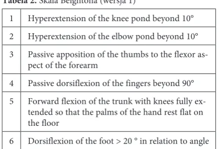
The excessive movement of joints is evaluated according to Beighton scale (Table 2). The scale has two versions. In the first version of the scale, there is an additional criterion for evaluating the
dorsi-flexion of foot. According to Beighton criteria, one can recognize the hypermobility of the joints when there are at least 3 to 6 possible symptoms [10].
For all the type of the Ehlers-Danlos syndrome, changes in the skin and joints are characteristic. The various types differ in intensity of cutaneous-articular symptoms. In the classical type the hy-permobility of joints occurs already in newborns, and the symptoms involve mainly the peripheral joints such as shoulder, knee, and a temporo-mandibular joint and spine. Children often have flat feet and scoliosis [10]. In the hypermobility type – they dominate, and skin symptoms are less pronounced. In the kyphoscoliotic type there is excessive, generalized loosening of joints, scolio-sis at birth, increasing with age, which may lead to respiratory and circulatory failure. The signifi-cant muscular hypotonia (called “floppy infant”) is also characteristic. However, in the vascular type excessive joint mobility affects only the joints of the fingers. Children with arthrochalasia type of-ten have congenital bilateral hip subluxation, there are also articular-musclar aches. In the classical type recurrent effusions in the ponds, floods to the articular cavities can be also observed [7, 11, 10]. Skin lesions include excessively stretch, thin, gauzy and atrophic skin. It easily comes to injuries and bruises. The healing of the wounds is disturbed. It comes to significant scarring. The wide scars
Table 1. Characteristics of each type of Ehlers-Danlos syndrome Tabela 1. Charakterystyka typów zespołu Ehlersa-Danlosa
Type (Typ) Previous naming
(Poprzednia nazwa)
Pattern of inheritance
(Wzór dziedziczenia) Abnormal protein (Nieprawidłowe białko) Abnormal gene (Nieprawidłowy gen)
Classical
(Klasyczny) type I/II AD collagen-type v and I COL5A1,COL5A2
Hypermobil-ity (Z nadmierną ruchomością stawów)
type III AD tenascin-X – in some unknown
vascular
(Naczyniowy) type Iv AD mainly collagen- type III COL3A1
Kyphoscoliotic
(Kifoskoliotyczny) type vI Ar lysyl hydroxylase defi-ciency PLOD
Arthrochalasia
(Z wiotkością stawów) type vII A/B AD collagen-type I COL1A1,COL1A2
Dematosparaxis
(Skórny) type vIIC Ar N-proteinase of type I procollagen ADAMTS2
with thin, parchment skin, in the Anglo-Saxon lit-erature known as “cigarette paper scars”, are very characteristic. They are mainly located in areas ex-posed to pressure such as elbows, knees, chin. The excessive extensibility of tissues may manifest as a symptom of Gorlin (the possibility of touching tip of the nose with the end of the tongue). There may be discoloration of the skin of the forehead, chin and legs. The dermatosparaxis type, in turn, is characterized by loss of elasticity of the skin and fascia. In the vascular type, symptoms affecting the skin are relatively poorly expressed [7, 11, 10].
Defects in collagen, other connective tissue proteins and related enzymes cause problems not only in the skin and joints, but also in many other tissues and organs in which connective tissue oc-curs. As is apparent from the available literature, in various types of EDS there are clinical symp-toms resulting from damage to other organs such as heart muscle, blood vessels, respiratory system, gastrointestinal tract, urinary bladder, skeletal sys-tem, eyes, periodontal tissue, amniotic membrane, and others.
In a classical, hypermobility, vascular and kyphoscloliotic type mitral valve prolapse flap can be observed. The expansion of the trunk of the aorta has been reported in the classical and hyper-mobility type [11, 10]. The symptoms associated with blood vessels are most dramatically expressed in the vascular type. It represents 5–10% of all the types of EDS [12]. Average survival of patients with this type of EDS is 50 years. Mainly large and medium-sized arteries are abnormal [12, 13]. Se-vere symptoms of arterial vessels and cavernosum organs are dominant in this type of EDS. These are mostly fatal hemorrhage from the aorta, iliac ar-tery, splenic arar-tery, liver, kidney, bleeding into the lung, the alveoli, to the chest as a result of rupture of the internal thoracic artery aneurysm and bleed-ing in pregnancy and childbirth due to rupture of the aorta, vena cava, uterus, intestines, liver, va-gina, perineum [10, 14–18]. The syndrome of
sud-den infant death due to subarachnoid hemorrhage has also been described [19] as well as the presence of spontaneous carotid-cavernosal fistulas, verte-bral artery and carotid artery dissections [20–25]. One of the symptoms of idiopathic carotid-cavern-ous fistula may be unilateral paralysis of the eyes and headache on the side of the fistula [24]. The vascular fragility occurs in the wall of the arteries but also intestines and uterus [7, 11, 10, 23, 26]. Symptoms affecting the skin and joints are here rather poorly expressed in comparison to other types of EDS. Spontaneous retroperitoneal bleed-ing is the most common cause of death in patients with type Iv EDS [27]. There is an increased risk of aneurysm [10]. There were some cases of abdomi-nal aortic aneurysms, intracranial arteries, reabdomi-nal arteries, hepatic artery [15, 20, 26, 28]. A patient developed hepatic artery aneurysms after splenec-tomy and an 8-month old child, giant aortic an-eurysm [29, 30]. The occurrence of hemorrhage is also possible in the kyphoscoliotic type of EDS. Wenstrup et al. described patients with rupture of the vertebral artery, multiple cracks in the femo-ral artery and two episodes of bleeding inside the chest [31]. In the classical and vascular type vari-cose veins can be observed [10, 32].
Spontaneous pneumothorax, spontaneous mediastinal emphysema, formation of the blis-ters under the pleura have been seen in the clas-sical and vascular type. In the vascular type the pneumothorax may be accompanied by bleeding into the pleural cavity and hemoptysis [10, 33]. In the vascular type the spontaneous rupture of the large intestine has been described and in the clas-sical type the diverticula of the bladder was found [34, 35]. In the Ehlers-Danlos syndrome urinary incontinence and prolapse of pelvic organ in wom-en has also bewom-en reported.
One of the skeletal-related changes observed in the kyphoscoliotic type is scoliosis that appears at an early age [31, 36, 37]. Distortion of the chest can cause respiratory and circulatory failure. Some cases of marfanoid habitus with arachnodactylia of the limbs have been also observed in this type. In the arthrochalasia type there are patients with short stature, round face and hypoplasia of the mandible [7, 10, 11, 36].
The pathological changes of the eye were ob-served in the kyphoscoliotic type. They included bleeding to the retina resulting in its detachment, to the vitreous humor, cracking of the cornea, rup-ture of the eyeball, subconjunctival lens disloca-tion after trauma. Such changes as a small cornea, cone of the cornea, cataracts, myopia, brown color of the sclera can also be observed [10, 36, 38–41]. The blue color of the sclera occurs in arthrochal-sia and kyphoscoliotic type [10, 40]. Badauy et al.
Table 2. Scale by Beighton (version I) Tabela 2. Skala Beightona (wersja 1)
1 Hyperextension of the knee pond beyond 10° 2 Hyperextension of the elbow pond beyond 10° 3 Passive apposition of the thumbs to the flexor
as-pect of the forearm
4 Passive dorsiflexion of the fingers beyond 90° 5 Forward flexion of the trunk with knees fully
ex-tended so that the palms of the hand rest flat on the floor
described the case of a 23-year old patient with vascular type, who lost 12 teeth. He also developed alveolar bone loss [32].
In the classical and kyphoscoliotic type, the absence of frenulum of the lower lip and tongue has been reported [10, 25, 42]. There are also char-acteristic facial features of infants with a vascu-lar type of Ehlers-Danlos syndrome, for example prominent eyes, caused by the deficiency of fat around the eyes. A small mouth, sharp nose, hol-low cheeks and a narrow auricle without petals are also characteristic [43].
There are few reports on the coexistence of the Ehlers-Danlos syndrome with other hamorrhagic diathesis [44, 45–50].
Actually in all types of EDS there is a risk as-sociated with pregnancy, including risk of uterine rupture, prematurity due to premature rupture of membranes [10, 17, 51]. In the vascular type of EDS, pregnancy-related mortality ranges from 11.5 to 25% [23]. All pregnancies should be treated as high-risk pregnancies. Cesarean section is recom-mended.
A case of difficult intubation in a patient with hypermobility type of EDS which probably was caused by the collapse of fibro-elastic tissues and cartilages of the trachea was described. The gen-eral anesthesia was performed for cesarean sec-tion due to prolonged second stage of labor [53]. Lind et al. studied a group of 46 pregnant women with Ehlers-Danlos syndrome. The most common complications were pelvic pain, instability of the pelvic, premature labor, postpartum hemorrhage, complicated perineal wounds [54]. It is also obvi-ous that during the pregnancy the pathology of the spine can increase and there can be difficulty in obtaining the vascular access.
Epidural anaethesia may be technically diffi-cult [55]. There is a risk of injury of the cervical spine during intubation due to lax ligaments of the neck and risk of pneumothorax during positive pressure ventilation [56]. various imaging stud-ies are used in diagnosing type Iv, for example CT scanning, MrI scanning, ultrasonography. In this type of EDS, classic arterography with cath-eterization should be avoided as it may cause an aneurysm or vessel rupture. Ultrastructural exami-nation of collagen fibrils is useful in diagnosis clas-sical and arthrochalasia type [7, 52].
The differential diagnosis should include dis-eases of the nervous system and muscles, Aarskog- -Scott syndrome, fragile X syndrome, congenital flaccidity of the skin (cutis laxa), osteogenesis im-perfecta and achondroplasia [7, 52]. It is possible to apply genetic testing in case of known molecular defects.
Currently there is no causal treatment. It is sug-gested that vascular interventions should be system-atically performed and not differed when it comes to acute complications such as rupture of the vessel. Brooke et al. described 40 patients with EDS who underwent 45 endovascular and 18 open procedures including embolization, angioplasty, arterial bypass and aortic aneurysm repair. 5-year survival free of complications was appropriately 85 and 54% [57]. In literature there are also cases of successful mitral and aortic valve replacement [58, 59].
It has been showed that desmopressin may be effective in bleeding associated with Ehlers-Danlos syndrome [60]. A case of bleeding in type Iv EDS which was successfully treated with rvIIa was de-scribed [61].
High dose of vitamin C (1–4 g/day) can be used in treatment the kyphoscoliotic type of EDS [7, 52].
References
[1] Szczeklik A: Choroby wewnętrzne. Medycyna praktyczna, Kraków 2006, 1765–1767.
[2] Dwie księgi Hipokratesa: I. O powietrzu, wodach i okolicach, przeł. Henryk Łuczkiewicz, Warszawa 1890.
[3] Czernogobow NA: Über einem Fall von Cutis laxa. Monatshefte für praktische Dermatologie (Hamburg) 1892, 14, 76.
[4] Ehlers EL: Cutis laxa. Neigung zu Heamorrhagien in der Haut. Lockering mehrerer Artikulationen. Dermatologische Zietschrift 1901, 173–174.
[5] Danlos H: Un cas de cutis laxa avec tumeurs par contusion des coudes et des genoux (xanthome juvénile pseudo-diabetique de MM Hallopeau et Macé de Lépinay). Biulletin de la Societé francaise de dermatologie et syphiligra-phie, Paris 1908, 19, 70–72
[6] Miranda CM, Navarette TL, Zúňiga NG: Niccolo Paganini: medical aspects of his life and work. rev Med Chil 2008, 136, 930–936.
[7] Beighton P, Paepe A, Steinman B, Tsipouras P, Wenstrup RJ: Ehlers-Danlose Syndromes: revised Nosology, villefranche, 1997. Am J Med Genet 1998, 77, 31–37.
[8] Chan TF, Poon A, Basu A, Addleman NR, Chen J, Phong A, Byers PH, Klein T, Kwok PY: Natural variation in four human collagen genes across an ethnically diverse population. Genomics 2008, 91, 307–314.
[10] Zimmermann-Górska I: Nadmierna ruchomość stawów a choroby reumatyczne. reumatologia 2007, 45, 397–403.
[11] Steiner RD: Ehlers-Danlos Syndrom. e-medicine 2008, Jun 30.
[12] Germain DP: Ehlers-Danlos syndrome type Iv. Orphanet J rare Dis 2007, 2, 32.
[13] Lee ST, Kim JA, Jang SY, Kim DK, Kim JW, Ki CS: A novel COL3A1 gene mutation in patient with aortic dis-sected aneurysm and cervical artery dissections. Heart vessels 2008, 23, 144–148.
[14] Yost BA, Vogelsang JP, Lie JT: Fatal hemoptysis in Ehlers-Danlos syndrome: old malady with a new curse. Chest 1995, 107, 1465–1467.
[15] Phan TG, Sakulsaengprapha A, Wilson M, Wing R: ruptured internal mammary artery aneurysm presenting as massive spontaneous haemothorax in a patient with Ehlers-Danlos syndrome (Letter). Aust New Zeal J Med 1998, 28, 210–211.
[16] Rudd NL, Nimrod C, Holbrook KA, Byers PH: Pregnancy complications in type Iv Ehlers-Danlos syndrome. Lancet 1983, 1, 1–3.
[17] Erez Y, Ezra Y, Rojansky N: Ehlers-Danlos type Iv in pregnancy. A case report and a literature review. Fetal Diagn Ther 2008, 23, 7–9.
[18] Combeer EL, Combeer AD: A rare cause of maternal death: liver and inferior vena cava rupture due to previously undiagnosed Ehlers-Danlos Syndrome type Iv. Eur J Anaesthesiol 2008, 25, 765–767.
[19] Byard RW, Keeley FW, Smith CR: Type Iv Ehlers-Danlos syndrome presenting as sudden infant death. Am J Clin Path 1990, 3, 579–582.
[20] North KN, Whiteman DAH, Pepin MG, Byers PH: Cerebrovascular complications in Ehlers-Danlos syndrome type Iv. Ann Neurol 1995, 38, 960–964.
[21] Fox R, Pope FM, Narcisi P, Nicholls AC, Kendall BE, Hourihan MD, Compston DAS: Spontaneous carotid-cavernous fistula in Ehlers-Danlos syndrome. J Neurol Neurosurg Psychiatry 1988, 51, 984–986.
[22] Hollands JK, Santarius T, Kirkpatrick PJ, Higgins JN: Treatment of a direct carotid-cavernous fistula in a patient with type Iv Ehlers-Danlos syndrome: a novel approach. Neuroradiology 2006, 48, 491–494.
[23] Pepin M, Schwarze U, Superti-Furga A, Byers PH: Clinical and genetic features of Ehlers-Danlose type Iv, the vascular type. N Eng J Med 2000, 673–680.
[24] Chuman H, Trobe JD, Petty EM, Scharze U, Pepin M, Byers PH, Deveikis JP: Spontaneous direct carotid-cavernous fistula in Ehlers-Danlos syndrome type Iv: two cases and a review of the literature. J Neuroophtalmol 2002, 22, 75–81.
[25] Perrinaud A, Matos M, Maruani A, Mondon K, Machet L: Absence of inferior labial or lingual frenula in Ehlers-Danlos syndrome: a new diagnostic criterion? Ann Dermatol venereol 2007, 134, 859–862.
[26] Ayuthaya RK, Patthamapasphong N, Sura T, Niumpradit N, Trachoo O: Ehlers-Danlos syndrome type Iv with gastric adenocarcinoma. J Med J Assoc Thai 2008, 91, 166–171.
[27] Soonawalla Z, Poe FM, Puntis M: Type Iv Ehlers-Danlos syndrome: a surgical emergency. Postgrad Med J 2002, 78, 922, 501–502, 506.
[28] Stone WM, Fowl RJ, Money SR: ruptured hepatic artery aneurysm: a novel approach to distal control. J vasc Surg 2007, 46, 574–575.
[29] Musumeci A, Minervini MI, Cintorino D, Gruttadauria S, Pipitone L, Alzetta M, Giovinetto A: Postoperative hepatic artery aneurysms development and remodeling in Ehlers-Danlos syndrome type Iv. Case report. Int Angiol 2008, 27, 166–169.
[30] Hetzar R, Delmo Walter EM, Meyer R, Alexi-Meskishvili V: Isolated giant aortic aneurysm in an infant: Ehlers-Danlos syndrome type Iv. Ann Thorac Surg 2008, 86, 632–634.
[31] Wenstrup RJ, Murad S, Pinnell SR: Ehlers-Danlos syndrome type vI: clinical manifestations of collagen lysyl hydroxylase deficiency. J Pediatr 1989, 115, 405–409.
[32] Badauy CM, Gomes SS, Sant’Ana Filho M, Chies JA: Ehlers-Danlos syndrome (EDS) type Iv: review of the lit-erature. Clin Oral Invest 2007, 11, 183–187.
[33] Ishiguro T, Takayanagi N, Kawabata Y, Mastsushima H, Yoshii Y, Harasawa K: Ehlers-Danlose Syndrome with recurrent spontaneous pneumothoraces and cavitary lesion on chest X-ray as the initial complications. Intern Med (Tokyo, Japan) 2009, 48, 717–722.
[34] Collins MH, Schwarze U, Carpentieri D, Kaplan P, Nathanson K, Meyer JS, Byers PH: Multiple vascular and Bowel ruptures in an Adolescent Male with Sporadic Ehlers-Danlos Syndrome Type Iv. Pediatr Dev Pathol 1999, 2, 86–93.
[35] Cuckow PM, Blackhall RJS, Mouriquand PDE: Huge bladder diverticula associated with Ehlers-Danlos syn-drome. J roy Soc Med 1994, 87, 290–291.
[36] Heim P, Raghunath M, Meiss L, Heise U, Myllyla R, Kohlschutter A, Steinmann B: Ehlers-Danlos syndrome type vI (EDS vI): problems of diagnosis and management. Acta Paediatr 1998, 87, 708–710.
[37] Krane SM, Pinnell SR, Erbe RW: Lysyl-protocollagen hydroxylase deficiency in fibroblasts from siblings with hydroxylysine-deficient collagen. Proc Nat Acad Sci 1972, 69, 2899–2903
[38] Chikamoto N, Teranishi S, Chikama T, Nishida T, Ohshima K, Hatsukawa Y: Abnormal retinal blood vessels in Ehlers-Danlos syndrome type vI. Jpn J Ophthalmol 2007, 51, 453–455.
[39] Cosar CB, Ceyhan N, Sevim S, Sakaoglu N, Sirvanci S, San T, Kurtkaya O: Corneal perforation with minor trauma: Ehlers-Danlos syndrome type vI. Acar Ophtalmic Surg Lasers Imaging 2005, 36, 3.
[41] Nowakowski T, Dembińska B, Ryś D, Musiał J: Ehlers-Danlos syndrome type vII-case report. Pol Arch Med Wewn 1997, 98, 446–451.
[42] De Felice C, Toti P, Di Maggio G, Parrini S, Bagnoli F: Absence of the inferior labial and lingual frenula in Ehlers-Danlos syndrome. Lancet 2001, 357, 10–12.
[43] Parapia LA, Jackson C: Ehlers-Danlos syndrome – a historical review. Br J Haematol 2008, 141, 32–35.
[44] Kaliyadan F, Namboothiri S: Type Iv Ehlers-Danlose syndrome associated with factor vIII deficiency. Indian J Dermatol. venereol Leprol 2008, 74, 664–666.
[45] Anstey A, Mayne K, Winter M, Van de Pette J, Pope FM: Platelet and coagulation studies in Ehlers-Danlos syn-drome. Br J Dermatol 1991, 125, 155–163.
[46] Bertin P, Gaillard S, Desproges-Gotteron R: Ehlers-Danlos syndrome, clotting disorders and muscular dystro-phy. Ann rheum Dis 1989, 48, 953–956.
[47] Cunnif C, Williamson-Kruse L: Ehlers-Danlos syndrome, type vIII presenting with periodontitis and prolonged bleeding time. Clin Dysmorphol 1995, 4, 145–149.
[48] Udén A: Collagen and bleeding diathesis in Ehlers-Danlos syndrome. Scand J Haematol 1982, 28, 425–430.
[49] Clough V, MacFarlane IA, O’Connor J, Wood JK: Acquired von Willebrand’s syndrome and Ehlers-Danlos syndrome presenting with gastro-intestinal bleeding. Scand J Haematol 1979, 22, 305–310.
[50] Suzuki T, Tsuchiyama Y, Kawaguchi Y, Sougawa T, Nakamura H, Kinoshita T: Anesthesia for a patient with Ehlers-Danlos syndrome and factor XIII deficiency. Masui 1989, 38, 1518–1521.
[51] Barabas AP: Ehlers-Danlos syndrome associated with prematurity and premature rupture of foetal membranes. Br Med J 1966, 2, 682–684.
[52] Levy HP: Ehlers-Danlose syndrome, Hypermobility type. Genereviews http://www.ncbi.nlm.nih.gov
[53] Sood V, Robinson DA, Suri J: Difficult intubation during rapid sequence induction in a partiurent with Ehlers-Danlose syndrome, hypermobility type. Int Obstet Anesth 2009, 18, 4, 408–412.
[54] Lind J, Wallenburg HC: Pregnancy and the Ehlers-Danlos syndrome: a retrospective study in Dutch population. Gynecol Scand 2002, 8, 4, 223–300.
[55] Sybert V: Selected hereditary diseases. In: Textbook of Neonatal Dermatology, eds. Eichenfield L, Frieden I, Esterly N. Philadelphia, Pa: Saunders, 2001, 458–459.
[56] Dill-Russell P, St. John Jones L: Anaesthesia for caeserean section in patient with Ehlers-Danlos syndrome and mitra valve prolapse. Int J Obstet Anesth 2001, 10, 192–197.
[57] Brooke BS, Arnaoutakis G, McDonnell NB, Black J.H.3rd: Contemporary management of vascular complications
associated with Ehlers-Danlos syndrome. J vasc Surg 2010, 51, 1, 131–138.
[58] Takano H, Miyamoto Y, Sawa Y, Fukushima N, Matsumiya G, Fujita T, Matsuda H: Successful mitral valve replacement in patient with Ehlers-Danlos syndrome type vI. Ann Thorac Surg 2005, 80, 320–322.
[59] Edmondson P, Nellen M, Ross DN: Aortic valve replacement in case of Ehlers-Danlos syndrome. Br Heart J 1979, 42, 1, 103–105.
[60] Mast KJ, Nunes ME, Ruyman FB, Kerlin BA: Desmopressin responsiveness in children with Ehlers-Danlos syn-drome associated bleeding symptoms. B J Haematol 2009, 144, 2, 230–233.
[61] Faber P, Craig WL, Duncan JL, Holliday K: The successful use of recombinant factor vIIa in a patient with vas-cular type Ehlers-Danlos syndrome. Acta Anaesthesiol Scand 2007, 51, 9, 1277–1279.
Address for correspondence:
Izabela Dereń-Wagemann
Department and Clinic of Hematology, Blood Neoplasms and Bone Marrow Transplantation Wroclaw Medical University
Pasteura 4 50-367 Wrocław Poland
Tel.: +48 71 784 26 04 E-mail: [email protected] Conflict of interests: None declared