DOI
10.17219/acem/69802
Copyright
© 2018 by Wroclaw Medical University This is an article distributed under the terms of the Creative Commons Attribution Non-Commercial License (http://creativecommons.org/licenses/by-nc-nd/4.0/)
Address for correspondence
Lobna Ben Mahmoud
E-mail: benmahmoud_lobna@medecinesfax.org
Funding sources
None declared
Conflict of interest
None declared
Received on November 13, 2016 Reviewed on February 19, 2017 Accepted on March 27, 2017
Abstract
Background. Methotrexate (MTX) is a key component of acute lymphoblastic leukemia (ALL) therapy, but it is associated with serious toxicities in a considerable number of patients.
Objectives. The aim of the current study was to determine which variables were associated with MTX toxicity in children, adolescents and young adults with ALL.
Material and methods. In this prospective study, 35 patients with newly diagnosed ALL, treated ac-cording to the 58951 European Organization for Research and Treatment of Cancer – Children’s Leukemia Group (EORTC-CLG) protocol, were prospectively enrolled. Toxicity data was collected objectively after each high-dose methotrexate (HD-MTX) course. The risk factors of MTX toxicity were determined using multiple linear regression analysis, with age, gender, immunophenotype, risk group, plasma MTX levels, plasma homocysteine (HCY) levels, and MTHFR C677T included as independent variables.
Results. Twenty-five (71.4%) patients experienced toxicity on at least 1 course of HD-MTX. In the univariate linear regression, the global toxicity score was associated with a significant rise in plasma HCY concentrations within 48 h after MTX administration (β = 0.4; R2 = 0.12; p = 0.02). In the multiple regression model, the global toxicity score was significantly associated with a higher MTX plasma levels at 48 h (β = 0.5; R2 = 0.38; p = 0.001) and CT 677 MTHFR genotype (β = 0.3; R2 = 0.38; p = 0.01).
Conclusions. Routine monitoring of plasma MTX concentrations is essential to detect patients at a high risk of MTX toxicity. MTHFR C677T genotyping may be useful for predicting MTX toxicity.
Key words: methotrexate, acute lymphoblastic leukemia, MTHFR C677T polymorphism, toxicity
Use of
MTHFR C677T
polymorphism and plasma
pharmacokinetics to predict methotrexate toxicity
in patients with acute lymphoblastic leukemia
Lobna Ben Mahmoud
1,A−F, Moez Mdhaffar
2,A−D,F, Rim Frikha
3,A,B, Hanen Ghozzi
1,A−C,
Ahmed Hakim
1,A, Zouheir Sahnoun
1,A, Moez Elloumi
2,A,E, Khaled Zeghal
1,A,B,E,F1 Department of Pharmacology, Faculty of Medicine, University of Sfax, Tunisia 2 Department of Hematology, Hedi Chaker University Hospital, Sfax, Tunisia 3 Department of Histology, Faculty of Medicine, University of Sfax, Tunisia
A – research concept and design; B – collection and/or assembly of data; C – data analysis and interpretation; D – writing the article; E – critical revision of the article; F – final approval of the article
Introduction
Acute lymphoblastic leukemia (ALL) is the most com-mon pediatric cancer; its survival rate has improved, with 5-year event-free survival (EFS) rates of 70–80% and overall cure rates of 80%.1,2Such an improvement in the treatment
outcome is largely due to the advances in chemotherapy. Methotrexate (MTX), an antifolate chemotherapeutic agent, plays an important role in the chemotherapy regi-men for ALL and has significantly reduced the recurrence rate of ALL in children.3
Methotrexate is predominantly taken up into cells via the reduced folate carrier (RFC).4 Inside the cell, MTX
is con-verted to its active polyglutamate forms (methotrexate poly-glutamates – MTXPGs).5 Both MTX and MTXPGs inhibit
dihydrofolate reductase, an enzyme that catalyzes the con-version of dihydrofolate to its active form tetrahydrofolate (THF), a substrate of thymidylate synthase (TS), to convert deoxyuridine monophosphate to deoxythymidine-5’-mo-nophosphate, resulting in DNA synthesis.6 Tetrahydrofolate
deficiency leads to the depletion of intracellular folates, and thereby to decreased synthesis of both purines and pyrimidines, contributing to the inhibition of nucleic acid synthesis and favoring cell death.7 Methotrexate
polyglu-tamates can also interfere with methylenetetrahydrofolate reductase (MTHFR), which converts 5,10-methylene-THF to 10-methyl-THF, the major circulating form of folate that provides a methyl group for homocysteine (HCY) methyla-tion to methionine, and channels the methyl group into DNA and protein methylation reactions.6
A high-dose methotrexate (HD-MTX) refers to infused MTX in doses of more than 1 g/m2.8 The use of HD-MTX
has shown great benefit in the treatment of childhood ALL and the prevention of extramedullary leukemia, i.e., central nervous system (CNS) leukemia and testicular leukemia.2
However, MTX is associated with various toxicities, includ-ing severe mucositis, myelosuppression, gastrointestinal toxicity, hepatic toxicity, neurotoxicity, and hematologi-cal toxicity, requiring a dose reduction and the interrup-tion of chemotherapy, and subsequently an increased risk of relapse.9 Methotrexate-related toxicity remains
a com-mon and often unpredictable clinical problem, because of a wide interindividual variation in pharmacokinetics and pharmacodynamics of this drug.8
The aim of the current study was, therefore, to deter-mine factors associated with the high risk of MTX toxicity that could help to develop personalized therapies in chil-dren, adolescents and young adults with ALL.
Material and methods
Patients and study design
From January 2013 to December 2014, 35 patients with newly diagnosed ALL were prospectively enrolled from
the Hematology Department of Hedi Chaker University Hospital (Sfax, Tunisia). The diagnosis of ALL was based on morphologic, cytochemical and immunophenotypical criteria.
Patients were selected according to the following in-clusion criteria: availability of clinical data, treatment according to the European Organization for Research and Treatment of Cancer – Children’s Leukemia Group (EORTC-CLG) 58951 protocol, administration of at least 1 course of intravenous MTX chemotherapy, and no his-tory of other active malignancies requiring a modification of chemotherapy regimen.10
Patients were stratified into 4 risk groups (low-risk − LR; average risk 1 − AR1; average risk 2 − AR2, and high-risk − HR) on the basis of their presenting clinical features, the biologic features of their leukemic cells, and their early response to remission-induction treatment.10,11
This study was conducted in accordance with the Hel-sinki Declaration and informed consent was given by all the persons participating in this study.
Treatment
All patients were treated according to the 58951 EORTC-CLG protocol, a Berlin−Frankfurt−Munster-like trial, with treatment phases including induction (IA), consolidation (IB/IB9), CNS prophylaxis without cranial irradiation, late intensification II, and maintenance.11,12
According to this protocol, the infusion of HD-MTX was given intravenously in each course at 5 g/m2 body
surface area (BSA) over a 4-hour period. Intravenous hy-dration and urinary alkalinization were performed 1 day before HD-MTX administration, and continued during and after MTX infusion. Leucovorin rescue (25 mg/m2)
was administered every 6 h, starting at 24 h after the ini-tiation of HD-MTX infusion.
High-dose methotrexate infusions were administered during the interval therapy to all patients, in the induc-tion phase to AR2/HR-ALL, in the consolidainduc-tion phase to HR-ALL and in the R1-R2 Bloc to HR-ALL.
Plasma methotrexate and homocysteine
levels determination
In each course of HD-MTX, blood samples were collect-ed in ethylencollect-ediaminetetraacetic acid (EDTA)-containing tubes at the following times: at 24, 48 and 72 h from the start of intravenous MTX infusion. Plasma was recuper-ated by centrifugation at 3000 rpm for 10 min and was stored at −20°C for the determination of MTX and HCY levels.
MTHFR C677T
genotyping
The MTHFR C677T polymorphism was determined
in 28 patients with ALL and 70 healthy subjects taken from the general population (35 males and 35 females, age range: 18–29 years). DNA was extracted from peripheral blood samples and polymerase chain reaction-restriction frag-ment length polymorphism (PCR-RFLP) was performed for the molecular diagnosis of the C677T MTHFR poly-morphism. The primers, lengths and restriction enzymes have been described previously.15 The 677C → T base pair
substitution creates a Hinf1 restriction site. Then, CC genotype would be reflected by a single band of 265 bp, CT genotype by 3 bands of 265, 171, and 94 bp, and TT genotype by 2 bands of 171 and 94 bp.
Toxicity
Toxicity data obtained from questionnaires and case records were prospectively collected objectively after each HD-MTX course, in the period from the end of HD-MTX infusion to the next HD-MTX course or until 14 days after HD-MTX infusion.
The toxicity was graded according to the Common Ter-minology Criteria for Adverse Events (CTCAE, v. 5.0).16
According to the modified method of Radtke et al., the global toxicity score for each patient was calculated in our study by adding up the grading of all adverse events that occurred during courses of MTX.17 This score integrates
both frequency and severity of MTX toxicity. In the study, the higher grade of toxicity in each HD-MTX course was considered.
Statistical analysis
All statistical analyses were performed using the IBM SPSS Statistics v. 20 software (Chicago, USA). Quantitative variables were expressed as means and standard error of the mean (SEM); they were compared using the t-test or the Wilcoxon-Mann-Whitney test according to the char-acteristics of the distribution. Qualitative variables were presented as a total number and proportion, and were compared using the χ2 test or Fisher’s exact test
accord-ing to sample sizes.
To identify the risk factors of MTX toxicity, a multi-variate linear regression model was constructed, using variables identified from the univariate analysis, which included age, gender, immunophenotype, risk group, MTX plasma levels, HCY plasma levels, and MTHFR C677T. A stepwise selection was used with probability value of p < 0.2 for entry and p < 0.05 for removal. For all com-parisons, differences were considered statistically signifi-cant at p < 0.05.
Results
Patient characteristics and plasma levels
of methotrexate and homocysteine
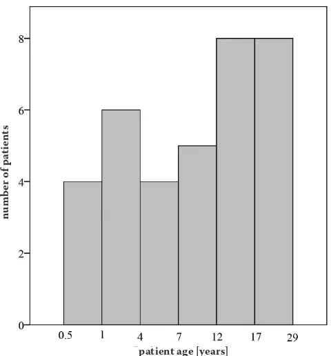
A total of 35 patients (21 males and 14 females) with newly diagnosed ALL treated with the 58951 EORTC-CLG protocol were enrolled in this study. The mean patient age at diagnosis was 11.7 ±9 years (median: 10.1 years; range: 0.5−29 years) (Fig. 1). Patients’ characteristics were shown in Table 1.
According to risk stratification, 12 patients were from AR1 group, 13 from AR2 group and 10 from HR group. A total of 173 courses of HD-MTX at 5 g/m2 were
administered.
In the study, 168 plasma MTX levels and 125 plasma HCY levels were analyzed. The results of these analyses are presented in Table 1.
Methotrexate levels ranged from 0.9 to 13 µmol/L at 24 h, from 0.02 to 6.7 µmol/L at 48 h and from 0.01 to 0.9 µmol/L at 72 h. Homocysteine levels ranged from 8 to 19 µmol/L at 24 h, from 5 to 15.5 µmol/L at 48 h and from 0.13 to 17 µmol/L at 72 h.
High-dose methotrexate-related toxicity
Of 35 patients, 25 (71.4%) experienced toxicity on at least 1 course of HD-MTX. The mean of global toxicity score for these patients was 5.1 ±2.1 (range: 1–12). A total of 59 cases of MTX-related toxicity were observed among 173 courses of HD-MTX (on average, 2 cases of toxicity per patient; range: 1–9) (Table 1).
Fig. 1. Age distribution of patients
patient age [years]
nu
m
be
r o
f p
at
ie
nt
Hepatic and gastrointestinal toxicities were the most fre-quently observed toxicities, accounting for 57.6% of the total number of observed toxicities. Methotrexate-induced hepa-totoxicity was expressed by elevated aspartate aminotrans-ferase/alanine aminotransferase (ASAT/ALAT). There were 55 cases of toxicity (93.2%) with grade 2 or greater, and only 4 cases of toxicity with grade 1 (6.8%) (Table 1).
Plasma methotrexate levels
and methotrexate-related toxicity
The univariate linear regression revealed that the global toxicity score was significantly associated with plasma levels of MTX at 24 h (β = 0.64; R2 = 0.41; p = 0.001),
48 h (β = 0.43; R2 = 0.19; p = 0.01) and 72 h (β = 0.37;
R2 = 0.14; p = 0.03) (Table 2).
Methotrexate plasma levels ≥10 µmol/L at 24 h were significantly associated with higher HCY plasma levels at 24 and 48 h after the initiation of MTX infusion (Table 3).
As shown in Table 3, the global toxicity score was sig-nificantly correlated with MTX levels >10 µmol/L at 24 h, 1 µmol/L at 48 h and 0.1 µmol/L at 72 h. Gastrointesti-nal and skin toxicity were significantly associated with high MTX plasma levels 48 and 72 h after MTX infusion (Table 3).
Plasma homocysteine levels
and methotrexate-related toxicity
The univariate linear regression revealed a positive cor-relation between global toxicity score and plasma HCY
Table 2. Risk factors for MTX toxicity (univariate linear regression)
Patients
p-value (Fisher's exact test,
global)
β β0 R² (%)
Age at diagnosis [years] 0.45 0.13 3.1 1.7
Gender (M vs F) 0.78 0.04 3.1 0.2
Immunophenotype (T vs B) 0.67 −0.07 4.5 0.6
Weight 0.37 0.15 2.7 2.4
Height 0.4 0.1 1.5 2.1
BSA 0.35 0.16 2.3 2.6
Risk group (AR1, AR2, HR) 0.14 0.2 0.5 0.6 Plasma levels of MTX
24 h 48 h 72 h 10–3 0.01 0.03 0.64 0.43 0.37 0.9 2.4 2.5 41.1 19.1 14.3 Plasma levels of HCY
24 h 48 h 72 h 0.14 0.02 0.18 0.28 0.4 –0.31 2.4 2.1 1.3 4.5 12.8 4.8
MTHFR C677T (CT vs CC) 0.02 0.4 1.8 19 BSA − body surface area; MTX − methotrexate; HCY − homocysteine; M − males; F − females; AR − average risk; HR − high risk; β − regression coefficient; β0 − intercept coefficient.
Table 1. Characteristics of patients, their clinical condition and toxicity experienced
Characteristics of patients Total number of patients (n = 35) Age at diagnosis [years],
mean ±SEM (min–max) <10 years, n (%) ≥10 years, n (%) 11.7 ±1.6 (0.5–29) 17 (48.6) 18 (51.4) Gender male, n (%)
female, n (%) 21 (60)14 (40)
Immunophenotype B-ALL, n (%)
T-ALL, n (%) 23 (65.7)12 (34.3)
Weight [kg], mean ±SEM
(min–max) 41.5 ±4 (11–60)
Height [cm], mean ±SEM (min–max)
138 ±5.5 (80–183) BSA [m2], mean ±SEM (min–max) 1.2 ±0.1 (0.5–2)
Risk group AR1, n (%) AR2, n (%) HR, n (%) 12 (34.3) 13 (37.1) 10 (28.6)
MTX-related toxicity Total number of patients (n = 35)* Total number of HD-MTX courses (n = 173) Hepatotoxicity, n (%) grade 2 grade 3 grade 4 13 (37.1) 7 4 2 17 (9.8) 9 6 2 Gastrointestinal toxicity, n (%)
grade 1 grade 2 grade 3 10 (28.6) 1 3 7 17 (9.8) 1 9 7 Mucositis, n (%) grade 3 grade 4 6 (17.1) 3 4 8 (4.6) 3 5 Neurotoxicity, n (%) grade 1 grade 3 grade 4 3 (8.6) 1 2 2 5 (2.9) 1 2 2 Skin toxicity, n (%)
grade 1 grade 2 grade 3 6 (17.1) 1 1 4 6 (3.4) 1 1 4 Hematotoxicity, n (%) grade 1 grade 4 2 (2.7) 1 1 2 (1.1) 1 1 Renal toxicity, n (%)
grade 2 1 (2.8)1 1 (0.6)1
Phlebitis, n (%) 2 (2.7) 3 (1.7)
Plasma MTX levels [µM] Total number (n = 168) 24 h (n = 66), mean ±SEM, 48 h (n = 54), mean ±SEM 72 h (n = 48), mean ±SEM 5.6 ±0.6 1.3 ±0.3 0.1 ±0.02 Plasma HCY levels [µM] Total number (n = 125) 24 h (n = 49), mean ±SEM 48 h (n = 48), mean ±SEM 72 h (n = 28), mean ±SEM 12.2 ±30.6 10 ±0.5 8 ±0.8
Table 3. Correlation between folate pathway and MTX toxicity
Plasma HCY and MTX toxicity
Plasma MTX
MTHFR
at 24 h (n = 31) at 48 h (n = 32) at 72 h (n = 30) <10 µM
(n = 25) ≥10 µM(n = 6) (n = 18)<1 µM (n = 14)≥1 µM <0.1 µM(n = 19) ≥0.1 µM(n = 11) (n = 22)CC CT (n = 6) Plasma HCY at 24 h [µM], mean ±SEM 11.6 ±0.6 15 ±1.8* 12.2 ±0.8 12.3 ±1.3 12 ±0.7 13 ±1.3 12 ±0.7 13.3±2 Plasma HCY at 48 h [µM], mean ±SEM 8.8 ±0.5 11.6 ±0.9* 9.2 ±0.6 10 ±0.7 9.1 ±0.6 10 ±0.8 8.2 ±0.5 10 ±0.9 Plasma HCY at 72 h [µM], mean ±SEM 8.5 ±0.8 6.3 ±4 9.6 ±1 7 ±1.1 8.7 ±1 8.2 ±1.7 8.1 ±0.9 5.9 ±3 Global toxicity, mean ±SEM 2.5 ±0.5 7.5 ±1.6* 1.6 ±0.5 6.3 ±0.7* 1.5 ±0.4 6.7 ±0.8* 2.5 ±0.6 6.1 ±1.7*
Gastrointestinal toxicity (n) 8 3 4 9† 4 7† 8 3
Mucositis (n) 2 2 1 4 1 5† 2 1
Nausea/vomiting/diarrhea (n) 4 3 2 7† 3 4 4 3
Abdominal pain (n) 3 1 2 3 1 3 4 1
Liver toxicity (n) 6 5 6 6 6 6 5 4
Renal toxicity (n) 0 1 0 1 0 1 0 1
Neurotoxicity (n) 2 2 1 3 1 1 1 2
Seizure (n) 1 0 0 1 0 0 1 0
Somnolence (n) 1 0 1 0 1 0 0 1
Paralysis (n) 0 1 0 1 0 1 − −
Agitation (n) 1 0 0 1 0 0 1 0
Anxiety/mood disorder (n) 0 1 0 1 0 0 0 1
Skin toxicity (n) 4 3 1 6† 2 5† 3 2
Hematotoxicity (n) 2 0 2 0 1 1 2 0
Thrombocytopenia (n) 1 0 1 0 1 0 1 0
Hemorrhage (n) 1 0 1 0 0 1 1 0
Phlebitis (n) 1 1 1 1 1 1 1 1
MTX − methotrexate; HCY − homocysteine; SEM − standard error of the mean; * statistically significant differences between groups estimated using Mann-Whitney U test; † statistically significant differences between groups estimated using Fisher’s exact test.
Fig. 2. Association of liver toxicity and gastrointestinal toxicity with plasma homocysteine levels Each column represents mean with standard deviation (±SD); HCY − homocysteine; * p = 0.02.
at 24 h
at 24 h at 48 h
at 48 h at 72 h
at 72 h
no no no
no no no
yes yes yes
yes yes
yes
pl
as
m
a l
ev
el
s o
f H
C
Y a
t 2
4 h [
m
m
ol
/L
]
pl
as
m
a l
ev
el
s o
f H
C
Y a
t 2
4 h [
m
m
ol
/L
]
pl
as
m
a l
ev
el
s o
f H
C
Y a
t 4
8 h [
m
m
ol
/L
]
pl
as
m
a l
ev
el
s o
f H
C
Y a
t 4
8 h [
m
m
ol
/L
]
pl
as
m
a l
ev
el
s o
f H
C
Y a
t 7
2 h [
m
m
ol
/L
]
pl
as
m
a l
ev
el
s o
f H
C
Y a
t 7
2 h [
m
m
ol
/L
]
gastrointestinal toxicity gastrointestinal toxicity
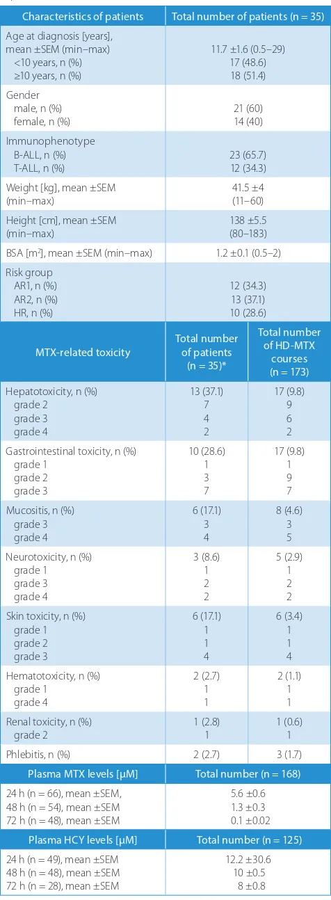
levels within 48 h after MTX administration (β = 0.4; R2 = 0.12; p = 0.02) (Table 2). Figure 2 shows that HCY
plasma levels at 48 h were significantly higher in patients with gastrointestinal toxicity (p = 0.02).
Gene polymorphisms
and methotrexate-related toxicity
The MTHFR C677T polymorphism was
in Hardy-Wein-berg equilibrium. MTHFR 677C>T was of the wild type (CC) in 22 patients (78.6%) and 70 controls (100%), and heterozygous (CT) in 6 (21.4%).
The influence of this polymorphism on MTX toxicity was analyzed in 28 patients with ALL included in this study. The mean global toxicity score was significantly higher in MTHFR 677 CT genotype than in wild genotype (p = 0.02) (Tables 2,3).
There was no significant association between MTHFR
677 C>T polymorphism and plasma MTX levels.
Risk factors for methotrexate toxicity
In a multiple regression model, the global toxicity score was significantly associated with 2 variables: higher MTX plasma levels at 48 h (≥1µmol/L) (β = 0.5; R2 = 0.38;
p = 0.001) and CT 677 MTHFR genotype (β = 0.3; R2 = 0.38;
p = 0.01).
Discussion
Intravenous HD-MTX is a key component in the therapy of ALL.3 However, despite leucovorin rescue with
hydra-tion and urinary alkalinizahydra-tion, MTX is associated with serious toxicities in a considerable number of patients.15
This could lead to the interruption of treatment, which may increase relapse risk.
This study identified several clinical variables that influ-ence MTX toxicity in patients with ALL treated according to the EORTC-CLG 58951 protocol. We used the global toxicity score that integrates both severity and frequency of MTX toxicity during 173 HD-MTX courses.
One of the major limitations of the current study was the small sample size; studies with a greater number of pa-tients would be necessary to confirm our results. However, a homogenous diagnosis, a standardized treatment pro-tocol followed by all patients, objective and well-recorded toxicity data make the results credible.
High-dose methotrexate was associated with toxicities in the majority of patients included in this study (71.1%). Consistent with previous studies,we found that the most common side effects following HD-MTX therapy were hepato-, skin and gastrointestinal toxicity, particularly the oral mucositis.18
This toxicity is unpredictable because of large in-ter-patient variability in the pharmacokinetics and
pharmacodynamics of this drug, even with the same treat-ment protocol.5,7,18 The mechanism of MTX-induced
toxic-ity could be mainly explained by an inhibition of normal cells and tissue adjacent to the target abnormal cells.8
In this study, acute MTX-induced hepatotoxicity was expressed by elevated ASAT/ALAT and was observed in 37.1% of patients. The pathophysiology of this side ef-fect remains unclear. Holmboe et al. suggested that 7-OH-MTX, a main metabolite of 7-OH-MTX, was involved in the de-velopment of HD-MTX hepatic toxicity in patients with osteosarcoma treated with HD-MTX.19
Oral mucositis was the most severe MTX-related toxic-ity observed in this study. Considerable effort has been expended to identify the etiopathophysiology of this side effect.20,21 Pico et al. reported that MTX may be secreted
in the saliva, leading to increased direct mucotoxicity.22
In the present study, we analyzed the relation between MTX pharmacokinetics and MTX-related toxicities during HD-MTX courses. In multiple linear regression analysis, we found that plasma MTX levels at 48 h were signifi-cantly correlated with the global toxicity score. Currently, few studies among patients with ALL have reported that the plasma levels of MTX may influence the risk of MTX toxicity.23−25
In the current study, acute MTX-induced hepatotoxicity, which was the most common side effect, was not associ-ated with plasma MTX levels. This can be explained by the fact that the small number of children in this study could influence its power to detect a significant association.
We found that the risk of oral mucositis was signifi-cantly associated with high MTX plasma levels 72 h after drug infusion (Table 3). This finding is consistent with those of Cheng, who revealed that 64% of children with oral mucositis had plasma MTX levels above the defined upper limit of the expected profile at 66 h.25 It is rather
remarkable to find a higher frequency of nausea/vomiting episodes in patients with high MTX plasma levels at 48 h. Although the exact mechanism is unclear, it was reported that nausea/vomiting can lead to dehydration, causing de-creased glomerular filtration rates, and thus limited renal clearance of MTX.24
It was also reported that MTX-related toxicity might be explained through the disruption of folate homeosta-sis.26 In this study, we determined the plasma HCY levels,
since it was considered a sensitive marker of deficient folate homeostasis.27
We found that elevated levels of HCY were associated with higher MTX plasma levels at 24 h, which is consistent with the previous study.28 This can be explained by the
in-terference of MTX with the metabolism of HCY by reduc-ing the level of 5-methyl-THF, which serves as the donor of the methyl group for the methylation of HCY to me-thionine. As a result, the levels of HCY increase, whereas the levels of methionine decrease.29
a significant rise in plasma HCY levels within 48 h after MTX administration. Although a strong association be-tween blood levels of HCY and the risk of the development of CNS disorders has been shown, there are few studies reporting such an association with gastrointestinal toxic-ity.30−32 Hyperhomocysteinemia may induce cell damage
through a number of complex mechanisms, including in-terference with the methylation process and disturbance of oxidative stress balance.33−35
Therefore, an increased level of HCY might be consid-ered a sensitive marker of MTX toxicity.
The MTHFR gene is located at the end of the short arm of chromosome 1 (1p36.3), and the encoded protein, MTH-FR, is a key enzyme in folate metabolism.36,37 The C677T
single nucleotide polymorphism (SNP) is the most studied polymorphism in the MTHFR gene and results in an ala-nine-to-valine substitution at codon 222. Its variant alleles cause a substantial reduction of the MTHFR enzyme ac-tivity in vitro compared with the wild type allele.38 People
with a heterozygous MTHFR 677 CT genotype have 60% enzyme activity compared with those with the wild-type allele.39 In the present study, we investigated whether
there exists an influence of MTHFR C677T polymor-phism on MTX-related toxicity. We found a significantly increased risk of MTX-related toxicity in patients with 677CT genotype compared with the wild genotype 677CC. This result was consistent with previous studies.40,41
A me-ta-analysis of studies concerning the toxicity of low-dose MTX (10–15 mg/week) in rheumatoid arthritis suggested that C677T polymorphism was significantly associated with increased toxicity.40 Another meta-analysis in ALL patients,
including 21 articles published before September 2010, sup-ported this association and suggested that the 677T allele serves as a toxicity predictor during treatment with MTX.41
In conclusion, the results of our study suggest that routine monitoring of plasma MTX levels during 48–72 h is essen-tial to detect patients at a high risk of developing toxicity and to adjust leucovorin rescue and hydration. Moreover, we suggest that MTHFR C677T genotyping may be use-ful for predictingMTX toxicity. Future studies with large sample sizes should be undertaken to verify current find-ings, which may provide further biomarkers of treatment efficacy and toxicity in patients with ALL.
References
1. Pui CH, Evans WE. Treatment of acute lymphoblastic leukemia. N Engl J Med. 2006;354:166–178.
2. Pui CH, Relling MV, Downing JR. Acute lymphoblastic leukemia.
N Engl J Med. 2004;350:1535–1548.
3. de Beaumais TA, Jacqz-Aigrain E. Intracellular disposition of metho-trexate in acute lymphoblastic leukemia in children. Curr Drug Metab. 2012;13:822–834.
4. Assaraf YG. Molecular basis of antifolate resistance. Cancer Metasta-sis Rev. 2007;26:153–181.
5. Mikkelsen TS, Thorn CF, Yang JJ, et al. PharmGKB summary: Metho-trexate pathway. Pharmacogenet Genomics. 2011;21:679–686. 6. Bagley PJ, Selhub J. A common mutation in the
methylenetetrahy-drofolate reductase gene is associated with an accumulation
of for-mylated tetrahydrofolates in red blood cells. Proc Natl Acad Sci USA. 1998;95:13217–13220.
7. Ongaro A, De Mattei M, Della Porta MG, et al. Gene polymorphisms in folate metabolizing enzymes in adult acute lymphoblastic leuke-mia: Effects on methotrexate-related toxicity and survival. Haema-tologica. 2009;94:1391–1398.
8. Treon SP, Chabner BA. Concepts in use of high-dose methotrexate therapy. Clin Chem. 1996;42:1322–1329.
9. Tantawy AA, El-Bostany EA, Adly AA, et al. Methylene tetrahydrofo-late reductase gene polymorphism in Egyptian children with acute lymphoblastic leukemia. Blood Coagul Fibrinolysis. 2010;21:28–34. 10. Vilmer E, Suciu S, Ferster A, et al.; Children Leukemia Cooperative
Group. Long-term results of three randomized trials (58831, 58832, 58881) in childhood acute lymphoblastic leukemia: A CLCG-EORTC report. Leukemia. 2000;14:2257–2266.
11. De Moerloose B, Suciu S, Bertrand Y, et al. Improved outcome with pulses of vincristine and corticosteroids in continuation therapy of children with average risk acute lymphoblastic leukemia (ALL) and lymphoblastic non-Hodgkin lymphoma (NHL): Report of the EORTC randomized phase 3 trial 58951. Blood. 2010;116:36–44. 12. Domenech C, Suciu S, De Moerloose B, et al. Dexamethasone (6 mg/m2/
day) and prednisolone (60 mg/m2/day) were equally effective as
induc-tion therapy for childhood acute lymphoblastic leukemia in the EORTC CLG 58951 randomized trial. Haematologica. 2014;99:1220–1227. 13. Nirenberg A, Mosende C, Mehta BM, Gisolfi AL, Rosen G.
High-dose methotrexate with citrovorum factor rescue: Predictive value of serum methotrexate concentrations and corrective measures to avert toxicity. Cancer Treat Rep. 1977;61:779–783.
14. Paci A, Veal G, Bardin C, et al. Review of therapeutic drug monitoring of anticancer drugs. Part 1: Cytotoxics. Eur J Cancer. 2014;50:2010–2019. 15. Ayad MW, El Naggar AA, El Naggar M. MTHFR C677T polymorphism: Association with lymphoid neoplasm and effect on methotrexate therapy. Eur J Haematol. 2014;93:63–69.
16. National Institutes of Health. National Cancer Institute CTEP CTCAE v. 5.0. 2016. http://ctep.cancer.gov/. Accessed November 27, 2017. 17. Radtke S, Zolk O, Renner B, et al. Germline genetic variations
in meth-otrexate candidate genes are associated with pharmacokinetics, toxicity, and outcome in childhood acute lymphoblastic leukemia.
Blood. 2013;121:5145–5153.
18. Schmiegelow K. Advances in individual prediction of methotrexate toxicity: A review. Br J Haematol. 2009;146:489–503.
19. Holmboe L, Andersen AM, Mørkrid L, Slørdal L, Hall KS. High dose methotrexate chemotherapy: Pharmacokinetics, folate and toxicity in osteosarcoma patients. Br J Clin Pharmacol. 2012;73:106–114. 20. Sonis ST. Mucositis as a biological process: A new hypothesis for the
development of chemotherapy-induced stomatotoxicity. Oral Oncol. 1998;34:39–43.
21. Sonis ST, Elting LS, Keefe D, et al.; Mucositis Study Section of the Mul-tinational Association for Supportive Care in Cancer, International Society for Oral Oncology. Perspectives on cancer therapy-induced mucosal injury: Pathogenesis, measurement, epidemiology, and consequences for patients. Cancer. 2004;100:1995–2025.
22. Pico JL, Avila-Garavito A, Naccache P. Mucositis: Its occurrence, con-sequences, and treatment in the oncology setting. Oncologist. 1998;3: 446–451.
23. Rask C, Albertioni F, Bentzen SM, Schroeder H, Peterson C. Clinical and pharmacokinetic risk factors for high-dose methotrexate-induced toxicity in children with acute lymphoblastic leukemia − a logistic regression analysis. Acta Oncol. 1998;37:277–284.
24. Relling MV, Fairclough D, Ayers D, et al. Patient characteristics associ-ated with high-risk methotrexate concentrations and toxicity. J Clin Oncol. 1994;12:1667–1672.
25. Cheng KK. Association of plasma methotrexate, neutropenia, hepatic dysfunction, nausea/vomiting and oral mucositis in children with cancer. Eur J Cancer Care (Engl). 2008;17:306–311.
26. Cole PD, Beckwith KA, Vijayanathan V, Roychowdhury S, Smith AK, Kamen BA. Folate homeostasis in cerebrospinal fluid dur-ing therapy for acute lymphoblastic leukemia. Pediatr Neurol. 2009;40:34–41.
28. Kubota M, Nakata R, Adachi S, et al. Plasma homocysteine, methio-nine and S-adenosylhomocysteine levels following high-dose meth-otrexate treatment in pediatric patients with acute lymphoblastic leukemia or Burkitt lymphoma: Association with hepatotoxicity. Leuk Lymphoma. 2014;55:1591–1595.
29. Tufekci O, Yilmaz S, Karapinar TH, et al. A rare complication of intra-thecal methotrexate in a child with acute lymphoblastic leukemia.
Pediatr Hematol Oncol. 2011;28:517–522.
30. Seshadri S, Wolf PA, Beiser AS, et al. Association of plasma total homo-cysteine levels with subclinical brain injury: Cerebral volumes, white matter hyperintensity, and silent brain infarcts at volumetric mag-netic resonance imaging in the Framingham Offspring Study. Arch Neurol. 2008;65:642–649.
31. Bottiglieri T. Homocysteine and folate metabolism in depression.
Prog Neuropsychopharmacol Biol Psychiatry. 2005;29:1103–1112. 32. Haagsma CJ, Blom HJ, van Riel PL, et al. Influence of sulphasalazine,
methotrexate, and the combination of both on plasma homocyste-ine concentrations in patients with rheumatoid arthritis. Ann Rheum Dis. 1999;58:79–84.
33. Crider KS, Yang TP, Berry RJ, Bailey LB. Folate and DNA methylation: A review of molecular mechanisms and the evidence for folate’s role.
Adv Nutr. 2012;3:21–38.
34. Ientile R, Curro’ M, Ferlazzo N, Condello S, Caccamo D, Pisani F. Homo-cysteine, vitamin determinants and neurological diseases. Front Biosci
(Schol Ed). 2010;2:359–372.
35. Papatheodorou L, Weiss N. Vascular oxidant stress and inflammation in hyperhomocysteinemia. Antioxid Redox Signal. 2007;9:1941–1958. 36. Goyette P, Sumner JS, Milos R, et al. Human methylenetetrahydro-folate reductase: Isolation of cDNA, mapping and mutation identi-fication. Nat Genet. 1994;7:195–200.
37. Kim YI. Folate and carcinogenesis: Evidence, mechanisms, and impli-cations. J Nutr Biochem. 1999;10:66–88.
38. Yamada K, Chen Z, Rozen R, Matthews RG. Effects of common polymorphisms on the properties of recombinant human meth-ylenetetrahydrofolate reductase. Proc Natl Acad Sci USA. 2001;98: 14853–14858.
39. Robien K, Ulrich CM. 5,10-methylenetetrahydrofolate reductase poly-morphisms and leukemia risk: A HuGE minireview. Am J Epidemiol. 2003;157:571–582.
40. Fisher MC, Cronstein BN. Metaanalysis of methylenetetrahydrofolate reductase (MTHFR) polymorphisms affecting methotrexate toxicity.
J Rheumatol. 2009;36:539–545.