RESEARCH ARTICLE
Physiological and molecular responses of the goldfish (
Carassius
auratus
) kidney to metabolic acidosis, and potential mechanisms
of renal ammonia transport
Michael J. Lawrence1,2,*, Patricia A. Wright3and Chris M. Wood1,4
ABSTRACT
Relative to the gills, the mechanisms by which the kidney contributes to ammonia and acid–base homeostasis in fish are poorly understood. Goldfish were exposed to a low pH environment ( pH 4.0, 48 h), which induced a characteristic metabolic acidosis and an increase in total plasma [ammonia] but reduced plasma ammonia partial pressure (PNH3). In the kidney tissue, total ammonia, lactate and intracellular pH
remained unchanged. The urinary excretion rate of net base under control conditions changed to net acid excretion under low pH, with contributions from both the NH4+(∼30%) and titratable acidity minus
bicarbonate (∼70%; TA–HCO3−) components. Inorganic phosphate
(Pi), urea and Na+ excretion rates were also elevated while Cl−
excretion rates were unchanged. Renal alanine aminotransferase activity increased under acidosis. The increase in renal ammonia excretion was due to significant increases in both the glomerular filtration and the tubular secretion rates of ammonia, with the latter accounting for ∼75% of the increase. There was also a 3.5-fold increase in the mRNA expression of renal Rhcg-b (Rhcg1) mRNA. There was no relationship between ammonia secretion and Na+ reabsorption. These data indicate that increased renal ammonia secretion during acidosis is probably mediated through Rhesus (Rh) glycoproteins and occurs independently of Na+transport, in contrast to
branchial and epidermal models of Na+-dependent ammonia transport in freshwater fish. Rather, we propose a model of parallel H+/NH
3
transport as the primary mechanism of renal tubular ammonia secretion that is dependent on renal amino acid catabolism.
KEY WORDS: Renal excretion, Renal tubule, Urine pH, Urine flow rate, Glomerular filtration rate, Amino acid catabolism, Rhesus glycoproteins, Na+transport, Acid–base regulation
INTRODUCTION
Under normal physiological conditions, the contribution of the kidney to whole-body ammonia excretion in freshwater teleost fish is minimal, typically representing <20% of total N-excretion, with >80% excreted at the gills (Smith, 1929; McDonald and Wood, 1981; Zimmer et al., 2014). However, metabolic acidosis dramatically elevates renal ammonia excretion in some species (McDonald and Wood, 1981; McDonald, 1983; King and Goldstein, 1983; Wood et al., 1999). Renal ammonia excretion
represents the shuttling of acid equivalents, in the form of NH4+(or
NH3+H+) from the body of the fish to the urine for subsequent
excretion, with the return of HCO3−to the extracellular fluid. This
mechanism may become quantitatively more important than titratable acid (TA) excretion in its contribution to total urinary acid excretion during metabolic acidosis in fish (McDonald and Wood, 1981; McDonald, 1983; King and Goldstein, 1983; Wood et al., 1999). In mammals, the majority of NH4+excreted in the urine
results not from filtration, but rather from elevated amino acid catabolism in the renal tubular cells, a process that produces equimolar amounts of NH4+for secretion and HCO3−for restoration
of extracellular pH (Knepper et al., 1989; Atkinson, 1992; Wright et al., 1992; Curthoys, 2001).
In teleosts, the limited renal studies to date suggest that similar mechanism(s) may be at play during metabolic acidosis. Activities of renal ammoniogenic enzymes increase while plasma ammonia concentrations rise only marginally in both trout and goldfish (King and Goldstein, 1983; Wood et al., 1999). However, elevated renal secretion of ammonia, and the cellular and molecular mechanisms by which it may occur, have not yet been directly demonstrated in teleosts.
In mammals, renal epithelial ammonia transport in the collecting duct (CD) of the nephron is facilitated by Rhesus (Rh) glycoproteins (Weiner, 2004), and in fish, recent evidence suggests that the same is true in the gills (Nakada et al., 2007b; Hung et al., 2007; Nawata et al., 2007; reviewed in Wright and Wood, 2009). These proteins appear to function as channels for the translocation of ammonia gas (NH3) along favourable partial pressure gradients (Knepper and
Agre, 2004; Javelle et al., 2007; Nawata et al., 2010). In the teleost gill, ammonia excretion is mediated by a Na+/NH
4+ exchange
complex or metabolon consisting of several key transporters (Rh proteins, V-type H+-ATPase, Na+/H+ exchanger), which excrete
NH3+H+while simultaneously facilitating active Na+uptake from
the water, thereby contributing to systemic ionic and acid–base homeostasis (Wright and Wood, 2009). However, information on the mechanisms of renal ammonia transport and acid–base regulation in fish is sparse. To date, mRNA transcripts of the Rh family glycoprotein Rhbg and multiple isoforms of Rhcg have been found in the kidney of the common carp (Wright et al., 2014) and mangrove killifish (Hung et al., 2007), while Rhbg expression has been reported in the trout kidney (Nawata et al., 2007). Immunohistochemical techniques have localized Rhcg-b (formerly termed Rhcg1 or Rhcg1b, see http://zfin.org/search? q=rhcg for latest terminology) to the CD and distal tubule of the zebrafish (Nakada et al., 2007a). Basolateral Na+/K+-ATPase
(NKA) and apical Rhcg-b have been co-localized in the distal tubule of zebrafish (Nakada et al., 2007a). Cooper et al. (2013) demonstrated a similar profile in the distal tubule of the mangrove killifish where Rhcg-b and a Na+/H+exchanger (NHE3) are on the
Received 27 January 2015; Accepted 5 May 2015 1
Department of Biology, McMaster University, Hamilton, ON, Canada L8S 4K1.
2
Department of Biology, Carleton University, Ottawa, ON, Canada K1S 5B6.
3
Department of Integrative Biology, University of Guelph, Guelph, ON, Canada N1G 2W1.4Department of Zoology, University of British Columbia, Vancouver, BC, Canada V6T 1Z4.
*Author for correspondence (m_lawrence27@live.ca)
The
Journal
of
Experimental
apical membrane with a basolateral NKA. Apical NHE3 also co-localizes with an apical V-type H+-ATPase (HAT) and basolateral
NKA in the proximal tubules of rainbow trout (Ivanis et al., 2008a). These observations suggest the presence of a Na+-coupled
mechanism of renal ammonia secretion similar to that of the gills (Wright and Wood, 2009). However, the only study to examine this issue found an elevation in urine [ammonia] and increased expression of Rhcg-a (formerly Rhcg1a or Rhcg3) and Rhcg-b mRNA in the kidney of the common carp during metabolic acidosis, yet there was no relationship between urinary ammonia excretion and Na+ excretion (Wright et al., 2014). But, interpretation was
confounded by a simultaneous decrease in urine flow rate under acid exposure such that there was no increase in urinary ammonia excretion in this species.
Therefore, the goal of the present study was to characterize the physiological, biochemical and molecular responses of the kidney to metabolic acidosis and develop a working model of renal ammonia transport in a freshwater teleost. We used the goldfish, Carassius auratus (Linnaeus 1758), the species in which renal ammonia excretion was first studied directly (King and Goldstein, 1983) and for which a suite of molecular probes are now available (Bradshaw et al., 2012; Sinha et al., 2013). We hypothesized that elevated urinary ammonia excretion during metabolic acidosis would be predominantly mediated by increased renal tubular secretion, rather than by increased glomerular filtration. Secondly, in light of the possible linkage of ammonia secretion via Rh proteins to Na+
reabsorption discussed earlier, we hypothesized that increased expression of apical Rh glycoprotein (Rhcg-a and Rhcg-b) mRNA, decreased urinary Na+ excretion, and increased renal tubular
Na+ reabsorption would occur. To test these hypotheses, low
environmental pH (4.0) was employed as a tool for inducing a sustained metabolic acidosis; in this circumstance the gills take up rather than excrete acidic equivalents, and the kidney becomes the sole route of net acid excretion (McDonald and Wood, 1981; McDonald, 1983; King and Goldstein, 1983; Wood et al., 1999; Wright et al., 2014). Animals were fitted with urinary bladder catheters and exposed to control pH 8.2 or acid pH 4.0 water. Urine was collected over successive 12 h intervals for 48 h, then blood and renal tissue were terminally sampled. The measurements yielded a detailed analysis of the response of the goldfish kidney to metabolic acidosis in vivo, including excretion, filtration, secretion and reabsorption rates of ammonia, TA–HCO3−, inorganic phosphate (Pi), Na+, Cl−and urea,
together with comparable plasma measurements, mRNA expression levels of potential transporters, and renal enzymatic activities.
RESULTS
Blood and tissue parameters
Exposure to pH 4.0 for 48 h lowered blood pH by almost 0.4 units, and plasma [HCO3−] by 40%, but had no significant effect on
plasma partial pressure of CO2 (PCO2) (Table 1). Plasma total
[ammonia] (Tamm) and [NH4+] were increased by about 60%, while
plasmaPNH3was lowered by 28%. Plasma lactate and glucose were
unresponsive to acid exposure, but plasma cortisol and urea were 81% and 78% higher, respectively, compared with control values (Table 1). Acid exposure also resulted in a significantly lower plasma Na+ concentration by approximately 23%, while both
plasma Piand Cl−were unchanged (Table 1).
In the kidney, intracellular pH was maintained at a constant value of about 6.9 (Table 2) despite the marked plasma acidosis. Similarly, renal tissue lactate, total ammonia and NH4+
concentrations, as well as tissue PNH3, were not significantly
altered (Table 2).
Urinary responses
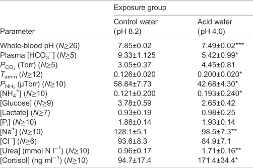
[image:2.612.312.563.79.246.2]In control fish, urine flow rate (UFR) increased significantly over 48 h. Fish exposed to acid demonstrated a higher UFR, relative to controls, over the first 36 h of exposure (Fig. 1A). Changes in UFR were significantly influenced by both time (P<0.001) and treatment (P=0.001) with a significant interaction (P<0.008) between the two. Table 1. Blood and plasma parameters for goldfish exposed to control and acidic water conditions for 48 h
Exposure group
Parameter
Control water (pH 8.2)
Acid water (pH 4.0)
Whole-blood pH (N≥26) 7.85±0.02 7.49±0.02*** Plasma [HCO3−] (N≥5) 9.33±1.125 5.42±0.99*
PCO2(Torr) (N≥5) 3.05±0.37 4.45±0.81
Tamm(N≥12) 0.126±0.020 0.200±0.020*
PNH3(µTorr) (N≥10) 58.84±7.73 42.68±4.30* [NH4
+
] (N≥10) 0.121±0.200 0.193±0.240* [Glucose] (N≥9) 3.78±0.59 2.65±0.42 [Lactate] (N≥7) 0.93±0.19 0.98±0.25
[Pi] (N≥10) 1.88±0.14 1.93±0.14
[Na+] (N≥10) 128.1±5.1 98.5±7.3**
[Cl−] (N≥6) 93.6±8.3 84.9±7.1
[Urea] (mmol N l−1) (N≥10) 0.96±0.17 1.71±0.16** [Cortisol] (ng ml−1) (N≥10) 94.7±17.4 171.4±34.4*
PCO2, partial pressure of CO2;Tamm, total ammonia;PNH3, partial pressure of ammonia; Pi, inorganic phosphate.
Means±1 s.e.m.; asterisks (*P<0.05, **P<0.01 and ***P<0.001) denote significant differences versus control fish. Unless otherwise noted, all values are expressed as mmol l−1.
List of symbols and abbreviations [3H]PEG-4000 polyethylene glycolMr4×10 3
CD collecting duct GFR glomerular filtration rate
HAT H+-ATPase
MS-222 tricaine methane sulfonate NHE Na+/H+-exchanger NKA Na+/K+-ATPase
PCO2 partial pressure of CO2 Pi inorganic phosphate
PNH3 partial pressure of ammonia gas
Rh Rhesus
RT-qPCR real-time quantitative PCR TA–HCO3− titratable acid minus bicarbonate
[image:2.612.311.566.634.726.2]Tamm total ammonia UFR urine flow rate UT urea transporter
Table 2. Acid–base status and nitrogen metabolism parameters in the kidney of goldfish exposed to control and acidic water conditions for 48 h
Renal parameter
Control water (pH 8.2)
Acid water (pH 4.0)
Intracellular pH (N≥6) 6.89±0.03 6.95±0.06 Whole-tissue ammonia
(mmol kg−1) (N=6)
1.38±0.14 1.26±0.23
PNH3(µTorr) (N=6) 72.39±9.65 84.98±24.11 [NH4
+
] (mmol kg−1) (N=6) 1.37±0.14 1.25±0.23 Whole-tissue lactate
(mmol kg−1) (N=6)
0.92±0.01 0.77±0.01
Means±1 s.e.m. No significant differences.
The
Journal
of
Experimental
Urine pH was significantly lower in the acid group, within the first 12 h, relative to the control group, and this difference was maintained throughout the 48 h acid exposure (Fig. 1B). Urine pH progressively decreased only in acid-exposed fish whereas this trend was not significant in the controls (Fig. 1B). Treatment (P<0.001) and time (P<0.001) both had a significant influence but no interaction was demonstrated.
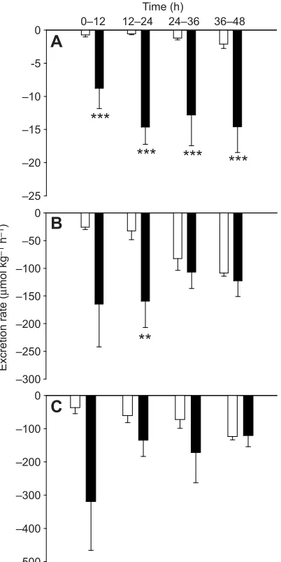
In Fig. 2A, by convention (see Materials and methods), negative values represent net acid excretion, and positive values net base excretion. In the controls, the kidney excreted base at a relatively constant rate, whereas acid excretion was apparent from 12 to 24 h of low pH exposure onwards (Fig. 2A). Treatment was the only significant influence (P<0.001).
Both ammonia (Fig. 2B) and TA–HCO3− excretion rates
(Fig. 2C) contributed to the increase in net acid excretion rate (Fig. 2A), but the change in the TA–HCO3−component made the
larger contribution (70% versus 30%). TA–HCO3−excretion was
reversed in acid-exposed fish relative to controls by 12–24 h of exposure (Fig. 2C). Urinary Pi excretion rate was significantly
elevated throughout the acid exposure, reaching values substantially higher than in the controls (Fig. 2D). Treatment (P<0.001) was the only contributing factor. Increases in TA–HCO3− excretion
correlated with elevations in Piexcretion (supplementary material
Fig. S1) (P<0.001,r2=0.282,N=37).
Urinary ammonia excretion increased significantly in the first 12 h of acid exposure, and this difference (4- to 10-fold higher than controls) was maintained throughout (Fig. 2B). In both groups, ammonia excretion increased over time. Both time (P<0.001) and treatment (P<0.001) had significant influences, but there was no interaction between the two.
Urinary urea excretion was elevated up to 12-fold over control rates throughout the 48 h acid-exposure period (Fig. 3A), an effect influenced by both treatment (P<0.001) and time (P=0.035). Urinary Na+excretion was variable over time in both control and
acid-exposed groups, but Na+excretion rates (influenced only by
treatment,P<0.001) were significantly higher in acid-exposed fish at 12–24 h (Fig. 3B). Urine [Na+] did not significantly correlate with
urinary [ammonia] (P=0.922, r2=0.00, N=51) (supplementary
material Fig. S2). Cl−excretion was not significantly affected by acid exposure (Fig. 3C).
Renal enzyme activity
Goldfish exposed to pH 4.0 exhibited significantly higher activity of alanine aminotransferase (2.7-fold) relative to control fish (Table 3). Despite trends towards higher aspartate aminotransferase (P=0.08) and glutamate dehydrogenase (P=0.06) activities, no other renal enzyme was significantly altered (Table 3). Glutaminase was undetectable.
Filtration, secretion and reabsorption
In the subset of fish employed for glomerular filtration rate (GFR) measurements, values over time were averaged to facilitate overall comparison of the control versus acid-exposure treatments (see Materials and methods). These data were used to determine mean rates of filtration, secretion and reabsorption (=negative secretion) of fluid and metabolites entering the urine. Increases in UFR and GFR in this subset were not significant (Fig. 4) so fluid filtration and reabsorption were not appreciably altered (Fig. 5A). Under control conditions, ammonia was both filtered and secreted at relatively low rates (Fig. 5B). In acid-exposed fish, there were significant elevations in the rates of both ammonia filtration (∼2-fold) and ammonia secretion (∼6-fold), and thus the latter made the larger contribution (∼75% versus 25%) to the elevation in ammonia excretion rate (Fig. 5B).
The filtration rates of Pi, Na+ and Cl−were not significantly
different between control and acid-exposed fish, and Na+and Cl−
reabsorption rates were also not affected (Fig. 5C–E). Urea filtration was elevated in acid-exposed fish (2.6-fold) relative to controls, while the reduction in urea reabsorption rate was not significant (Fig. 5F). Together, these accounted for the large increase in urea excretion rate during metabolic acidosis. Pi
reabsorption was significantly reduced to approximately zero under acidosis, explaining the substantial elevation in Pi
excretion rate. Renal Na+ reabsorption did not correlate with
renal ammonia secretion (P=0.644, r2=0.00, N=9; data not
shown).
mRNA expression
Rhcg-a, Rhcg-b and Rhbg were all expressed at the mRNA level in goldfish kidney. Rhcg-b mRNA was significantly elevated by 3.5-fold under acidosis relative to control conditions, while Rhcg-a and Rhbg did not change significantly (Fig. 6). While the expression of NKA and urea transporter (UT) were unaltered, HAT declined by
∼50%. The mRNA transcripts of NHE3 and NHE2 were below the limit of detection.
Time (h)
pH
6.0 6.5 7.0 7.5 8.0 8.5 9.0
b
b
a,b
a
**
***
***
***
0–12 12–24 24–36
B
Flow rate (ml kg
–1
h
–1
)
0 2 4 6 8 10 12
*
***
***
a a
b
b
A
36–48
[image:3.612.62.287.337.661.2]0–12 12–24 24–36 36–48
Fig. 1. Urine flow rate and urine pH of goldfish exposed to control and low environmental pH over 48 h.Control, pH 8.2 (white bars); low environmental pH, pH 4.0 (black bars). (A) Urine flow rate (N≥5); (B) urine pH (N≥5). Means+1 s.e.m. Asterisks denote significant differences (***P<0.001, **P<0.01, *P<0.05) between control and acid-exposed fish at a specific time interval whereas different letters denote significant differences (P<0.05) within a
treatment group.
The
Journal
of
Experimental
DISCUSSION Overview
Goldfish exposed to water of pH 4.0 exhibited a classic metabolic acidosis with renal compensation by increased urinary excretion of acidic equivalents in the form of both TA–HCO3− and NH4+. In
contrast to previous studies in which interpretation was confounded by a decrease in UFR (Wright et al., 2014) and/or an absence of ammonia filtration and secretion measurements (McDonald and Wood, 1981; Wood et al., 1999; King and Goldstein, 1983), the present study clearly revealed the pathways of increased urinary ammonia excretion. In confirmation of our first hypothesis, the elevation in urinary NH4+excretion was predominately mediated
through increased renal tubular secretion (∼75%), though increased glomerular filtration of ammonia also made a significant contribution (∼25%). However, our second hypothesis was only partially supported. As predicted, there was an upregulation of Rhcg-b mRNA expression and increased ammoniogenic enzyme activity in the kidney, but there was no evidence linking increased NH4+ secretion to increased Na+ reabsorption. Thus, elevated
tubular ammonia secretion is probably mediated through Rh glycoproteins in goldfish, similar to the mammalian CD (reviewed in Weiner and Verlander, 2014), but unlike the fish gill, there may be no obligatory coupling to Na+counter-transport. Fig. 7
provides an overview diagram.
Secretion of ammonia
In contrast to the common carp, which shut down UFR during acid exposure (Wright et al., 2014), goldfish tended to increase UFR, though by 36–48 h, the overall increase was not significant relative to simultaneous controls (Fig. 1). A similar situation prevailed in the subset of fish in which UFR and GFR were measured, with both increasing non-significantly (Fig. 4). Thus, our estimates of the magnitude of changes in both filtration and secretion/reabsorption rates during acid exposure are if anything conservative. Renal ammonia secretion was markedly elevated (∼6-fold), whereas there was a smaller increase (∼2-fold) in ammonia filtration rate
(Fig. 5B). Our results help clarify those of King and Goldstein (1983) who observed increased urinary ammonia excretion during pH 4.0 exposure in the same species, but did not partition it into filtration versus secretion components. In our study, unlike in that of King and Goldstein (1983), plasma [Tamm] did increase significantly
(Table 1), and this, in combination with the non-significant increase in GFR (Fig. 4), was sufficient to approximately double the ammonia filtration rate at the glomeruli (Fig. 5B). Further, in support of the role of increased filtration in mediating ammonia excretion, we found a higher UFR during acid exposure, which has been previously shown to correlate with ammonia excretion in vertebrates (Orloff and Berliner, 1956; MacKnight et al., 1962; King and Goldstein, 1983). Nevertheless, King and Goldstein (1983) suggested that elevated urinary ammonia output reflected an increase in ammonia secretion, and the present results demonstrate that this was the major component (Fig. 5B). Rainbow trout experiencing metabolic acidosis exhibited elevated urinary [ammonia] and ammonia excretion rates (McDonald and Wood, 1981; Wood et al., 1999) while UFR did not change greatly, again suggesting a largely secretion-based mechanism of ammonia transport.
In mammals, elevated ammonia secretion during metabolic acidosis occurs in the proximal tubule and the CD, but Rh proteins appear to be involved only in the latter (Glabman et al., 1963; Sajo et al., 1981; Simon et al., 1985; reviewed in Weiner and Verlander, 2010, 2014). Given that Rh proteins localize to the distal tubule and CD in teleosts (see Introduction; Nakada et al., 2007a; Cooper et al., 2013; Wright et al., 2014), we suggest that secretion is probably occurring in the CD as well in freshwater fish. However, isolated nephron perfusion experiments would be required to confirm this proposition.
Renal ammonia transport mechanisms
Rh protein involvement in increased renal ammonia secretion by the goldfish kidney seems very probable. Rhbg, Rhcg-a and Rhcg-b were expressed in the kidney (Fig. 6), and as predicted, metabolic –30
–20 –10 0 10 20 30
*
***
***
A
–50 -40 –30 –20 –10 0
***
***
***
**
D
–30 –25 –20 –15 –10 –5 0B
***
***
***
*
A
A,B A,B
B a
b,c a,b
c
B
Excretion rate (
μ
mol kg
–1
h
–1
)
–30 –20 –10 0 10 20 30
***
***
C
**
Time (h)
0–12 12–24 24–36 36–48
Time (h)
[image:4.612.51.426.56.322.2]0–12 12–24 24–36 36–48
Fig. 2. Components of urinary acid excretion in goldfish exposed to control and low environmental pH over 48 h.
Control, pH 8.2 (white bars); low environmental pH, pH 4.0 (black bars). (A) Net acid excretion (negative values indicate net acid excretion whereas positive values denote net base excretion,
N≥3), (B) total ammonia (Tamm)
excretion (N≥5), (C) titratable acid–HCO3− (TA–HCO3−) excretion (N≥4), and (D) inorganic phosphate (Pi) excretion (N≥3). Means±1 s.e.m. Asterisks denote significant differences (***P<0.001, **P<0.01, *P<0.05) between control and acid-exposed fish at a specific time interval whereas different letters denote significant differences (P<0.05) within a treatment group.
The
Journal
of
Experimental
acidosis resulted in a concurrent elevation in renal Rhcg-b mRNA and in both urinary ammonia secretion and excretion. Wright et al. (2014) reported that acidotic carp similarly experience concurrent increases in renal Rhcg-b expression (at the mRNA and protein level) and urinary [ammonia], but increased urinary ammonia excretion did not occur, probably because UFR declined greatly, in contrast to goldfish. In both mammals (Biver et al., 2008; Lee et al., 2009, 2010) and fish (Braun et al., 2009; Shih et al., 2008), the knockdown/knockout of Rh glycoproteins severely impaired the whole organism’s ability to excrete ammonia. However, in the fish study by Braun et al. (2009), it was not clear whether this effect occurred only in the gills, or in the kidney as well. Furthermore, the knockout of Rhcg impaired mammalian ammonia excretion and acid–base regulatory capacity under both control (Biver et al., 2008) and acidotic conditions (Biver et al., 2008; Lee et al., 2009, 2010). Assuming that changes in Rhcg mRNA levels were associated with
corresponding changes in protein levels, as observed in the closely related common carp (Wright et al., 2014), then Rhcg is probably involved in facilitating renal ammonia transport and, therefore, is important in renal acid–base balance in the goldfish.
We had also predicted that increased ammonia secretion would be coupled with increased Na+reabsorption. However, in opposition to
this hypothesis, metabolic acidosis slightly increased urinary Na+
excretion (Fig. 3B) and had no significant influence on urinary Na+
reabsorption (Fig. 5D), and these parameters did not correlate with urinary ammonia parameters. These data suggest that NH4+
secretion is not directly coupled to Na+ reabsorption. This is in
stark contrast to the coupling of these ions typically observed in the gills and skin (Tsui et al., 2009; Wu et al., 2010; Kumai and Perry, 2011; Shih et al., 2012; Kwong et al., 2014). Furthermore, the mRNAs of NHE2 and NHE3, the two transport proteins implicated in Na+/NH4+coupling (Zimmer et al., 2010; Ito et al., 2014), were
below detectable levels in the kidney (note that they were detectable in goldfish gill tissue in our hands), and there was no NKA response (Table 3, Fig. 6). However, it remains possible that tissue-specific –500
–400 –300 –200 –100 0
Excretion rate (
μ
mol kg
–1
h
–1
)
–300 –250 –200 –150 –100 –50 0
**
–25 –20 –15 –10 -5 0
***
***
***
***
Time (h)
0–12 12–24 24–36 36–48
[image:5.612.77.278.58.460.2]C
B
A
Fig. 3. Urinary urea excretion, Na+excretion and Cl−excretion of goldfish exposed to control and low environmental pH over 48 h.Control, pH 8.2 (white bars); low environmental pH, pH 4.0 (black bars). (A) Urea excretion (measured asμmol N,N≥4), (B) Na+excretion (N≥4) and (C) chloride excretion (N≥3). Means±1 s.e.m. Asterisks denote significant differences (***P<0.001, **P<0.01) between control and acid-exposed fish at a specific time interval whereas different letters denote significant differences (P<0.05) within a treatment group.
Flow rate (ml kg
–1
h
–1
)
0 2 4 6 8 10
Control GFR Control UFR Acid GFR Acid UFR
[image:5.612.311.563.79.249.2]GFR UFR
Fig. 4. Comparison of the mean glomerular filtration rate (GFR) and mean urinary flow rate (UFR) of goldfish exposed to control and low
[image:5.612.324.558.519.683.2]environmental pH over 48 h.Control, pH 8.2 (white bars); low environmental pH, pH 4.0 (grey bars). GFR,N=5; UFR,N=5. Means±1 s.e.m. There were no significant effects.
Table 3. Renal enzyme activities of goldfish exposed to control and acidic water conditions for 48 h
Renal enzyme
Control water (pH 8.2)
Acid water (pH 4.0)
Alanine aminotransferase (N≥6)
26.98±4.32 73.70±17.32*
Glutamate dehydrogenase (N≥7)
1.86±0.26 3.22±0.66
Glutamine synthetase (N≥12) 0.51±0.12 0.29±0.06 Aspartate aminotransferase
(N≥6)
32.41±8.59 55.55±8.77
Arginase (N≥7) 4.04±0.66 6.24±2.03
Glutaminase (N≥9) ND ND
Na+/K+-ATPase (µmol ADP mg−1protein h−1) (N≥7)
3.70±0.73 4.38±1.02
H+-ATPase (µmol ADP mg−1 protein h−1) (N≥7)
1.21±0.18 1.12±0.14
Carbonic anhydrase (1 mg−1 protein) (N≥7)
0.15±0.02 0.12±0.02
Means±1 s.e.m. All activities are expressed in µmol min−1g−1unless otherwise noted. Asterisks denote significant differences (*P<0.05) versus control fish. ND, not detected.
The
Journal
of
Experimental
splice variants of NHE are present in the goldfish kidney as in other species (Inokuchi et al., 2008; Ivanis et al., 2008b; Watanabe et al., 2008; Hwang, 2009; Li et al., 2013) and were not amplified by the primer sets used in the present study. Regardless, the data together provide evidence against a renal Na+/NH
4+exchange complex in the
goldfish kidney similar to that thought to be present in the gills (Wright and Wood, 2009).
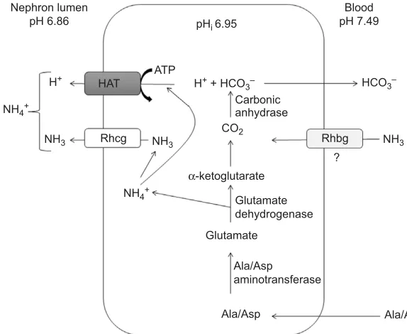
We suggest a model of renal ammonia transport (Fig. 7) involving parallel H+/NH
3 transport independent of Na+ uptake,
similar to the protein arrangement observed in the mammalian CD (Weiner and Verlander, 2014). This is supported by the expression of Rhcg-a, Rhcg-b and HAT mRNA coupled with relatively high activity of renal HAT. In support of this model, previous work has demonstrated the localization of Rhcg-b to the distal tubule and CD in teleosts (see Introduction), with additional weak expression in the proximal tubules, at least in carp (Wright et al., 2014). HAT has been localized to the apical membranes of the proximal tubules (Perry and Fryer, 1997; Ivanis et al., 2008a). HAT activity was not affected by acidosis in the present study (Table 3), but its activity in the goldfish kidney was much higher than previously reported in the goldfish gill (Sinha et al., 2013) where ammonia excretion rates were∼20- to 50-fold greater (Maetz, 1972; Sinha et al., 2013) than observed here. Furthermore, the lower urinary pH during acidosis would tend to inhibit operation of NHE in kidney tubules (Parks et al., 2008), favouring HAT as a more likely route of H+secretion
for NH3trapping under these conditions. Despite the constant HAT
activity (Table 3), HAT mRNA expression decreased during acid exposure (Fig. 6). The cause is unknown; perhaps this represents an energy conservation strategy to cope with the long-term energetic costs of chronic acidosis (Butler et al., 1992; Kalinin and Gesser, 2002; Deigweiher et al., 2010).
Ammonia production
Under acidosis, plasma PNH3 decreased despite the elevation in
plasma [Tamm], thereby reducing available NH3 for
plasma-to-tubule cell ammonia translocation (Table 1). In goldfish, the basal expression level of Rhbg (the basolateral Rh protein) was low relative to the Rhcg proteins and decreased non-significantly during acidosis (Fig. 6), whereas in carp, it decreased significantly during acidosis (Wright et al., 2014). Together, these data suggest that diffusive input of ammonia through the basolateral membranes of tubular cells is potentially limited, and imply that the increased ammonia secreted into the urine during metabolic acidosis (Fig. 5B) was probably produced mainly in the kidney, mediated through renal amino acid catabolism. It remains possible, however, that ammonia may be produced in cells lacking apical Rhcg, and enters basolaterally to some degree into tubular cells possessing apical Rhcg. This possibility has been included in our model (Fig. 7). In other teleost studies, aspartate and alanine have been identified as the primary substrates (King and Goldstein, 1983; Wood et al., 1999). In agreement with this model (Fig. 7), acidotic goldfish exhibited a significant increase in renal alanine aminotransferase
–4 –2 0 2 4 6 8 10
Control GFR
Acid GFR Acid TSR
A
0 1 2 3
4
B
*
*
–1000 –800 –600 –400 –200 0 200 400 600 800
1000
D
Rate (
μ
mol kg
–1
h
–1
)
–15 –10 –5 0 5 10 15
20
C
*
–800 –600 –400 –200 0 200 400 600
800
E
–10 –5 0 5 10 15
20
F
*
[image:6.612.53.478.55.432.2]Control TSR
Fig. 5. Mean glomerular filtration rate and mean tubular secretion rate in the kidney of goldfish exposed to control and low environmental pH over 48 h.
Control, pH 8.2 (white bars); low environmental pH, pH 4.0 (grey bars). Glomerular filtration rate (GFR), solid bars; mean tubular secretion rate (TSR), hatched bars. (A) Fluid (N=5),
(B) ammonia (N≥4), (C) Pi(N=5), (D) Na+(N=5), (E) Cl−(N=5) and (F) urea (measured asμmol N;N≥4). Note that negative net secretion rates represent net reabsorption rates. Means±1 s.e.m. Asterisks (*P<0.01) denote differences between control and acid-exposed fish for a particular parameter.
The
Journal
of
Experimental
activity, and non-significant increases in aspartate aminotransferase (P=0.08) and glutamate dehydrogenase (P=0.06) (Table 3). Similar effects have been reported in mammals (Schoolwerth et al., 1978; Wright et al., 1992; Schroeder et al., 2003; Nowik et al., 2008).
We propose that the goldfish kidney catabolizes glutamate for NH4+synthesis by glutamate dehydrogenase and that alanine and
potentially aspartate, but not glutamine, are the primary substrates (Fig. 7). This conclusion reflects the high levels of both alanine aminotransferase and aspartate aminotransferase seen in both the present study and in that of King and Goldstein (1983), plus the lack of detectable glutaminase activity (Table 3) in the kidney of goldfish. This enzyme has been detected in the kidneys of other teleosts, but at low activity (King and Goldstein, 1983; Wood et al., 1999; Wright et al., 2014). King and Goldstein (1983) also reported that incubation of intact renal tubule cells with alanine or aspartate resulted in a ∼2–4 times greater ammonia production than with glutamine. The transdeamination process that liberates ammonia produces α-ketoglutarate, which is subsequently metabolized via α-ketoglutarate dehydrogenase to yield CO2. This is quickly
converted to H+and HCO
3− through carbonic anhydrase (Wood
et al., 1999; Wright, 1995), with HCO3−moving back to the blood
compartment to help restore the pH imbalance (Fig. 7).
General acid–base and ion responses
Goldfish urinary net acid secretion responses (Fig. 2A) were qualitatively similar to those seen in previous studies on metabolic acidosis in teleosts (McDonald and Wood, 1981; King and Goldstein, 1983; Wood et al., 1999) and mammals (Sartorius et al., 1949; Hills, 1973; Hamm and Simon, 1987), but differ from most in that the increased TA–HCO3−component made a larger
contribution (∼70%; Fig. 2C) than the NH4+component (∼30%;
Fig. 2B). This may simply reflect the fact that the TA–HCO3−
secretion was substantially negative in goldfish under control conditions such that the HCO3− component was lost during
acidosis, giving a larger absolute change. The increase in TA– HCO3−excretion was associated with a large rise in Piexcretion
(Fig. 2D). Unlike ammonia, the urinary Piexcretion, and by proxy
TA–HCO3− excretion (Wheatly et al., 1984), appeared to result
from decreased tubular reabsorption (Fig. 5C) rather than direct secretion of Pi. In mammals (Strickler et al., 1964; Agus et al.,
1971), changes in Pireabsorption similarly regulate the degree of
urinary Piexcretion.
Rhcg-a Rhcg-b Rhbg
HAT NKA UT
*
*
Normalized mRNA
expression
[image:7.612.68.282.53.338.2]0 1 2 3 4 5 0 1 2 3 4 5
Fig. 6. Normalized mRNA expression of various transport proteins found in the kidney of goldfish exposed to control and low environmental pH over 48 h.Control, pH 8.2 (white bars); low environmental pH, pH 4.0 (black bars). Rhcg-a,N=8; Rhcg-b,N=8; Rhbg,N=5; H+-ATPase (HAT),N=8; Na+/K+-ATPase (NKA),N=8; urea transporter (UT),N≥7. Means±1 s.e.m. Asterisks denote a significant difference (*P<0.05) between control and acid-exposed fish.
Glutamate dehydrogenase
Ala/Asp
aminotransferase
Ala/Asp Glutamate
Carbonic anhydrase CO2 NH4+
HCO3– H+ + HCO
3– H+
NH3
NH3 NH3
Ala/Asp NH4+
Rhbg Rhcg
HAT
α-ketoglutarate
? Blood pH 7.49 Nephron lumen
pH 6.86 pH
i 6.95
ATP
Fig. 7. A proposed model for ammonia transport in the kidney of the goldfish under metabolic acidosis.Alanine (Ala) and/or aspartate (Asp) enter the tubule cell and are catalysed via aminotransferases to form glutamate. Glutamate is subsequently catabolized by glutamate dehydrogenase to form α-ketoglutarate, simultaneously liberating ammonia.
α-Ketoglutarate is further metabolized to succinate and CO2via α-ketoglutarate dehydrogenase. Carbonic anhydrase mediates the reaction of the CO2and H2O to form H
+
+HCO3−. Newly synthesized HCO3−is transported to the extracellular fluid while H+is translocated to the urine via the V-type H+-ATPase (HAT) along with the ammonia via Rhesus (Rh) protein Rhcg. It is also possible that ammonia may translocate through the basolateral surface.
The
Journal
of
Experimental
[image:7.612.46.333.499.734.2]The decline of plasma Na+ and Cl− in acid-exposed goldfish
(Table 1) has been seen previously in other acid-exposed teleosts (McDonald et al., 1980; McDonald and Wood, 1981; Ultsch et al., 1981; Fugelli and Vislie, 1982; Wright et al., 2014). It reflects the effect of low pH inhibiting active branchial ion uptake and promoting increased diffusive losses (McDonald and Wood, 1981; Ultsch et al., 1981). Renal Na+and Cl− reabsorption rates
were unaffected by acid exposure (Fig. 5D,E), so there was no renal compensation. Therefore, goldfish appear to acid–base regulate rather than ionoregulate under acidosis, in contrast to carp, as part of the‘acid/base–ion balance compromise’discussed by Wright et al. (2014).
A role for cortisol?
The almost 2-fold increase in plasma cortisol in acid-exposed goldfish (Table 1) may have promoted renal ammoniogenesis by augmenting the supply of amino acids through a stimulation of proteolysis (Milligan, 1997; Wiseman et al., 2007; reviewed in Mommsen et al., 1999), as well as upregulation of ammoniogenic enzymes (Chan and Woo, 1978; Wood et al., 1999; Ortega et al., 2005). Increased cortisol is associated with metabolic acidosis (Brown et al., 1986; Wood et al., 1999) and not respiratory acidosis in fish (Wood and LeMoigne, 1991; Wood et al., 1999), and the former is accompanied by a much larger change in renal ammonia excretion (Wood et al., 1999). Additionally, the hypothalamic-pituitary–interrenal (HPI) axis is believed to play a pivotal role in mediating the expression of Rh glycoproteins (Nawata and Wood, 2009; Tsui et al., 2009) and therefore may be regulating the increased levels of Rhcg-b mRNA observed here. Stress has been implicated in causing diuresis in teleost fish (Hunn, 1982). Indeed, blocking of glucocorticoid receptors significantly reduced UFR in trout (McDonald and Wood, 2004). Here, the stress of surgery and confinement may have contributed to the temporal rise in UFR in controls, as well as in experimental fish (Fig. 1A).
Urea metabolism
The elevation in plasma urea (Table 1) and urinary urea excretion (Fig. 3A) during acidosis probably represents a detoxification mechanism to prevent ammonia toxicity, thereby maintaining circulating [ammonia] within a homeostatic range (Fromm and Gillette, 1968; Olson and Fromm, 1971; Arillo et al., 1981; Mommsen and Walsh, 1992; Ip et al., 2004). This action has been demonstrated previously in goldfish exposed to high environmental ammonia (Olson and Fromm, 1971; Sinha et al., 2013). Renal arginase activity was not affected by acidosis in the present study (Table 3), and thus, urea synthesis probably occurs outside the goldfish kidney. In accordance with this idea, the marked elevation in urinary urea excretion was largely the product of increased glomerular filtration of urea, with the kidney performing urea reabsorption (Fig. 5F) similar to that seen in trout (McDonald and Wood, 1998), a very different situation from the renal handling of ammonia. This conclusion is in accord with the unchanged expression of UT mRNA (Fig. 6), as UT would be involved in urea reabsorption, not urea filtration (Mistry et al., 2005; reviewed in McDonald et al., 2006).
Conclusions
For the first time, we have shown that increased renal ammonia excretion during metabolic acidosis in goldfish is primarily the result of tubular secretion, and the mechanism does not seem to involve a Na+/NH
4+exchange system. We propose that ammonia
transport is probably mediated through a parallel H+/NH
3transport
facilitated by Rhcg-b and HAT in concert with enhanced endogenous renal cell ammonia synthesis from amino acid metabolism (Fig. 7). While this study lays the foundation for understanding teleost renal ammonia transport, further work is needed to address specific sites of renal ammonia secretion as well as the localization of renal transporter proteins.
MATERIALS AND METHODS
All procedures were approved by the McMaster University Animal Research Ethics Board (animal utilization protocol AUP 12-12-45) in accordance with the Canadian Council on Animal Care. Unless otherwise noted, all chemicals were purchased from the Sigma-Aldrich Corporation (Oakville, ON, Canada).
Animal care and cannulation
Goldfish (C. auratus; 33.9±0.8 g) were obtained from Aquality Inc. (Mississauga, ON, Canada) and held at McMaster University at 18–20°C under a 12 h light:12 h dark photoperiod in 200 l tanks served with recirculating filters and dechlorinated, moderately hard Hamilton tapwater (in mequiv l−1): Na+0.6, Cl−0.8, Ca2+1.8, Mg2+0.3, K+0.05; titration alkalinity 2.1, pH∼8.2, hardness∼140 mg l−1as CaCO3equivalents. Fish
were fed to satiation 3 times per week with flaked food (Big Al’s Canada, Woodbridge, ON, Canada), but fasted for 7 days prior to experimentation to avoid confounding effects of feeding on ammonia excretion (Zimmer et al., 2010).
For urinary cannulation, fish were anaesthetized [200 mg l−1tricaine methane sulfonate (MS-222) solution (Syndel Laboratories, Qualicum Beach, BC, Canada) neutralized with KOH] and weighed. During cannulation, the gills were artificially ventilated with a dilute MS-222 solution (100 mg l−1). An indwelling catheter (Clay-Adams PE-50 tubing; Becton, Dickinson and Co., Franklin Lakes, NJ, USA) was placed in the urinary bladder. The technique of Wood and Patrick (1994) was modified such that the distal 2.5 cm of the tubing was bent at 90 deg so as to conform to the fish’s urinary tract. In a subset of fish used for GFR measurements, 1 µCi of [3H]polyethylene glycol-4000 (PEG-4000; Sigma-Aldrich, St Louis, MO, USA) in 140 mmol l−1NaCl was injected into the caudal haemal arch at 0.003 ml g−1fish. PEG-4000 is generally considered to be the best GFR marker in fish (discussed by Wood and Patrick, 1994).
Goldfish were allowed to recover in individual experimental chambers. Each comprised a small plastic box (∼1 l) fitted with a perforated piece of 3.8 cm diameter PVC pipe (to prevent the animal from turning around), an aeration device and an independent water source (1000 ml min−1) from a pH-controlled ( pH 8.2) 250 l reservoir. Urine was collected continuously by gravity (head∼3 cm) into a glass vial.
Experimental protocol
Control fish were exposed to water pH 8.2±0.1, and experimental fish to acidic water ( pH 4.0±0.1) for 48 h. This water was continually pumped through each of the chambers from the pH-controlled reservoir. Water pH was maintained within the desired range using a pH-stat system consisting of a PHM82 pH meter (Radiometer-Copenhagen, Brønshøj, Denmark) coupled with a Radiometer GK24O1C combination glass pH electrode and an auto-titration controller (Radiometer TTT-80) which metered the addition of a 0.1 mol l−1 HCl solution via a solenoid valve into the continuously mixed reservoir.
After at least 18 h of recovery at pH 8.2, exposure to pH 4.0 or 8.2 was initiated (time 0 h). Urine was collected over successive 12 h intervals. UFR, pH and TA–HCO3−were measured immediately, and remaining urine was
frozen at−20°C for subsequent analysis of urinaryTamm, urea, Na+, Cl−, Pi
and [3H]PEG-4000 radioactivity (if applicable).
In the subset of fish used for GFR measurements, it was necessary to quantify the loss rate of [3H]PEG-4000 radioactivity through the gills. This was achieved by stopping the water flow to each chamber from 44 h to 48 h. Mixing was maintained by aeration. As the chambers were isolated from the pH-stat system, the appropriate pH was maintained over this period through manual titration (0.1 mol l−1HCl) in combination with a handheld pH meter (SympHony SP70C, VWR Inc., Edmonton, AB, Canada). At the start and
The
Journal
of
Experimental
end of this closed period, 4 ml water samples were collected and frozen at
−20°C for later analysis of [3H]PEG-4000 radioactivity.
At 48 h, fish were quickly killed with MS-222 (750 mg l−1) that was pH-adjusted with KOH to match the experimental pH. A terminal blood sample was drawn by caudal puncture into a 1 ml syringe rinsed with lithium heparinized (300 mg l−1) Cortland saline (Wolf, 1963), quickly transferred to a 0.5 ml centrifuge tube and blood pH was measured at the experimental temperature (18±1°C) with a microelectrode (Orion PerpHecT ROSS, Thermo Fisher Scientific, Toronto, ON, Canada) coupled to a pH meter (SympHony SP70C, VWR Inc.), taking care to minimize air exposure. The blood was immediately spun at 1500g(1 min). The plasma was decanted and flash-frozen in liquid N2for later analysis of plasmaTamm, urea, Na+,
Cl−, Pi, HCO3−, lactate, glucose, cortisol and [3H]PEG-4000 concentrations.
Lastly, the kidney tissue was removed, and flash-frozen in liquid N2, for later
measurement of enzymatic activity and mRNA expression. Samples were stored at−80°C.
Urinary analyses
UFR was determined gravimetrically. Urine pH was measured as for blood pH. Urinary [TA–HCO3−] was determined within 24 h using
double-endpoint titration (Hills, 1973) and standardized procedures for fish urine (McDonald and Wood, 1981; Wood, 1988). A glass pH electrode (Radiometer-Copenhagen pHC2005-8) coupled to a H160 pH meter (Hach, Mississauga, ON, Canada) and 2 ml microburettes (Gilson, Middleton, WI, USA) filled with standardized solutions, 0.02 mol l−1
HCl or 0.02 mol l−1NaOH, were employed. All samples were titrated first to
below pH 4.0 and aerated with CO2-free air to eliminate HCO3−, then back
to a control blood pH of 8.00 (interpolated from Tzaneva et al., 2011 for arterially cannulated goldfish).
Total urinary ammonia and urea were determined through the
colorimetric salicylate (Verdouw et al., 1978) and monoxime
(Rahmatullah and Boyde, 1980) methods, respectively. Urinary Na+was
determined by flame spectrophotometry (Spectra AA 220FS, Varian, Palo Alto, CA, USA) and Cl−by a mercury thiocyanate-based colorimetric assay (Zall et al., 1956). [3H]PEG-4000 radioactivity was determined by
scintillation counting (Tri-Carb 2900TR Liquid Scintillation Analyzer, PerkinElmer Inc., Waltham, MA, USA). Urine samples (100 µl) and experimental water samples (4 ml) were incubated in a 1:4 ratio (sample: fluor) with Optiphase HiSafe fluor (PerkinElmer Inc.). Total Piin the urine
was measured with a commercial kit (Pointe Scientific, Canton, MI, USA).
Plasma analyses
Whole-blood pH was measured at experimental temperature (18±1°C) using the same pH microelectrode system as for urine. PlasmaTammwas assayed
using a commercial enzymatic kit (Raichem, Cliniqa, San Marcos, CA, USA). Plasma ions and urea were measured as for urinary parameters. Plasma cortisol was determined with a commercial radioimmunoassay kit (Gammacoat, DiaSorin, Mississauga, ON, Canada). Lactate and glucose were measured with a handheld lactate meter (Lactate Pro, Arkray Inc., Kyoto, KP, Japan) and a commercially available reagent kit (Infinity Glucose Hexokinase Liquid Stable Reagent, Thermo Fisher Scientific), respectively. Total plasma bicarbonate was measured by the double-endpoint titration method described earlier using the mean control blood pH ( pH 7.85) as measured in this study by caudal puncture. Plasma [3
H]PEG-4000 radioactivity (100 µl– made up to 1 ml with distilled water) was determined by scintillation counting as described above, but using Optima Gold scintillation fluor (PerkinElmer Inc.; 1:2 sample:fluor ratio). The external standard ratio method was used to quench-correct to the same efficiency as water samples.
Enzymatic analyses
The activities of glutamate dehydrogenase (EC 1.4.1.2), alanine aminotransferase (EC 2.6.1.2), aspartate aminotransferase (EC 2.6.1.1), arginase (EC 3.5.3.1), glutamine synthetase (EC 6.3.1.2) and glutaminase (EC 3.5.1.2) were assayed using a common homogenization buffer. Frozen kidney tissue was weighed to an appropriate mass (∼40 mg) and immediately sonicated on ice in 200 µl of glycerol buffer (50% glycerol,
20 mmol l−1 K2HPO4, 10 mmol l−1 Hepes, 0.5 mmol l−1 EDTA,
1 mmol l−1 DTT; pH 7.5). The homogenate was centrifuged at 4°C at 11,500g for 3 min, and the supernatant was assayed. Specific assay conditions for glutamate dehydrogenase, aspartate aminotransferase, alanine aminotransferase and arginase followed Mommsen et al. (1980) and those for glutamine synthetase and glutaminase were similar to those outlined in Webb and Brown (1976) and Walsh et al. (1990), respectively. NKA (EC 3.6.3.9) and HAT (EC 3.6.3.6) activities were determined according to McCormick (1993) and Nawata et al. (2007), respectively, using a Molecular Devices microplate reader (SpectraMax 340PC, Sunnyvale, CA, USA) at room temperature. Samples were homogenized (Power Gen 125, Thermo Fisher Scientific) in imidazole buffer (50 mmol l−1 imidazole, 125 mmol l−1 sucrose, 5 mmol l−1 EGTA; pH 7.5) at 4°C. Activity was normalized to total protein content as measured with Bradford’s reagent.
Carbonic anhydrase (EC 4.2.1.1) activity was assayed according to Henry (1991). Frozen tissues were homogenized in buffer (10 mmol l−1NaH2PO4,
225 mmol l−1mannitol, 10 mmol l−1Tris, 75 mmol l−1 sucrose; pH 7.4 using H3PO4), and the supernatant obtained by centrifugation (4°C,
11,500g, 1 min). Changes in pH during the reaction were recorded using the Radiometer-Copenhagen pHC2005-8 glass pH electrode and Hach H160 pH meter described earlier. Activity was normalized to total protein content.
Whole-tissue analyses
Frozen kidney tissue was ground under liquid nitrogen with a chilled mortar and pestle. Whole-tissue ammonia was assayed on tissues that had been deproteinized (8% perchloric acid, 1 mmol l−1 EDTA) and returned to
physiological pH (1 mol l−1KOH; pH∼7.5). Samples were centrifuged
(4°C, 11,500g) for 3 min; supernatant was assayed for total ammonia content using the same Raichem kit as for plasma. Whole-tissue lactate was measured on the same supernatant using the lactate:hydrazine sink method of Walsh (1987) but with a slightly modified reaction buffer (0.4 mol l−1
hydrazine, 2 mmol l−1EDTA, 1 mol l−1glycine; pH 9.5).
Intracellular pH was measured according to Pörtner et al. (1990). Tissue powdered under liquid nitrogen (0.1 g) was incubated in buffer (1 ml: 6 mmol l−1sodium nitrilotriacetate, 150 mmol l−1 KF), then centrifuged
(11,500g, 4°C) for 3 min. The same pH microelectrode system, at experimental temperature, was used as for blood pH.
mRNA expression
Total RNA was extracted from previously frozen renal tissue using a TRIzol (Invitrogen, Burlington, ON, Canada)-based extraction method and stored at
−80°C. Samples were then assessed optically for total RNA purity (Nanodrop ND-1000, Nanodrop Technologies, Wilmington, DE, USA) in which the 260 nm/280 nm and 260 nm/230 nm ratios were determined. Samples were acceptable if both ratios were 2.00±0.1. RNA quality was further evaluated by gel electrophoresis (1% agarose). cDNA was generated from total RNA (1 µg) by incubation with SuperScript II reverse transcriptase (Invitrogen), oligo (dT17) primers (Invitrogen) and excess deoxyribonucleotide triphosphate (dNTP). Genomic DNA was removed from each sample using DNase I (Invitrogen). cDNA was stored at−20°C. mRNA transcript expression of candidate genes was ascertained using quantitative real-time RT-PCR (qPCR). Primer sequences were derived from Sinha et al. (2013), A. K. Sinha, H. J. Liew, C. M. Nawata, R. Blust, C.M.W. and G. De Boeck (unpublished) and Bradshaw et al. (2012) (supplementary material Table S1). qPCR reactions consisted of a total volume of 10 µl: 4 µl of cDNA sample, 5 µl of 2× SSoFast EvaGreen Supermix (Bio-Rad Laboratories Inc., Hercules, CA, USA), 0.8 µl (10 µmol l−1) of both the reverse and forward primers (Mobix, Hamilton, ON, Canada) and 0.2 µl of RNase-free water. Reactions were conducted in an RT-PCR unit (CFX Connect Real-Time PCR detection system, Bio-Rad Laboratories Inc.). The following protocol was utilized: polymerase activation (98°C, 2 min), followed by a two-stage amplification (1st: 98°C, 2 s; 2nd: 60°C, 5 s) ×39 cycles. A melt curve was generated by heating samples from 75 to 95°C in 0.2°C increments every 10 s to ensure the production of a single gene product. No-template controls (RNase-free
The
Journal
of
Experimental
water) and no-reverse transcriptase samples were run on every plate. The efficiency of each primer set reaction was determined through a standard curve derived from a pool of all cDNA samples (supplementary material Table S1). mRNA expression data were normalized against the expression of two housekeeping genes, EF1α(elongation factor-1α) andβ-actin, using the GeNorm algorithm (Primer Design Ltd, Southampton, Hampshire, UK; Vandesompele et al., 2002).
Calculations
UFR was calculated as the total urine produced (V) divided by fish mass (m) and the collection duration (t):
UFR¼V=ðmtÞ: ð1Þ
The excretion rate of a metabolite (M) was calculated as the product of UFR and the concentration in the urine ([M]u):
Excretion rate¼UFR½Mu: ð2Þ
Negative values indicate a net efflux of a metabolite via the urine. GFR was calculated as the product of the urinary [3H]PEG-4000
excretion rate for a given period ([PEG-4000]u) divided by the estimated
mean plasma [3H]PEG-4000 ([PEG-4000]
p) for the same period:
GFR¼UFR½PEG-4000u=½PEG-4000p: ð3Þ
Mean plasma [3H]PEG-4000 radioactivity ([PEG-4000]p) represents an
average of the two values bracketing the urine collection interval. Other than terminal measurements (48 h), radioactivity was estimated at each time as follows. First, branchial [3H]PEG-4000 losses (JPEG-4000,gill) were tabulated
as the product of [[3H]PEG-4000] in the water ([PEG-4000H2O]) and the
volume of the flux chamber (VE) while also accounting for the mass of the
fish (m) and the flux time (tflux) (Eqn 4). In each fish,JPEG-4000,gillwas less
than 10% of the urinary efflux, as previously observed in the Amazonian oscar (Wood et al., 2009). The percentage value (X% of measured urinary
3
[H]PEG-4000 efflux) measured for each individual was assumed to be constant over the whole experiment and was used to calculate the particular value ofJPEG-4000,gillasX% of the measured urinary loss rate in each period.
This calculated value ofJPEG-4000,gillwas then used in Eqn 5 to estimate the
absolute branchial loss (PEG-4000gill) for each time frame (tcollect):
JPEG-4000;gill¼ ð½PEG - 4000H2OVEÞ=m=tflux; ð4Þ
PEG - 4000gill¼JPEG4000;gilltcollectm: ð5Þ
The radioactivity in the plasma (in cpm), at any given time point (PEG-4000t), was calculated as the sum of the plasma radioactivity in the next time step (PEG-4000t+1), the branchial loss (PEG-4000gill) and urinary loss
(PEG-4000urine) (Eqn 6). The two plasma values were then averaged and
expressed as a concentration using the extracellular fluid volume (193 ml kg−1fish) given by Munger et al. (1991) (Eqn 7):
PEG - 4000t¼ ðPEG - 4000tþ1þPEG - 4000gillþPEG - 4000urineÞ; ð6Þ
½PEG - 4000p¼ ½ðPEG - 4000tþPEG - 4000tþ1Þ1=2=ð193mÞ: ð7Þ
While GFR values were calculated for individual periods for each fish, values were subsequently averaged across time for each fish for use in the subsequent filtration, secretion and reabsorption calculations.
The filtration rate of a metabolite was calculated as the product of the GFR and concentration in the plasma ([M]p):
Filtration rate¼GFR½Mp: ð8Þ
The secretion rate was calculated as the difference between the excretion and filtration rates:
Secretion rate¼ ðUFR½MuÞ GFR½Mp: ð9Þ
Here, positive values represent net secretion, and negative values net reabsorption.
Net acid excretion is the sum of two components, titratable acid (e.g. Pi−,
organic acids) and non-titratable acid where the latter effectively represents the total ammonia excretion (Hills, 1973). As such, total urinary acid
excretion rate was calculated as the sum of urinary total ammonia (Tamm)
excretion rate and the TA–HCO3−excretion rate.
The dissociation constant ( pK) and the solubility of ammonia (αNH3),
from Cameron and Heisler (1983), at the experimental temperature, were used in ammonia partitioning calculations. Plasma ammonium ion concentrations (NH4+) and the partial pressure of ammonia gas (PNH3) were calculated using
the Henderson–Hasselbalch equation, with blood pH or tissue ( pHi) and the
measured total plasma or tissue ammonia concentration ([Tamm]).
Partial pressures of CO2(PCO2) and HCO3
−concentrations in plasma were
also determined with the Henderson–Hasselbalch equation, using measured blood plasma pH and [HCO3−]pvalues, and apparent dissociation ( pK1) and
solubility constants (αCO2) at the experimental temperature from Boutilier
et al. (1984).
Statistical analyses
Data are reported as means±1 s.e.m. (N) throughout. All statistical analyses were performed using SigmaPlot v10.0 (Systat Software Inc., San Jose, CA, USA). Significance was accepted at 5%. A two-way ANOVA model (factors=time, treatment) combined with a Tukey’s post hoc test was employed for urinary excretion parameters. Differences in plasma parameters, enzymatic activities, and filtration and secretion/reabsorption rates between control and acid-exposed groups were evaluated by Student’s unpairedt-tests.
Acknowledgements
The authors thank Pat Walsh for advice on enzyme measurements, Alex Zimmer for advice on molecular techniques, and two anonymous reviewers for constructive comments.
Competing interests
The authors declare no competing or financial interests.
Author contributions
M.J.L., P.A.W. and C.M.W. designed the project. The experiments, assays and data analysis were performed by M.J.L. with the help of C.M.W. The manuscript was written by M.J.L., and revised by P.A.W. and C.M.W.
Funding
This work was supported by Natural Sciences and Engineering Research Council of Canada (NSERC) Discovery grants to C.M.W. and P.A.W. C.M.W. was supported by the Canada Research Chairs Program. M.J.L. received an Ontario Graduate Scholarship.
Supplementary material
Supplementary material available online at
http://jeb.biologists.org/lookup/suppl/doi:10.1242/jeb.117689/-/DC1
References
Agus, Z. S., Puschett, J. B., Senesky, D. and Goldberg, M.(1971). Mode of action of parathyroid hormone and cyclic adenosine 3′,5′-monophosphate on renal tubular phosphate reabsorption in the dog.J. Clin. Invest.50, 617-626.
Arillo, A., Margiocco, C., Melodia, P., Mensi, P. and Schenone, G.(1981). Ammonia toxicity mechanism in fish: studies on rainbow trout (Salmo gairdneri
Rich.).Ecotoxicol. Environ. Safe.5, 316-328.
Atkinson, D. E.(1992). Functional roles of urea synthesis in vertebrates.Physiol. Zool.65, 243-267.
Biver, S., Belge, H., Bourgeois, S., Van Vooren, P., Nowik, M., Scohy, S., Houillier, P., Szpirer, J., Szpirer, C., Wagner, C. A. et al.(2008). A role for Rhesus factor Rhcg in renal ammonium excretion and male fertility.Nature456, 339-343.
Boutilier, R. G., Heming, T. A. and Iwama, G. K. (1984). Appendix: physicochemical parameters for use in fish physiology. In Fish Physiology Volume X Gills Part A(ed. W. S. Hoar and D. J. Randall), pp. 403-430. Orlando: Academic Press.
Bradshaw, J. C., Kumai, Y. and Perry, S. F.(2012). The effects of gill remodeling on transepithelial sodium fluxes and the distribution of presumptive sodium-transporting ionocytes in goldfish (Carassius auratus).J. Comp. Physiol. B182, 351-366.
Braun, M. H., Steele, S. L., Ekker, M. and Perry, S. F.(2009). Nitrogen excretion in developing zebrafish (Danio rerio): a role for Rh proteins and urea transporters.
Am. J. Physiol. Renal Physiol.296, F994-F1005.
Brown, S. B., Eales, J. G. and Hara, T. J.(1986). A protocol for estimation of cortisol plasma clearance in acid-exposed rainbow trout (Salmo gairdneri).Gen. Comp.
Endocrinol.62, 493-502.
The
Journal
of
Experimental
Butler, P. J., Day, N. and Namba, K.(1992). Interactive effects of seasonal temperature and low pH on resting oxygen uptake and swimming performance of adult brown troutSalmo trutta.J. Exp. Biol.165, 195-212.
Cameron, J. N. and Heisler, N.(1983). Studies of ammonia in the rainbow trout: physico-chemical parameters, acid–base behaviour and respiratory clearance.
J. Exp. Biol.105, 107-125.
Chan, D. K. O. and Woo, N. Y. S.(1978). Effect of cortisol on the metabolism of the eel,Anguilla japonica.Gen. Comp. Endocrinol.35, 205-215.
Cooper, C. A., Wilson, J. M. and Wright, P. A.(2013). Marine, freshwater and aerially acclimated mangrove rivulus (Kryptolebias marmoratus) use different strategies for cutaneous ammonia excretion.Am. J. Physiol. Regul. Integr. Comp. Physiol.304, R599-R612.
Curthoys, N. P.(2001). Role of mitochondrial glutaminase in rat renal glutamine metabolism.J. Nutr.131, 2491S-2495S.
Deigweiher, K., Hirse, T., Bock, C., Lucassen, M. and Pörtner, H. O.(2010). Hypercapnia induced shifts in gill energy budgets of Antarctic notothenioids.
J. Comp. Physiol. B180, 347-359.
Fromm, P. O. and Gillette, J. R.(1968). Effect of ambient ammonia on blood ammonia and nitrogen excretion of rainbow trout (Salmo gairdneri).Comp. Biochem. Physiol.26, 887-896.
Fugelli, K. and Vislie, T.(1982). Physiological response to acid water in brown trout (Salmo trutta L.): cell volume regulation in heart ventricle tissue.J. Exp. Biol.101, 71-82.
Glabman, S., Klose, R. M. and Giebisch, G.(1963). Micropuncture study of ammonia excretion in the rat.Am. J. Physiol.205, 127-132.
Hamm, L. L. and Simon, E. E.(1987). Roles and mechanisms of urinary buffer excretion.Am. J. Physiol. Renal Physiol.253, F595-F605.
Henry, R. P.(1991). Techniques for measuring carbonic anhydrase activity in vitro. InThe Carbonic Anhydrases(ed. S. J. Dodgson, R.E. Tashian, G. Gros and N.D. Carter), pp. 119-125. New York: Springer.
Hills, A. G.(1973).Acid-Base Balance Chemistry, Physiology, Pathophysiology. Baltimore: The Williams & Wilkins Company.
Hung, C. Y. C., Tsui, K. N. T., Wilson, J. M., Nawata, C. M., Wood, C. M. and Wright, P. A. (2007). Rhesus glycoprotein gene expression in the mangrove killifishKryptolebias marmoratusexposed to elevated environmental ammonia levels and air.J. Exp. Biol.210, 2419-2429.
Hunn, J. B.(1982). Urine flow rate in freshwater salmonids: a review.Prog. Fish Cult.44, 119-125.
Hwang, P.-P.(2009). Ion uptake and acid secretion in zebrafish (Danio rerio).J. Exp. Biol.212, 1745-1752.
Inokuchi, M., Hiroi, J., Watanabe, S., Lee, K. M. and Kaneko, T.(2008). Gene expression and morphological localization of NHE3, NCC and NKCC1a in branchial mitochondria-rich cells of Mozambique tilapia (Oreochromis mossambicus) acclimated to a wide range of salinities. Comp. Biochem. Physiol. A Mol. Integr. Physiol.151, 151-158.
Ip, Y. K., Chew, S. F., Wilson, J. M. and Randall, D. J.(2004). Defences against ammonia toxicity in tropical air-breathing fishes exposed to high concentrations of environmental ammonia: a review.J. Comp. Physiol. B174, 565-575.
Ito, Y., Kato, A., Hirata, T., Hirose, S. and Romero, M. F.(2014). Na+/H+and Na+/ NH4+exchange activities of zebrafish NHE3b expressed inXenopusoocytes.
Am. J. Physiol. Regul. Integr. Comp. Physiol.306, R315-R327.
Ivanis, G., Braun, M. and Perry, S. F.(2008a). Renal expression and localization of SLC9A3 sodium/hydrogen exchanger and its possible role in acid-base regulation in freshwater rainbow trout (Oncorhynchus mykiss).Am. J. Physiol. Regul. Integr. Comp. Physiol.295, R971-R978.
Ivanis, G., Esbaugh, A. J. and Perry, S. F.(2008b). Branchial expression and localization of SLC9A2 and SLC9A3 sodium/hydrogen exchangers and their possible role in acid-base regulation in freshwater rainbow trout (Oncorhynchus mykiss).J. Exp. Biol.211, 2467-2477.
Javelle, A., Lupo, D., Li, X.-D., Merrick, M., Chami, M., Ripoche, P. and Winkler, F. K.(2007). Structural and mechanistic aspects of Amt/Rh proteins.J. Struct. Biol.158, 472-481.
Kalinin, A. and Gesser, H.(2002). Oxygen consumption and force development in turtle and trout cardiac muscle during acidosis and high extracellular potassium.
J. Comp. Physiol. B Biochem. Syst. Environ. Physiol.172, 145-151.
King, P. A. and Goldstein, L.(1983). Renal ammonia excretion and production in goldfish,Carassius auratus, at low environmental pH.Am. J. Physiol. Regul. Integr. Comp. Physiol.245, R590-R599.
Knepper, M. A. and Agre, P.(2004). Structural biology: the atomic architecture of a gas channel.Science305, 1573-1574.
Knepper, M. A., Packer, R. and Good, D. W.(1989). Ammonium transport in the kidney.Physiol. Rev.69, 179-249.
Kumai, Y. and Perry, S. F.(2011). Ammonia excretion via Rhcg1 facilitates Na+ uptake in larval zebrafish,Danio rerio, in acidic water.Am. J. Physiol. Regul. Integr. Comp. Physiol.301, R1517-R1528.
Kwong, R. W. M., Kumai, Y. and Perry, S. F.(2014). The physiology of fish at low pH: the zebrafish as a model system.J. Exp. Biol.217, 651-662.
Lee, H.-W., Verlander, J. W., Bishop, J. M., Igarashi, P., Handlogten, M. E. and Weiner, I. D.(2009). Collecting duct-specific Rh C glycoprotein deletion alters
basal and acidosis-stimulated renal ammonia excretion.Am. J. Physiol. Renal Physiol.296, F1364-F1375.
Lee, H.-W., Verlander, J. W., Bishop, J. M., Nelson, R. D., Handlogten, M. E. and Weiner, I. D.(2010). Effect of intercalated cell-specific Rh C glycoprotein deletion on basal and metabolic acidosis-stimulated renal ammonia excretion.
Am. J. Physiol. Renal Physiol.299, F369-F379.
Li, S., Kato, A., Takabe, S., Chen, A.-P., Romero, M. F., Umezawa, T., Nakada, T., Hyodo, S. and Hirose, S.(2013). Expression of a novel isoform of Na+/H+ exchanger 3 in the kidney and intestine of banded houndshark,Triakis scyllium.
Am. J. Physiol. Regul. Integr. Comp. Physiol.304, R865-R876.
Linnaeus, C.(1758).Systema Naturae, Vol. 1, 10th edn. Holmiae: Salvii.
Macknight, A. D. C., Macknight, J. M. and Robinson, J. R.(1962). The effect of urinary output upon the excretion of‘ammonia’in man.J. Physiol.163, 314-323.
Maetz, J.(1972). Branchial sodium exchange and ammonia excretion in the goldfish
Carassius auratus. Effects of ammonia loading and temperature changes.J. Exp. Biol.56, 601-620.
McCormick, S. D.(1993). Methods for nonlethal gill biopsy and measurement of Na+, K+ATPase activity.Can. J. Fish. Aquat. Sci.50, 656-658.
McDonald, D. G.(1983). The interaction of environmental calcium and low pH on the physiology of the rainbow trout,Salmo gairdneri: I. branchial and renal net ion and H+ fluxes.J. Exp. Biol.102, 123-140.
McDonald, D. G. and Wood, C. M.(1981). Branchial and renal acid and ion fluxes in the rainbow trout,Salmo gairdneri, at low environmental pH.J. Exp. Biol.93, 101-118.
McDonald, M. D. and Wood, C. M.(1998). Reabsorption of urea by the kidney of the freshwater rainbow trout.Fish Physiol. Biochem.18, 375-386.
McDonald, M. D. and Wood, C. M.(2004). The effect of chronic cortisol elevation on urea metabolism and excretion in the rainbow trout (Oncorhynchus mykiss).
J. Comp. Physiol. B Biochem. Syst. Environ. Physiol.174, 71-81.
McDonald, D. G., Hobe, H. and Wood, C. M.(1980). The influences of calcium on the physiological responses of the rainbow trout, Salmo gairdneri, to low environmental pH.J. Exp. Biol.88, 109-132.
McDonald, M. D., Smith, C. P. and Walsh, P. J.(2006). The physiology and evolution of urea transport in fishes.J. Membr. Biol.212, 93-107.
Milligan, C. L.(1997). The role of cortisol in amino acid mobilization and metabolism following exhaustive exercise in rainbow trout (Oncorhynchus mykiss Walbaum).
Fish Physiol. Biochem.16, 119-128.
Mistry, A. C., Chen, G., Kato, A., Nag, K., Sands, J. M. and Hirose, S.(2005). A novel type of urea transporter, UT-C, is highly expressed in proximal tubule of seawater eel kidney.Am. J. Physiol. Renal Physiol.288, F455-F465.
Mommsen, T. P. and Walsh, P. J. (1992). Biochemical and environmental perspectives on nitrogen metabolism in fishes.Experientia48, 583-593.
Mommsen, T. P., French, C. J. and Hochachka, P. W.(1980). Sites and patterns of protein and amino acid utilization during the spawning migration of salmon.
Can. J. Zool.58, 1785-1799.
Mommsen, T. P., Vijayan, M. M. and Moon, T. W.(1999). Cortisol in teleosts: dynamics, mechanisms of action, and metabolic regulation.Rev. Fish. Biol. Fish. 9, 211-268.
Munger, R. S., Reid, S. D. and Wood, C. M.(1991). Extracellular fluid volume measurements in tissues of the rainbow trout (Oncorhynchus mykiss)in vivoand their effects on intracellular pH and ion calculations.Fish Physiol. Biochem.9, 313-323.
Nakada, T., Hoshijima, K., Esaki, M., Nagayoshi, S., Kawakami, K. and Hirose, S.(2007a). Localization of ammonia transporter Rhcg1 in mitochondrion-rich cells of yolk sac, gill, and kidney of zebrafish and its ionic strength-dependent expression.Am. J. Physiol. Regul. Integr. Comp. Physiol.293, R1743-R1753.
Nakada, T., Westhoff, C. M., Kato, A. and Hirose, S.(2007b). Ammonia secretion from fish gill depends on a set of Rh glycoproteins.FASEB J.21, 1067-1074.
Nawata, C. M. and Wood, C. M. (2009). mRNA expression analysis of the physiological responses to ammonia infusion in rainbow trout.J. Comp. Physiol. B 179, 799-810.
Nawata, C. M., Hung, C. C. Y., Tsui, T. K. N., Wilson, J. M., Wright, P. A. and Wood, C. M.(2007). Ammonia excretion in rainbow trout (Oncorhynchus mykiss): evidence for Rh glycoprotein and H+-ATPase involvement.Physiol. Genomics31, 463-474.
Nawata, C. M., Wood, C. M. and O’Donnell, M. J. (2010). Functional characterization of Rhesus glycoproteins from an ammoniotelic teleost, the rainbow trout, using oocyte expression and SIET analysis.J. Exp. Biol.213, 1049-1059.
Nowik, M., Lecca, M. R., Velic, A., Rehrauer, H., Brändli, A. W. and Wagner, C. A.
(2008). Genome-wide gene expression profiling reveals renal genes regulated during metabolic acidosis.Physiol. Genomics32, 322-334.
Olson, K. R. and Fromm, P. D.(1971). Excretion of urea by two teleosts exposed to different concentrations of ambient ammonia.Comp. Biochem. Physiol. A Physiol. 40, 999-1007.
Orloff, J. and Berliner, R. W.(1956). The mechanism of the excretion of ammonia in the dog.J. Clin. Invest.35, 223-235.
Ortega, V. A., Renner, K. J. and Bernier, N. J.(2005). Appetite-suppressing effects of ammonia exposure in rainbow trout associated with regional and temporal
The
Journal
of
Experimental
activation of brain monoaminergic and CRF systems. J. Exp. Biol. 208, 1855-1866.
Parks, S. K., Tresguerres, M. and Goss, G. G.(2008). Theoretical considerations underlying Na+ uptake mechanisms in freshwater fishes. Comp. Biochem. Physiol. C148, 411-418.
Perry, S. F. and Fryer, J. N.(1997). Proton pumps in the fish gill and kidney.Fish Physiol. Biochem.17, 363-369.
Pörtner, H. O., Boutilier, R. G., Tang, Y. and Toews, D. P.(1990). Determination of intracellular pH and PCO2after metabolic inhibition by fluoride and nitrilotriacetic acid.Respir. Physiol.81, 255-273.
Rahmatullah, M. and Boyde, T. R. C.(1980). Improvements in the determination of urea using diacetyl monoxime; methods with and without deproteinisation.Clin. Chim. Acta107, 3-9.
Sajo, I. M., Goldstein, M. B., Sonnenberg, H., Stinebaugh, B. J., Wilson, D. R. and Halperin, M. L.(1981). Sites of ammonia addition to tubular fluid in rats with chronic metabolic acidosis.Kidney Int.20, 353-358.
Sartorius, O. W., Roemmelt, J. C. and Pitts, R. F.(1949). The renal regulation of acid-base balance in man. IV. The nature of the renal compensations in ammonium chloride acidosis.J. Clin. Invest.28, 423-439.
Schoolwerth, A. C., Nazar, B. L. and LaNoue, K. F. (1978). Glutamate dehydrogenase activation and ammonia formation by rat kidney mitochondria.
J. Biol. Chem.253, 6177-6183.
Schroeder, J. M., Liu, W. and Curthoys, N. P.(2003). pH-responsive stabilization of glutamate dehydrogenase mRNA in LLC-PK1-F+cells.Am. J. Physiol. Renal Physiol.285, F258-F265.
Shih, T.-H., Horng, J.-L., Hwang, P.-P. and Lin, L.-Y.(2008). Ammonia excretion by the skin of zebrafish (Danio rerio) larvae.Am. J. Physiol. Cell. Physiol.295, C1625-C1632.
Shih, T.-H., Horng, J.-L., Liu, S.-T., Hwang, P.-P. and Lin, Y.-H.(2012). Rh